XRD Identification of TiO2 Polymorphs: A Comprehensive Guide for Anatase, Rutile, and Brookite Analysis
This article provides a comprehensive guide for researchers and scientists on identifying the polymorphs of Titanium Dioxide (TiO2)—Anatase, Rutile, and Brookite—using X-ray Diffraction (XRD).
XRD Identification of TiO2 Polymorphs: A Comprehensive Guide for Anatase, Rutile, and Brookite Analysis
Abstract
This article provides a comprehensive guide for researchers and scientists on identifying the polymorphs of Titanium Dioxide (TiO2)—Anatase, Rutile, and Brookite—using X-ray Diffraction (XRD). It covers the foundational crystal structures and properties of each phase, detailed methodologies for XRD sample preparation and analysis, troubleshooting for common issues like phase overlap and preferred orientation, and validation techniques for pure and mixed-phase samples. The content is tailored to support advanced material characterization in fields ranging from photocatalysis and drug development to energy applications, emphasizing practical protocols for accurate phase identification and quantification.
Understanding TiO2 Polymorphs: Crystal Structures, Properties, and Phase Stability
Fundamental Properties and Structural Differences
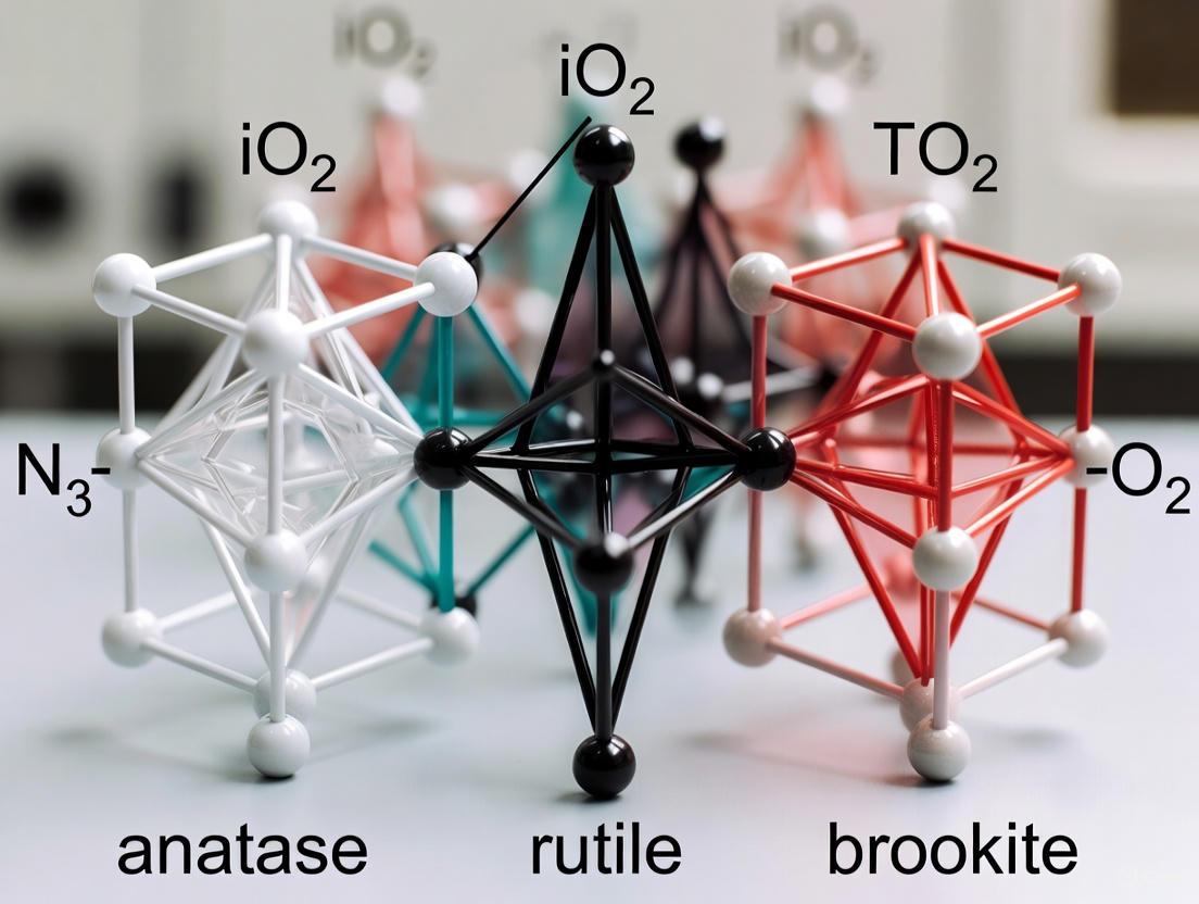
Titanium Dioxide (TiO₂) is a pivotal semiconductor material that exists naturally in three primary crystalline forms, or polymorphs: anatase, rutile, and brookite [1]. A fourth synthetic polymorph, TiO₂(B), also exists but is less common [1]. These polymorphs are all based on TiO₆ octahedra but differ in how these octahedral units are distorted and assembled, leading to distinct crystal structures and properties [2]. Rutile is the thermodynamically stable phase, while both anatase and brookite are metastable and can transform to rutile at elevated temperatures [3].
The table below summarizes the key characteristics of these polymorphs:
| Property | Anatase | Rutile | Brookite |
|---|---|---|---|
| Crystal System | Tetragonal [1] | Tetragonal [1] | Orthorhombic [1] |
| Space Group | I4₁/amd [1] | P4₂/mnm [1] | Pbca [1] |
| Density (g/cm³) | 3.89 [1] | 4.25 [1] | 4.12 [1] |
| Band Gap Energy (eV) | 3.20 – 3.23 [1] | 3.02 – 3.04 [1] | 3.14 – 3.31 [1] |
| General Photocatalytic Activity | Often considered the most active single phase [1] [3] | Generally lower activity; high charge carrier recombination [4] | Highly dependent on specific surface area; can be comparable to anatase [4] |
Photocatalytic Performance and Mechanisms
The photocatalytic activity of TiO₂ polymorphs is primarily governed by their ability to generate and separate photo-excited charge carriers—electrons (e⁻) and holes (h⁺) [1]. When a photon with energy equal to or greater than the material's band gap is absorbed, it excites an electron from the valence band (VB) to the conduction band (CB), creating a hole in the valence band [1] [3]. These charge carriers then migrate to the surface to drive redox reactions, such as the formation of superoxide radicals (•O₂⁻) from oxygen and hydroxyl radicals (•OH) from water [1] [3].
The key difference in activity between the polymorphs lies in the dynamics of these charge carriers. Anatase, an indirect band gap semiconductor, typically exhibits a longer charge carrier lifetime, which allows more time for the holes to participate in surface reactions [4]. Brookite possesses shallow electron traps, which also extends the "lifetime" of the generated holes, making it efficient in systems where this is the limiting factor [4]. In contrast, rutile has deep electron traps, leading to a high recombination rate of electrons and holes, which diminishes its photocatalytic efficiency [4].
Experimental Characterization by X-ray Diffraction (XRD)
X-ray Diffraction (XRD) is an indispensable tool for identifying and quantifying the phases present in a TiO₂ sample. Each polymorph has a unique crystal structure that produces a characteristic diffraction pattern.
The table below outlines the primary diagnostic peaks for each polymorph, which can be used for their identification in XRD research:
| Polymorph | Major XRD Peaks (2θ) | Corresponding Lattice Planes (hkl) |
|---|---|---|
| Anatase | 25.4° [2] | (011) [2] |
| 48.2° [2] | (020) [2] | |
| Rutile | 27.4° [2] | (110) [2] |
| 36.1° [2] | (101) [2] | |
| Brookite | 25.3° [2] | (120) [2] |
| 30.8° [2] | (121) [2] |
For accurate phase identification, it is crucial to use the full diffraction pattern and reference databases like the Inorganic Crystal Structure Database (ICSD) [2]. The phase composition in mixed-phase samples can be quantified using the relative intensity ratios of these characteristic peaks through reference intensity ratio (RIR) methods or more advanced Rietveld refinement.
Synthesis Methods and Phase Control
The synthesis of TiO₂ polymorphs can be achieved through various methods, with the choice of technique and parameters significantly influencing the resulting phase, crystallite size, and morphology.
The flowchart above illustrates a generalized hydrolytic synthesis pathway, such as the sol-gel or hydrothermal method [2] [5]. Key synthesis parameters include:
- pH: A higher pH (e.g., 1 to 3) favors anatase formation, while an intermediate pH (~0.7-0.8) promotes brookite. A lower pH, combined with high titanium precursor concentration, leads to rutile formation [5].
- Calcination Temperature: This is a critical postsynthesis parameter. Lower temperatures (<500°C) often yield anatase or anatase-brookite mixtures with high surface area. Higher temperatures (650–700°C) promote the transformation to the stable rutile phase [3] [6].
The Superiority of Mixed-Phase (Heterophase) Systems
A significant advancement in the field is the use of mixed-phase TiO₂, which often demonstrates superior photocatalytic performance compared to its single-phase counterparts [1] [2]. The enhanced activity is attributed to the formation of a heterojunction at the interface between different polymorphs, which facilitates the efficient transfer of photo-generated electrons from one phase to another [1]. This process leads to a more effective separation of electrons and holes, thereby drastically reducing their recombination rate and increasing the quantum efficiency of the photocatalytic process [1] [2].
Common and effective mixed-phase systems include:
- Anatase/Rutile (A/R): The most famous example is the commercial photocatalyst Evonik P25 (~80% anatase, ~20% rutile), known for its excellent activity [1] [5].
- Anatase/Brookite (A/B): This combination has shown high activity for reactions like the degradation of methyl orange and recovery of silver from industrial effluent [5] [6].
- Triphase (A/R/B): Recent studies show that a combination of all three polymorphs can be even more effective. For instance, a triphase TiO₂ with 76% anatase, 7% rutile, and 17% brookite demonstrated a 75.4% degradation efficiency of metformin, significantly higher than single or biphasic catalysts [2].
The Scientist's Toolkit: Key Research Reagents and Materials
| Reagent/Material | Function in TiO₂ Polymorph Research | Example Use Case |
|---|---|---|
| Titanium(IV) Isopropoxide (TTIP) | A common alkoxide precursor for sol-gel synthesis of TiO₂ nanoparticles [2]. | Synthesis of triphase anatase-rutile-brookite via ultrasound-assisted sol-gel [2]. |
| Titanium Tetrachloride (TiCl₄) | An inorganic precursor used in hydrolytic synthesis routes [7]. | Green synthesis of anatase TiO₂ nanoparticles using plant extracts [7]. |
| Ethanol / Isopropanol | Solvents for titanium precursors; also used as sacrificial hole scavengers in photocatalytic reactions [2] [6]. | Used in photocatalytic silver recovery to consume holes, enhancing electron availability for reduction reactions [6]. |
| Nitric Acid (HNO₃) / Sodium Hydroxide (NaOH) | pH control agents during synthesis, critical for directing the nucleation and growth of specific polymorphs [2] [5]. | Tuning the anatase-rutile-brookite ratio in sol-gel synthesis [2]. |
| Evonik P25 | A commercial benchmark mixed-phase (80% A / 20% R) photocatalyst used for comparative activity studies [1] [5]. | Served as a reference material to evaluate the performance of newly synthesized mixed-phase catalysts [5]. |
Titanium dioxide (TiO₂) is a material of paramount importance in materials science, with its photocatalytic, photovoltaic, and sensing properties heavily dependent on its crystalline phase. The three primary polymorphs—anatase, rutile, and brookite—exhibit distinct crystal structures and lattice parameters that directly influence their functional characteristics. For researchers working on polymorph identification, understanding these structural differences is fundamental. This guide provides a comparative analysis of TiO₂ polymorphs, focusing on their crystal systems, lattice parameters, and the experimental methodologies used for their characterization, particularly through X-ray diffraction (XRD). The ability to accurately distinguish between these phases is critical for optimizing TiO₂-based materials for applications in environmental remediation, energy generation, and sensing technologies [8] [9].
Comparative Structural Analysis of TiO₂ Polymorphs
The crystalline phase of TiO₂ determines its electronic and optical properties, making accurate phase identification a crucial step in material development. The following sections provide a detailed comparison of the primary TiO₂ polymorphs.
Crystal Structures and Lattice Parameters
Table 1: Structural Parameters of TiO₂ Polymorphs
| Polymorph | Crystal System | Space Group | Lattice Parameters (nm) | Density (g/cm³) | Band Gap (eV) |
|---|---|---|---|---|---|
| Anatase | Tetragonal | I4₁/amd | a = 0.3785, c = 0.9514 [10] | ~3.9 | 3.2 [10] |
| Rutile | Tetragonal | P4₂/mnm | a = 0.458, c = 0.295 [10] | ~4.2 | 3.0 [10] |
| Brookite | Orthorhombic | Pbca | a = 0.916, b = 0.544, c = 0.514* | ~4.1 | ~3.3-3.4 |
Note: Brookite parameters are representative values from established literature, as they were not explicitly detailed in the search results.
Anatase and rutile, both tetragonal, are the most studied phases. Anatase has a more open structure with a larger c parameter compared to its a parameter, while rutile has a more compact structure with a smaller c parameter. Brookite's orthorhombic structure is more complex, with three unequal lattice parameters. Anatase is generally preferred for photocatalytic applications due to its higher conduction band edge and lower charge carrier recombination rate, whereas rutile's narrower band gap allows for better visible light absorption [10]. The band gap is not a fixed property and can be engineered; for instance, co-doping with Al³⁺/Al²⁺ and S⁶⁺ ions can reduce the band gap of anatase from 3.23 eV to as low as 1.98 eV, enhancing visible-light activity [9].
X-ray Diffraction (XRD) Fingerprints
XRD is the primary technique for phase identification and quantification. Each polymorph produces a characteristic diffraction pattern.
Table 2: Characteristic XRD Peaks for TiO₂ Polymorphs
| Polymorph | Primary Peaks (hkl indices) | Key Distinguishing Features |
|---|---|---|
| Anatase | (101), (004), (200), (105), (211) [10] | The (101) peak is the most intense. The (004) peak intensity can increase with film thickness and preferred orientation [10]. |
| Rutile | (110), (101), (111), (211) | The (110) peak is the most intense. |
| Brookite | (120), (111), (121) | The (120) peak is often the most intense. Overlap with anatase and rutile peaks can make identification challenging. |
For mixed-phase samples, the ratio of the intensities of the anatase (101) and rutile (110) peaks is commonly used to evaluate their relative proportions [10]. It is important to note that structural defects, microstrain, and crystallite size can cause peak broadening and shifts in angular position, complicating analysis [8] [10]. For example, compressive stress in thin films can shift anatase peaks to higher angles, indicating lattice distortion [10].
Experimental Protocols for Polymorph Identification
A multi-technique approach is essential for reliable characterization of TiO₂ nanostructures, as each method provides complementary information [8].
Sample Preparation and Synthesis
TiO₂ nanostructures can be synthesized via various methods, including liquid phase deposition (LPD) [11], reactive magnetron sputtering [10], and hydrothermal synthesis [9]. The LPD method, for instance, involves a two-step process to achieve uniform films. A precursor solution is first prepared by reacting TiO₂ powder with hydrofluoric acid (HF) to form hexafluorotitanic acid (H₂TiF₆). This solution is then deposited onto substrates like fluorine-doped tin oxide (FTO) glass. Post-deposition annealing (e.g., at 450°C, 550°C, or 650°C) is critical for crystallizing the amorphous films into specific polymorphs and can significantly influence functional properties like UV photoresponse [11].
Multi-Technique Characterization Workflow
The following diagram illustrates a standard workflow for the comprehensive characterization of TiO₂ polymorphs.
Characterization Workflow for TiO₂ Polymorphs
- X-ray Diffraction (XRD): This is the first step for phase identification. XRD determines the crystal structure, identifies present polymorphs, and estimates crystallite size using the Scherrer equation or Williamson-Hall analysis. It can also quantify microstrain and dislocation density, which often correlate inversely with crystallite size [8].
- Raman Spectroscopy: This technique provides complementary phase confirmation based on the unique vibrational fingerprints of each polymorph. It is highly sensitive to local symmetry and can detect phases that might be present in amounts too small for XRD detection. Raman spectra also reveal peak broadening and shifts induced by lattice strain from dopants [8] [9].
- Electron Microscopy (SEM/TEM): These techniques visualize the morphology, grain size, and distribution of particles. A consistent dimensional hierarchy is observed: crystallite size (XRD) < grain size (TEM) < particle size (SEM). The deviations between these sizes can range from ~3% to over 130%, influenced by synthesis temperature and agglomeration [8].
- UV-Vis Spectroscopy: This method determines the optical band gap, a key property for applications in photocatalysis and photodetectors, via Tauc plot analysis. For pure anatase, this is typically around 3.2 eV, but it can be modulated by doping or lattice strain [11] [10] [9].
The Scientist's Toolkit: Essential Research Reagents and Materials
Table 3: Key Reagents and Materials for TiO₂ Research
| Item | Function/Application | Example from Literature |
|---|---|---|
| Fluorine-doped Tin Oxide (FTO) Glass | Conductive substrate for thin film deposition and device fabrication. | Used as a transparent substrate for LPD-synthesized TiO₂ thin films for UV photodetectors [11]. |
| Hydrofluoric Acid (HF) | Reactant for precursor formation in solution-based synthesis. | Used in a 1:6 molar ratio with TiO₂ powder to create the H₂TiF₆ precursor for LPD [11]. |
| Titanium Target (99.6% pure) | Source material for physical vapor deposition methods. | Used in reactive DC magnetron sputtering to deposit TiO₂ coatings on glass fiber wafers [10]. |
| Aluminum and Sulfur precursors | Dopant sources for band gap engineering and modifying phase stability. | Aluminum nitrate nonahydrate and sodium sulfate were used as sources of Al³⁺/Al²⁺ and S⁶⁺ to reduce the band gap and enhance visible-light photocatalysis [9]. |
| Computational Tools (e.g., XRDlicious) | Online calculation of theoretical diffraction patterns from crystal structures for comparison with experimental data. | A web-based tool for calculating powder XRD patterns from crystal structures, useful for phase identification and educational purposes [12]. |
Advanced Analysis: Phase Stability and Transformation
The stability and transformation between anatase and rutile are critical for material performance. The anatase-to-rutile transformation is influenced by factors such as particle size, microstrain, and the presence of dopants [8]. Introducing dopants like Al³⁺ and S⁶⁺ can induce oxygen vacancies and alter phase stability by reducing the transformation energy, thereby facilitating the transition from anatase to rutile [9]. Furthermore, lattice strain, often present in thin films, can significantly impact material properties. For anatase TiO₂, a direct relationship exists between lattice volume strain (∆V) and band gap energy (∆Eg), described by ∆Eg (eV) = -6∆V. Compressive strain (negative ∆V) leads to an increase in band gap energy [10]. This principle allows for the tuning of the optoelectronic properties of TiO₂ thin films for specific applications.
The distinct crystal systems and lattice parameters of TiO₂ polymorphs define their unique structural and functional properties. Anatase, with its larger c lattice parameter and band gap of ~3.2 eV, is often preferred for photocatalysis, while rutile's narrower band gap allows for broader light absorption. Accurate identification requires a multi-technique approach that combines XRD for primary phase analysis, Raman spectroscopy for confirmation, electron microscopy for morphology, and UV-Vis for optical properties. Understanding factors such as dopant-induced phase transitions, lattice strain, and the effects of post-synthesis treatments like annealing is essential for tailoring TiO₂ nanomaterials for enhanced performance in environmental and energy applications.
Within titanium dioxide (TiO₂) research, the precise identification of its primary polymorphs—anatase, rutile, and brookite—is a fundamental step in materials science and catalysis. These polymorphs, though chemically identical, possess distinct crystal structures that impart unique physical and electronic properties. Understanding these differences is crucial for selecting the appropriate TiO₂ phase for specific applications, from photocatalysis to pigment development. This guide provides a structured comparison of the key physical properties of these polymorphs, supported by experimental data and methodologies relevant for researcher-level analysis.
Comparative Physical Properties of TiO₂ Polymorphs
The following table summarizes the key differentiating physical properties of the main TiO₂ polymorphs, collated from experimental data.
Table 1: Key Physical Properties of TiO₂ Polymorphs
| Property | Anatase | Rutile | Brookite |
|---|---|---|---|
| Crystal System | Tetragonal [13] [14] | Tetragonal [13] [14] | Orthorhombic [13] [14] |
| Density (g/cm³) | 3.89 [13] [14] | 4.25 [13] [14] | 4.12 [13] [14] |
| Band Gap Energy (eV) | 3.20 - 3.23 [13] [14] | 3.02 - 3.04 [13] [14] | 3.14 - 3.31 [13] [14] |
| General Photocatalytic Activity | High [13] [14] | Variable, often lower [4] [13] | Comparable to anatase (with adequate surface area) [4] |
Note: A comprehensive, quantitative value for Mohs hardness was not explicitly available in the search results for all polymorphs.
Essential Experimental Protocols for Polymorph Identification
A critical aspect of working with TiO₂ is the accurate identification and characterization of its phases. The following experiments are central to this process.
X-Ray Diffraction (XRD) for Phase Identification
XRD is the primary technique for identifying and quantifying TiO₂ polymorphs based on their unique crystal structures.
- Objective: To unambiguously identify the presence of anatase, rutile, and/or brookite phases in a sample and estimate crystallite size.
- Key Reagents: The sample of TiO₂ nanoparticles or powder.
- Methodology:
- The TiO₂ sample is irradiated with a monochromatic X-ray beam, and the intensity of the diffracted rays is measured as a function of the diffraction angle (2θ).
- The resulting diffraction pattern is compared to standard reference patterns for anatase, rutile, and brookite. Each phase has a unique set of characteristic peaks.
- The average crystallite size can be estimated from the peak broadening using the Scherrer equation [15]. More advanced models like the Williamson-Hall plot and the Halder-Wagner Model can further refine size estimates and also compute intrinsic parameters like strain and energy density [15].
- Expected Outcome: A diffraction pattern where peak positions and intensities confirm the identity of the polymorph(s) present. The crystallite size is typically in the nanometer range for high-activity photocatalysts [15] [4].
Band Gap Determination via Diffuse Reflectance Spectroscopy (DRS)
The optical band gap is a decisive property for photocatalytic applications and can be determined using DRS.
- Objective: To determine the band gap energy of TiO₂ samples.
- Key Reagents: The sample of TiO₂ powder.
- Methodology:
- The TiO₂ sample is placed in a spectrometer equipped with an integrating sphere to measure its diffuse reflectance spectrum.
- The reflectance data is converted to a Kubelka-Munk function.
- The band gap energy is estimated by plotting (F(R) * hν)^n against the photon energy (hν), where n depends on the nature of the optical transition (direct or indirect). The extrapolation of the linear region of the plot to the x-axis gives the band gap value.
- Expected Outcome: Experimental band gap values that align with the known ranges for the identified polymorphs (e.g., ~3.2 eV for anatase, ~3.0 eV for rutile) [13] [14]. Doping with elements like Al and S can reduce the band gap to as low as 1.98 eV, enhancing visible-light absorption [9].
Visualizing the Phase Transition
The transformation between TiO₂ polymorphs is a thermally driven process. The following diagram illustrates the typical pathway from the metastable anatase phase to the stable rutile phase, a transition directly observed via in-situ TEM [16].
Diagram Title: Anatase to Rutile Phase Transition Pathway.
The Scientist's Toolkit: Key Research Reagents
Table 2: Essential Reagents for TiO₂ Polymorph Synthesis and Doping
| Reagent | Function in Research |
|---|---|
| Titanium Tetraisopropoxide (TTIP) | A common titanium alkoxide precursor used in sol-gel and electrospinning synthesis of TiO₂ nanostructures [16]. |
| Titanium(III) Chloride Hexahydrate | A titanium salt used as a precursor in hydrothermal synthesis methods [9]. |
| Polyvinylpyrrolidone (PVP) | A polymer used as a structure-directing agent and to control viscosity in electrospinning of TiO₂ nanofibers [16]. |
| Aluminum Nitrate Nonahydrate | A source of Al³⁺ ions used as a dopant to modify TiO₂'s phase stability, introduce oxygen vacancies, and modulate its band gap [9]. |
| Thiourea | A source of S⁶⁺ ions used as a non-metal dopant, typically co-doped with metals to enhance visible-light absorption by band gap narrowing [9]. |
| Sodium Hydroxide (NaOH) | Used as a precipitating agent in hydrothermal synthesis to control the pH and facilitate the formation of TiO₂ nanoparticles [9]. |
Thermodynamic Stability and Irreversible Phase Transitions
Titanium dioxide (TiO₂) is a material of paramount importance in numerous scientific and industrial applications, from photocatalysis to drug development. Its functionality is intrinsically governed by the stability and interactions between its primary crystalline polymorphs: anatase, rutile, and brookite. A deep understanding of their thermodynamic stability and the often irreversible nature of phase transitions is critical for designing materials with tailored properties. This guide objectively compares the stability and transition behaviors of these polymorphs, framing the discussion within the broader context of identifying and characterizing them via X-ray diffraction (XRD) and other analytical techniques. The comparative data and experimental protocols herein are designed to assist researchers in predicting material behavior under thermal stress and synthesizing stabilized polymorphic configurations for advanced applications.
Fundamental Properties of TiO₂ Polymorphs
The three main polymorphs of TiO₂—anatase, rutile, and brookite—differ in their crystal structure, thermodynamic stability, and electronic properties. These fundamental differences dictate their practical applications and behavior under thermal treatment.
Table 1: Fundamental Characteristics of TiO₂ Polymorphs [2] [13]
| Property | Anatase | Rutile | Brookite |
|---|---|---|---|
| Crystal System | Tetragonal | Tetragonal | Orthorhombic |
| Thermodynamic Stability | Metastable | Stable (Bulk) | Metastable |
| Typical Band Gap (eV) | 3.20 – 3.23 | 3.02 – 3.04 | 3.14 – 3.31 |
| Density (g/cm³) | 3.89 | 4.25 | 4.12 |
| Primary Synthesis Challenge | Stabilization against transformation to rutile | Facilitating formation at low temperatures | Difficulty in obtaining pure phase |
A critical concept is that thermodynamic stability is size-dependent. While rutile is the stable phase in bulk materials, anatase becomes more stable than rutile at the nanoscale when the particle size is below a critical diameter, reported to be between 6.9 and 22.7 nm [17]. This inversion of stability is a crucial consideration for nanoparticle synthesis and application. The metastable brookite phase is notoriously difficult to synthesize in its pure form and is less studied, though it plays a significant role in enhancing the photocatalytic activity of heterophase systems [2] [13].
Comparative Analysis of Stability and Phase Transitions
The irreversible drive towards the rutile phase defines the thermal processing window for TiO₂ materials. The following table summarizes key transition data and stabilization strategies.
Table 2: Phase Transition Behavior and Stabilization Data [2] [18] [17]
| Parameter | Anatase-to-Rutile Transition | Biphasic/Triphasic System Performance | Stabilization Strategies |
|---|---|---|---|
| Typical Onset Temperature Range | 450 – 850 °C | N/A | Heteroelement doping (e.g., Si, P, Al) |
| Reported Stabilization Efficacy | > 900 °C (Si, P); > 700 °C (Al, Ge) | N/A | Particle size control (< ~14 nm) |
| Photocatalytic Degradation Efficiency (Metformin) | 75.4% (Triphasic A76R7B17) | 75.4% (Triphasic A76R7B17) | N/A |
| Key Stability Mechanism | N/A | Heterojunction effect reduces electron-hole recombination | "Glass effect" from amorphous oxide phase inhibiting crystal growth |
The anatase-to-rutile transformation is a complex process influenced by multiple factors. Research indicates that rutile nucleation is favored at specific interfaces, such as {112} twin boundaries in anatase, which can decrease the thermal stability of the anatase phase [17]. The transition temperature is not a fixed value but is highly dependent on the synthesis method, precursor materials, presence of dopants, and the particle size and morphology of the initial material [17] [19].
Stabilization against this transformation is commonly achieved through two primary strategies:
- Doping with Heteroelements: The introduction of ions like Si⁴⁺, PO₄³⁻, Al³⁺, or Ge⁴⁺ can significantly enhance thermal stability. These elements typically form an amorphous oxide phase that embeds the anatase particles, acting as a diffusion barrier and inhibiting their growth and subsequent transformation. Silicon and phosphorus offer the most potent effect, raising the mesostructure collapse temperature by about two hundred degrees Celsius [18].
- Morphology and Size Control: As predicted by size-dependent stability theory, maintaining a small crystallite size is an effective barrier to transformation. One study demonstrated that anatase thin films with a crystallite size of about 17-21 nm remained stable without transforming to rutile even after annealing at 1000°C [17].
Experimental Data from Key Studies
Quantitative data from recent studies provides a clear comparison of the performance of different polymorphic configurations.
Table 3: Photocatalytic Performance Comparison [2]
| Photocatalyst Type | Specific Phase Composition | Degradation Efficiency (Metformin, 120 min UV) | Key Reason for Performance |
|---|---|---|---|
| Triphasic TiO₂ | 76% Anatase, 7% Rutile, 17% Brookite (A76R7B17) | 75.4% | Optimal heterojunction, effective electron transfer, reduced recombination |
| Biphasic TiO₂ | Mixed Anatase-Rutile (e.g., P25) | Lower than Triphasic | Good charge separation, but less effective than optimal triphasic |
| Single-Phase TiO₂ | Pure Anatase or Pure Rutile | Lowest | High electron-hole recombination rate |
The data underscores a critical finding: multiphase TiO₂ systems often exhibit superior photocatalytic activity compared to their single-phase counterparts. This enhancement is attributed to the heterojunction effect, where the energy level differences between polymorphs create an internal electric field that drives the separation of photogenerated electrons and holes, thereby reducing their recombination probability [2] [13]. For instance, the triphasic A76R7B17 sample demonstrated a significant improvement in degrading metformin, a pharmaceutical pollutant, under UV light [2].
Essential Methodologies and Protocols
Synthesis of Triphasic TiO₂ Polymorphs
The synthesis of well-defined multiphase TiO₂ requires precise control over reaction conditions. The following protocol, adapted from a study on metformin degradation, details the synthesis of triphasic anatase-rutile-brookite TiO₂ [2].
- Objective: To synthesize triphasic TiO₂ (anatase-rutile-brookite) via an ultrasound-assisted sol-gel method.
- Materials:
- Precursor: Titanium(IV) tetraisopropoxide (TTIP, 97%).
- Solvent: Isopropyl alcohol (IPA, 99%).
- Reaction Medium: Nitric acid (HNO₃, 65%) or Sodium hydroxide (NaOH, 98%) for pH/phase control.
- Equipment: Ultrasonic cleaner.
- Procedure:
- Dilute TTIP in IPA to form the precursor solution.
- Add the solution dropwise to deionized water under continuous stirring. The volume and concentration of the acid (e.g., HNO₃) or base (e.g., NaOH) added are critical for tuning the final anatase-rutile-brookite ratio [2].
- Subject the resulting mixture to ultrasound irradiation for a specific duration. The acoustic cavitation phenomenon promotes high crystallinity and homogeneity.
- Age the formed gel, then dry and calcine it at a predetermined temperature to achieve the desired crystallization.
- Characterization: The resulting powder should be characterized by XRD to determine phase composition and crystallite size, BET analysis for specific surface area, and electron microscopy (SEM/TEM) for morphology.
Protocol for Investigating Phase Stability
To evaluate the thermal stability of a TiO₂ polymorph, a controlled annealing experiment coupled with XRD analysis is essential [18] [17].
- Objective: To determine the temperature of anatase-to-rutile phase transformation.
- Materials: Anatase-phase TiO₂ sample (e.g., as-synthesized nanoparticles or thin film).
- Procedure:
- Divide the sample into several aliquots.
- Using a furnace or rapid thermal annealer, heat each aliquot at different temperatures (e.g., from 400°C to 1100°C) for a fixed duration (e.g., 1 hour) in an oxygen or air atmosphere [17].
- Allow the samples to cool to room temperature under controlled conditions.
- Characterization & Analysis:
- Perform XRD on each heat-treated sample.
- Identify the phases present (anatase vs. rutile) based on their characteristic diffraction peaks.
- Use the Scherrer equation on the (101) peak of anatase to calculate the crystallite size as a function of annealing temperature [17].
- The transformation temperature can be defined as the point at which the first diffraction peaks of rutile are detected. The fraction of rutile can be quantified using reference intensity ratio methods.
The Scientist's Toolkit: Research Reagent Solutions
Table 4: Essential Materials for TiO₂ Polymorph Research [2] [18] [20]
| Reagent / Material | Function in Research | Example Application |
|---|---|---|
| Titanium(IV) Isopropoxide (TTIP) | High-purity alkoxide precursor for sol-gel synthesis. | Primary titanium source for synthesizing mesostructured and nanoparticulate TiO₂ [2] [18]. |
| Pluronic P123 Triblock Copolymer | Structure-directing agent (soft template). | Used in Evaporation-Induced Self-Assembly (EISA) to create ordered mesoporous TiO₂ films and powders [18]. |
| Silicon (e.g., TMOS) & Phosphorus (e.g., H₃PO₄) precursors | Dopants for enhancing thermal stability. | Incorporated into the TiO₂ matrix to delay anatase crystallization and rutile transformation via a "glass effect" [18]. |
| Glucose and other Polyols | Green complexing agent and crystal phase modifier. | Forms glucose-Ti complexes (GTCs) to precisely control the anatase/rutile mass ratio in heterophase junctions [20]. |
| Nitric Acid (HNO₃) / Sodium Hydroxide (NaOH) | Catalysts and phase-control agents in sol-gel. | Adjusting hydrolysis and condensation rates; acid conditions favor anatase, while base can promote brookite formation [2]. |
Signaling Pathways and Workflow Diagrams
The following diagrams visualize the logical relationships in phase stability and the experimental workflow for synthesis and analysis.
Diagram 1: Logic of TiO2 polymorph stability.
Diagram 2: Experimental workflow for synthesis and analysis.
The interplay between the thermodynamic stability and irreversible phase transitions of TiO₂ polymorphs is a fundamental aspect that dictates their application in research and industry. While rutile is the ultimate thermodynamic sink, the metastable anatase and brookite phases can be kinetically stabilized and often provide superior functional properties, especially in nanoscale and multiphase formulations. The experimental data clearly demonstrates that triphasic anatase-rutile-brookite systems can leverage the heterojunction effect to achieve performance metrics that surpass single-phase and even commercial biphasic benchmarks. For researchers, the strategic application of stabilization methods—such as heteroelement doping and precise size control—combined with rigorous characterization via XRD and other techniques, is indispensable for mastering the phase chemistry of TiO₂ and harnessing its full potential in material design.
The Significance of Polymorph Identification in Material Performance
Titanium dioxide (TiO₂) is a foundational material in modern technology, serving critical roles in applications ranging from photocatalysis for environmental remediation to solar energy conversion and biomedical coatings. Its functionality, however, is not governed by chemical composition alone. A paramount factor determining its performance is the identification and control of its crystalline forms, or polymorphs. The three primary polymorphs of TiO₂—anatase, rutile, and brookite—exhibit distinct crystallographic structures and electronic properties, which directly dictate their efficiency in specific applications. Within the context of materials science research, accurately identifying these polymorphs, typically through X-ray Diffraction (XRD) analysis, is not merely a procedural step but a critical determinant of material performance. This guide provides an objective comparison of the TiO₂ polymorphs, underpinned by experimental data, to equip researchers and scientists with the knowledge to select the optimal phase for their specific developmental goals.
TiO₂ Polymorphs: A Crystallographic Perspective
At the nanoscale, the various TiO₂ polymorphs are assemblies of TiO₆ octahedra, with the specific distortion and connectivity of these octahedra defining each unique crystal structure [14] [3]. Anatase and rutile both possess a tetragonal crystal structure, whereas brookite crystallizes in an orthorhombic structure [21]. The metastable anatase and brookite phases irreversibly transform to the thermodynamically stable rutile phase upon heating [3]. The synthesis conditions, such as the hydrolysis pH of the precursor, temperature, and use of additives, are powerful tools for tailoring the predominant polymorph formed, enabling researchers to target specific phases for their experiments [22].
Table 1: Fundamental Crystallographic Properties of TiO₂ Polymorphs.
| Property | Anatase | Rutile | Brookite |
|---|---|---|---|
| Crystal System | Tetragonal [21] | Tetragonal [21] | Orthorhombic [21] |
| Characteristic XRD Plane (hkl) | (101) [23] [21] | (110) [21] | (121) [21] |
| Primary XRD Peak (~2θ) | 25.288° [23] | Information Missing | Information Missing |
| Lattice Parameters | a = b = 3.7882 Å, c = 9.5143 Å [23] | Information Missing | Information Missing |
| Band Gap (eV) | ~3.20 – 3.23 [21] [3] | ~3.00 – 3.03 [21] [3] | ~3.34 – 3.40 [21] |
| Thermodynamic Phase Stability | Metastable [3] | Stable [3] | Metastable [3] |
Performance Comparison: Photocatalytic Activity
The photocatalytic activity of a semiconductor like TiO₂ is governed by its ability to generate and separate electron-hole pairs upon light absorption, and to facilitate subsequent redox reactions with surface-adsorbed species [14] [3]. The distinct electronic properties of each polymorph lead to significant differences in this process.
- Anatase: Generally regarded as the most photocatalytically active single phase, anatase is an indirect band gap semiconductor, which contributes to a longer lifetime for its photogenerated charge carriers (electrons and holes) [4] [14]. This extended lifetime increases the probability that the charge carriers will migrate to the surface and participate in reactions instead of recombining.
- Rutile: While rutile has a narrower band gap, allowing it to absorb a broader spectrum of UV light, it typically exhibits lower activity. This is attributed to a much higher recombination rate of photogenerated electrons and holes, a consequence of deep electron traps that prevent electrons from engaging in surface reactions [4].
- Brookite: The photocatalytic activity of brookite is highly dependent on its specific surface area. It features shallow electron traps, which extend the number and "lifetime" of the generated holes, making it highly effective in reactions where hole availability is a limiting factor [4]. However, its activity drops significantly with decreasing surface area [4].
Table 2: Comparative Photocatalytic Performance and Key Characteristics.
| Polymorph | Charge Carrier Dynamics | Key Advantage | Key Disadvantage |
|---|---|---|---|
| Anatase | Long-lived charge carriers due to indirect band gap; moderate recombination rate [4]. | High photocatalytic activity per unit surface area [3]. | Wider band gap limits light absorption to lower-wavelength UV [21]. |
| Rutile | High recombination rate due to deep electron traps; electrons often cannot participate in surface reactions [4]. | narrower band gap enables wider UV absorption [4] [3]. | Low density of surface-adsorbed radicals and high recombination reduces activity [3]. |
| Brookite | Shallow electron traps extend hole lifetime; performance is highly surface-area dependent [4]. | Can outperform anatase with adequate specific surface area [4]. | Activity plummets with low surface area; difficult to synthesize in pure form [4] [3]. |
The Heterophase Synergy
A powerful strategy to enhance photocatalytic performance is the creation of heterophase (or mixed-phase) systems. The most famous example is the commercial benchmark P25, which comprises approximately 80% anatase and 20% rutile [14]. The "synergistic effect" in such mixtures arises from the efficient electron transfer from one phase to another, which leads to superior separation of charge carriers and suppresses their recombination [14]. For instance, electrons can be transferred from the conduction band of anatase to that of rutile, leaving the holes in anatase to drive oxidation reactions. This effect is not automatic; it depends on particle size and interfacial contact. Studies show that for a synergistic effect triggered by particle collisions, the particles of different polymorphs must have close diameters [4]. Other effective biphase combinations include anatase/brookite and rutile/brookite, while triphase systems (anatase/rutile/brookite) are also emerging as high-performance materials [14].
The diagram above illustrates the charge separation mechanism in a mixed-phase anatase/rutile system. Upon UV light absorption, both phases generate electron-hole pairs. The key step is the transfer of a photogenerated electron from the conduction band of anatase to a trapping site in rutile. This physical separation of the electron and the remaining hole in anatase prevents their recombination, allowing the hole to migrate to the surface and generate reactive oxygen species (e.g., hydroxyl radicals, •OH) that drive the degradation of organic pollutants [14].
Experimental Protocols for Polymorph Identification and Synthesis
Identification via X-ray Diffraction (XRD)
XRD is the primary technique for identifying and quantifying TiO₂ polymorphs. The Whole Powder Pattern Fitting (WPPF) method, including Rietveld refinement, is used for a precise quantitative phase analysis [22] [23].
- Sample Preparation: The TiO₂ powder sample is ground to a fine consistency and packed uniformly into a sample holder to ensure a flat surface for analysis.
- Data Collection: XRD analysis is performed using a diffractometer with Cu Kα radiation. A typical scan might cover a 2θ range from 20° to 80° with a small step size (e.g., 0.02°).
- Phase Identification: The resulting diffraction pattern is analyzed by matching the observed peaks to reference patterns. The dominant peak for anatase is typically the (101) plane at ~25.3° [23], for rutile the (110) plane at ~27.4°, and for brookite the (121) plane at ~30.8° [21].
- Quantitative Analysis: Using WPPF software, the weight fractions of anatase, rutile, and brookite present in a mixed-phase sample can be determined. For example, one study refined a sample to contain 86.70% anatase and 13.30% rutile [23].
Synthesis via pH-Controlled Hydrolysis
The hydrolysis of a titanium precursor, such as titanium isopropoxide (TTIP), in a controlled pH medium is a common and effective route for the synthesis of different polymorphs [22].
- Materials: Titanium isopropoxide (TTIP, precursor), isopropyl alcohol (solvent and peptizing agent), and acidic/basic solutions (e.g., HNO₃ or NaOH to adjust hydrolysis pH).
- Procedure: The TTIP precursor is added to an isopropyl alcohol solution. The hydrolysis medium's pH is precisely adjusted to a value between 2.0 and 9.5 using the acidic or basic solutions. The hydrolysis reaction is carried out at low temperature. The resulting precipitate is then filtered, washed, and calcined at a desired temperature to crystallize the TiO₂.
- Outcome: The pH of the hydrolysis medium directly influences the predominant polymorph formed. Studies have successfully yielded samples consisting of 65.0% anatase, 68.0% brookite, or 45.0% rutile in weight fraction by tailoring the hydrolysis pH [22].
The Scientist's Toolkit: Essential Research Reagents and Materials
Table 3: Key Reagents and Materials for TiO₂ Polymorph Research.
| Reagent/Material | Function in Research | Example Application |
|---|---|---|
| Titanium Isopropoxide (TTIP) | A common alkoxide precursor for the sol-gel synthesis of TiO₂ nanoparticles [22]. | Hydrolysis under varied pH conditions to form anatase, brookite, or rutile [22]. |
| Isopropyl Alcohol | Serves as a solvent and peptizing agent to prevent agglomeration of forming particles [22]. | Used as the reaction medium for the hydrolysis of TTIP [22]. |
| XRD Reference Patterns | Crystallographic standards (e.g., ICDD PDF cards) for anatase, rutile, and brookite. | Used as a benchmark for phase identification by matching diffraction peaks from experimental data [21]. |
| Model Pollutant (e.g., Bisphenol A) | An organic compound used to benchmark and quantify photocatalytic performance [4]. | Degradation experiments under UV light to test the efficacy of synthesized polymorphs [4]. |
The performance of titanium dioxide as a functional material is intrinsically linked to its crystalline phase. Anatase, rutile, and brookite each offer a unique set of electronic and structural properties that make them suited for different applications, with anatase generally leading in photocatalytic performance among the single-phase materials. The emergence of heterophase systems, which leverage synergistic charge transfer between phases, presents a robust pathway for designing superior photocatalysts. For researchers in drug development and material science, a rigorous approach to polymorph identification through techniques like XRD is not optional—it is fundamental. The ability to synthesize, identify, and quantify these polymorphs empowers scientists to move beyond a one-size-fits-all approach and rationally design TiO₂-based materials with tailored performance characteristics.
XRD in Practice: Protocols for Sample Preparation and Data Collection of TiO2
Principles of X-Ray Diffraction for Phase Identification
X-ray diffraction (XRD) is a powerful non-destructive analytical technique that provides unparalleled insights into the atomic and molecular structure of crystalline materials [24]. The fundamental principle behind XRD is that every crystalline material possesses a unique atomic arrangement that produces a distinctive diffraction pattern, effectively serving as a fingerprint for that specific phase [25] [26]. When a monochromatic X-ray beam interacts with a crystalline material, it is diffracted by parallel atomic planes, and under specific geometrical conditions described by Bragg's Law, this results in constructive interference and detectable diffraction peaks [26].
Phase identification represents the most important application of X-ray powder diffraction (XRPD), enabling the identification of major and minor single or multiple phases in unknown samples [25]. This technique is indispensable across numerous fields, including materials science, pharmaceuticals, geology, and nanotechnology, where understanding crystalline composition is critical for determining material properties and functionality [25] [24]. For TiO₂ polymorph research, XRD provides the definitive method for distinguishing between anatase, rutile, and brookite phases, each exhibiting distinct diffraction patterns despite identical chemical composition [27].
Fundamental Principles of X-Ray Diffraction
Bragg's Law: The Foundation of XRD
The entire theoretical framework of X-ray diffraction rests on Bragg's Law, formulated in 1913 by physicists William Henry and William Lawrence Bragg, who received the Nobel Prize in Physics in 1915 for this groundbreaking work [26]. This fundamental relationship describes the precise conditions required for constructive interference of X-rays scattered by crystalline lattices [24].
The mathematical expression of Bragg's Law is:
nλ = 2d sin θ
Where:
- n = order of diffraction (integer: 1, 2, 3...)
- λ = X-ray wavelength, typically 1.5418 Å for copper Kα radiation
- d = interplanar spacing, the perpendicular distance between parallel crystal planes
- θ = Bragg angle, the angle between the incident X-ray beam and the crystal plane [24]
This relationship demonstrates that each set of atomic planes in a crystal will diffract X-rays at specific angles dependent on their interplanar spacing, creating a unique pattern that reveals the material's structural identity [26].
The XRD Pattern: A Crystalline Fingerprint
An XRD pattern displays diffraction intensity versus diffraction angle (2θ), where each peak corresponds to a specific set of parallel crystal planes characterized by Miller indices (hkl) [24]. The pattern provides comprehensive structural information through several key characteristics:
- Peak Position: The angular position directly relates to d-spacing through Bragg's law, determining lattice parameters and enabling phase identification [24].
- Peak Intensity: The height or integrated area indicates the atomic arrangement within the crystal structure and the relative abundance of different phases [24] [28].
- Peak Width: The breadth reveals crystal quality, including crystallite size and microstrain effects [24].
- Peak Shape: The detailed shape provides insights into crystal defects, stacking faults, and other structural imperfections [24].
For phase identification, the measured diffraction peak positions and intensities are compared with reference database entries using search-match algorithms, a process also known as qualitative phase analysis [25].
XRD Instrumentation and Experimental Geometries
X-Ray Diffractometer Components
A modern X-ray diffractometer consists of several essential components working in coordination to produce precise diffraction data [24] [26]:
- X-ray Source: Generates monochromatic X-rays through electron bombardment of a metal target, typically copper (Cu Kα, λ = 1.5418 Å) or cobalt [24] [26].
- Incident Beam Optics: Conditions the X-ray beam using Soller slits for controlling beam divergence, monochromators for wavelength selection, and focusing mirrors for beam concentration [24].
- Sample Stage: Holds the specimen and allows precise positioning and rotation during measurement, providing accurate angular positioning [24] [26].
- Detector System: Records the diffracted radiation and converts it into digital data, producing a pattern known as a diffractogram; modern systems employ position-sensitive detectors (PSDs) or area detectors [24] [26].
- Goniometer: The precision mechanical system controlling angular relationships between X-ray source, sample, and detector with exceptional accuracy [24].
Measurement Geometries for Different Sample Types
Different sample types require optimized measurement geometries to obtain high-quality diffraction data [25]:
- Bragg-Brentano Reflection Geometry: Most commonly used for inorganic powder samples [25].
- Transmission Geometry: Normally preferred for organic materials (pharmaceuticals and polymers), liquid crystalline materials, and suspensions [25].
- Grazing Incidence Setup: Most appropriate for thin films, allowing characterization of coatings and surface layers with nanometric precision [25] [26].
The instrument operates by directing X-rays at the sample while rotating both sample and detector according to θ-2θ geometry, ensuring the detector captures diffracted beams at the correct angle for constructive interference [24].
TiO₂ Polymorph Identification: Experimental Data and Comparison
Characteristic XRD Patterns of TiO₂ Polymorphs
The three main TiO₂ polymorphs—anatase, rutile, and brookite—each produce distinctive XRD patterns enabling clear identification. The crystalline phases exhibit different lattice parameters and space group symmetries, resulting in unique diffraction fingerprints [27].
Table 1: Characteristic XRD Peaks of TiO₂ Polymorphs
| Polymorph | Crystal System | Major Peak Positions (2θ, Cu Kα) | Relative Intensities |
|---|---|---|---|
| Anatase | Tetragonal | 25.3° (101), 37.8° (004), 48.0° (200) | Strong, Medium, Medium |
| Rutile | Tetragonal | 27.4° (110), 36.1° (101), 54.3° (211) | Strong, Medium, Weak |
| Brookite | Orthorhombic | 25.3° (120), 30.8° (121), 36.2° (031) | Strong, Medium, Medium |
Experimental Phase Composition and Photocatalytic Performance
Research has demonstrated that mixed-phase TiO₂ compositions often exhibit enhanced photocatalytic performance compared to single-phase materials, with the specific phase composition significantly influencing material properties [27].
Table 2: Phase Composition and Properties of Experimental TiO₂ Samples
| Sample | Anatase (wt%) | Rutile (wt%) | Brookite (wt%) | Surface Area (m²/g) | Band Gap (eV) | Photocatalytic Activity* |
|---|---|---|---|---|---|---|
| M | 100 | 0 | 0 | 112 | 3.2 | 1.00 |
| RM | 54 | 46 | 0 | 60 | 3.1 | 1.85 |
| TF-200 | 20 | 0 | 80 | 197 | 3.4 | 2.30 |
| TF-600 | 86 | 14 | 0 | 22 | 3.2 | 0.45 |
| P25 | 80 | 20 | 0 | 50 | 3.2 | 1.00 (reference) |
*Relative to standard Degussa P25 under simulated solar light [27]
The data reveals that the TF-200 sample with high brookite content (80%) exhibited superior photocatalytic activity, attributed to its high surface area and favorable heterojunction formation that reduces electron-hole pair recombination [27]. In contrast, TF-600 calcined at 600°C showed significantly reduced activity despite high anatase content, likely due to dramatic surface area reduction and brookite phase destruction [27].
Experimental Protocols for TiO₂ Polymorph Analysis
Sample Preparation Methodologies
Proper sample preparation is critical for obtaining high-quality XRD data. For TiO₂ polymorph analysis, several synthesis approaches have been employed:
- Micelle Template-Assisted Sol-Gel (M-TASG): Uses tri-block copolymer forming micelles in water/ethanol to obtain TiO₂ nanoparticles with intra- and inter-particle mesoporosity [27].
- Reverse-Micelle Template Assisted Sol-Gel (RM-TASG): Employs di-block surfactant in cyclohexane/water mixture to produce TiO₂ nanoparticles with inter-particle mesoporosity [27].
- Template-Free Sol-Gel (TF-SG): Conducted under pH control with calcination at different temperatures (200°C or 600°C) to tune brookite content and nanoparticle size [27].
For powder XRD measurements, the sample should be finely ground and homogenized to minimize preferred orientation effects, then mounted on a glass slide or in a capillary depending on the measurement geometry [25] [28].
Data Collection Parameters
Standard data collection parameters for TiO₂ polymorph identification using a laboratory X-ray diffractometer with Cu Kα radiation include [27] [29]:
- X-ray Source: Copper anode (λ = 1.5406 Å) operated at 40 kV and 40 mA
- Scan Range: 5° to 90° 2θ
- Step Size: 0.008° to 0.02°
- Counting Time: 0.5-2 seconds per step
- Divergence Slits: Automatic variable divergence slits to maintain constant illuminated area
- Detector: Solid-state PIXcel or similar position-sensitive detector
For enhanced resolution, synchrotron X-ray sources may be employed, providing higher intensity and better angular resolution [30] [28].
Phase Identification and Quantification Protocol
The phase analysis workflow involves sequential steps to ensure accurate identification and quantification:
- Data Preprocessing: Background subtraction, Kα₂ stripping, and smoothing if necessary [30].
- Peak Identification: Automatic or manual identification of diffraction peak positions and intensities [28].
- Database Search: Comparison with reference patterns from ICDD PDF-2 or Crystallography Open Database (COD) using search-match algorithms [28].
- Phase Identification: Matching experimental pattern with reference patterns based on peak position, relative intensity, and profile characteristics [25].
- Quantitative Analysis: Using Rietveld refinement or reference intensity ratio (RIR) methods to determine phase abundances [27] [28].
For mixed-phase TiO₂ samples, Rietveld refinement provides the most accurate quantification, as it uses the entire diffraction pattern rather than individual peaks, properly accounting for peak overlap between polymorphs [27].
Computational Methods and Machine Learning in XRD Analysis
Recent advances have incorporated machine learning and automated algorithms to accelerate XRD analysis, particularly for high-throughput materials discovery [31] [30] [29]. These approaches are especially valuable for complex multi-component systems where traditional analysis becomes time-consuming [30].
The SIMPOD (Simulated Powder X-ray Diffraction Open Database) dataset, comprising 467,861 crystal structures and their corresponding simulated powder X-ray diffractograms, has enabled the development of machine learning models for space group prediction and crystal parameter determination [29]. Similarly, the opXRD database provides an openly available collection of labeled and unlabeled experimental powder diffractograms to guide machine learning research toward fully automated analysis of pXRD data [31].
Advanced automated phase mapping algorithms like AutoMapper integrate domain-specific knowledge including crystallography, thermodynamics, and kinetics to solve high-throughput XRD patterns in combinatorial libraries [30]. These approaches encode materials science constraints into optimization algorithms, enabling robust performance across diverse material systems [30].
Research Reagent Solutions for XRD Analysis
Table 3: Essential Materials and Reagents for TiO₂ XRD Research
| Item | Function/Application | Examples/Specifications |
|---|---|---|
| XRD Instrument | Data collection for phase identification | Malvern Panalytical Empyrean, Aeris; GNR AreX, Explorer, EDGE [25] [26] |
| Reference Databases | Phase identification reference | ICDD PDF-2, Crystallography Open Database (COD) [28] |
| Analysis Software | Data processing and phase quantification | HighScore Plus, DIFFRAC.EVA, JADE [25] [28] |
| TiO₂ Reference Materials | Calibration and method validation | NIST standard reference materials, commercial high-purity anatase/rutile/brookite [27] |
| Sample Preparation Tools | Powder homogenization and mounting | Mortar and pestle, sample holder, back-loading preparation for minimal preferred orientation [28] |
Workflow Visualization
Figure 1: TiO₂ Polymorph XRD Analysis Workflow
Figure 2: TiO₂ Polymorph Relationships and Properties
X-ray diffraction remains the definitive technique for phase identification of TiO₂ polymorphs, providing unambiguous discrimination between anatase, rutile, and brookite phases based on their unique diffraction fingerprints. The principles of Bragg's Law establish the theoretical foundation for interpreting diffraction patterns, while advanced quantification methods like Rietveld refinement enable precise determination of phase abundances in mixed systems.
Experimental evidence demonstrates that phase composition significantly influences TiO₂ photocatalytic performance, with mixed-phase systems often exhibiting enhanced activity due to heterojunction formation that reduces charge carrier recombination. Recent advancements in machine learning and automated phase analysis promise to accelerate materials discovery, though these approaches still require integration of domain-specific knowledge to ensure physically meaningful results.
For researchers investigating TiO₂ polymorphs, careful attention to sample preparation, data collection parameters, and appropriate reference materials is essential for obtaining reliable phase identification and quantification results that accurately reflect material structure-property relationships.
Sample Preparation Techniques for Optimal XRD Analysis
Titanium dioxide (TiO2) is a versatile material that exists naturally in three primary crystalline forms, or polymorphs: anatase, rutile, and brookite [13]. These polymorphs, along with a synthesized form known as TiO2 (B), exhibit distinct crystalline structures and properties that directly influence their photocatalytic performance and other functional applications [13]. X-ray diffraction (XRD) analysis serves as a critical characterization technique for identifying these phases, determining phase purity, and understanding crystallite size. However, the reliability of XRD data is profoundly dependent on sample preparation techniques, which must be optimized for each TiO2 polymorph and synthesis method. This guide systematically compares various preparation methodologies, providing experimental protocols and data to enable researchers to obtain optimal XRD results for TiO2 polymorph analysis.
The fundamental challenge in TiO2 polymorph analysis stems from their structural similarities and the prevalence of mixed-phase systems. While rutile is the thermodynamically stable phase, both anatase and brookite are metastable, with brookite being particularly difficult to synthesize in pure form [32] [13]. Furthermore, heterophase systems (combinations of two or more polymorphs) often exhibit enhanced photocatalytic activity due to improved electron-hole separation, making accurate phase identification and quantification via XRD essential for correlating structure with function [13].
Table 1: Fundamental Properties of TiO2 Polymorphs [13]
| Polymorph | Crystal System, Space Group | Density (g/cm³) | Band Gap Energy (eV) |
|---|---|---|---|
| Brookite | Orthorhombic, Pbca | 4.12 | 3.14–3.31 |
| Rutile | Tetragonal, P4₂/mnm | 4.25 | 3.02–3.04 |
| Anatase | Tetragonal, I4₁/amd | 3.89 | 3.20–3.23 |
| TiO2 (B) | Monoclinic, C2/m | 3.73 | 3.09–3.22 |
Synthesis Methods and XRD Sample Preparation
The synthesis pathway directly determines the resulting TiO2 phase, crystallite size, and morphology, all of which influence XRD sample preparation and the resulting diffraction pattern. Below is a comparative analysis of prominent synthesis methods.
Phase Inversion Temperature (PIT)-Nano-Emulsion Method
The PIT-nano-emulsion method is a low-energy approach that provides excellent control over particle size and phase purity by confining the hydrolysis-condensation reaction of TiO2 within aqueous nanodroplets [32].
Experimental Protocol for Brookite Synthesis [32]:
- Emulsion System Preparation: Combine 9.75 mL of nanopure water, 1.25 mL of isopropyl alcohol (IPA), 3 mL of heptane, and 0.9 mL of the surfactant BrijL4 in a 20 mL scintillation vial. This creates a moderately acidic environment (pH ~4.5).
- Homogenization: Homogenize the mixture using a high-speed homogenizer (e.g., IKA T10 Basic Ultra Turrax) for 30 seconds.
- PIT Determination and Nano-emulsion Formation: Place the vial in a temperature-controlled bath with magnetic stirring. Heat the emulsion while monitoring conductivity to identify the Phase Inversion Temperature, where the system changes from an oil-in-water (O/W) microemulsion to a water-in-oil (W/O) nano-emulsion.
- TiO2 Formation: Within the nano-emulsion, add the titanium precursor (Titanium(IV) isopropoxide) to form amorphous TiO2 particles.
- Crystallization: Recover the amorphous particles via centrifugation and thermally treat them at 200 °C to obtain the crystalline brookite phase.
XRD Preparation Notes: For PIT-derived samples, ensure thorough removal of organic surfactants and solvents before XRD analysis to avoid interference with the diffraction pattern. Gentle grinding of the centrifuged and calcined powder is essential to obtain a representative, homogeneous sample for the XRD holder.
Low-Temperature Dissolution-Precipitation (LTDRP) on Membranes
This method enables the direct synthesis of non-agglomerated, mixed-phase TiO2 nanoparticles on a polymer membrane, which is ideal for applications but presents challenges for direct XRD analysis [33].
Experimental Protocol for Mixed-Phase Nanoparticles [33]:
- Reaction Setup: A porous polyethersulfone (PES) membrane is placed in a solution containing a titanium precursor (Titanium(IV) isopropoxide) and hydrochloric acid (HCl, concentration varied from 0.1–1 M).
- Hydrolysis-Precipitation: The reaction is conducted at temperatures between 25–130 °C. The acidity and temperature are key parameters controlling the phase composition.
- Crystallization: The nanoparticles precipitate and crystallize directly on the membrane surface without requiring high-temperature calcination.
XRD Preparation Notes: A significant challenge is that the amount of TiO2 on the membrane is often too low, and the membrane surface too rough, for direct XRD measurement [33]. Researchers typically collect the "non-attached TiO2 nanoparticles" that remain in the solution or are lightly scrubbed from the membrane surface for analysis [33]. This highlights the importance of designing a synthesis that produces sufficient excess powder for characterization.
Extraction–Pyrolytic Method (EPM)
The EPM produces storage-stable liquid precursors that yield impurity-free, nanocrystalline TiO2 powders, suitable for highly reliable XRD analysis [34].
Experimental Protocol [34]:
- Precursor (Extract) Preparation: Perform liquid-liquid extraction using an aqueous solution of Titanium(III) chloride (TiCl3) as the titanium source and valeric acid (C4H9COOH) as the extractant. A sodium hydroxide (NaOH) solution is added stepwise until the organic phase turns deep blue, indicating the formation of titanium valerate.
- Phase Separation and Filtration: Separate the organic phase (the extract) and filter it through a cotton or paper filter to remove water droplets.
- Pyrolysis: Heat the extracted precursor in a furnace at defined temperatures (e.g., 400-600 °C) to decompose the organic components and form the crystalline TiO2 phase. The pyrolysis temperature can be tuned to obtain anatase, rutile, or mixed phases.
XRD Preparation Notes: The EPM produces pristine powders that are ideal for XRD. A key preparatory step is the use of simultaneous TGA-DSC to determine the optimal pyrolysis temperature that ensures complete removal of organic residues, which could otherwise create a broad amorphous hump in the XRD baseline [34].
Table 2: Comparison of TiO2 Synthesis Methods and XRD Implications
| Synthesis Method | Key Controlling Parameters | Typical Crystallite Size/Phase | Critical XRD Preparation Step |
|---|---|---|---|
| PIT-Nano-Emulsion [32] | Aqueous phase pH, Thermal treatment temperature | ~80 nm brookite (200°C), ~60 nm anatase (400°C), ~140 nm rutile (850°C) | Complete removal of surfactants via calcination |
| LTDRP [33] | HCl concentration (0.1-1 M), Reaction temperature (25-130°C) | Mixed-phase anatase/brookite/rutile nanoparticles | Analysis of non-attached powder due to low loading on membrane |
| Extraction-Pyrolytic [34] | Pyrolysis temperature, Type of carboxylic acid | Nanocrystalline powders; phase depends on temperature | TGA-DSC analysis to define complete pyrolysis temperature |
| Microwave [35] | Microwave irradiation time, Calcination temperature | Anatase; crystallite size increases with irradiation time | Control of calcination temperature (e.g., 500°C) to avoid anatase-to-rutile transformation |
The Scientist's Toolkit: Key Research Reagents
The following reagents are fundamental for the synthesis and preparation of TiO2 samples for XRD analysis.
Table 3: Essential Reagents for TiO2 Polymorph Synthesis and Analysis
| Reagent | Function in Synthesis | Example Use Case |
|---|---|---|
| Titanium(IV) Isopropoxide (TTIP) | Common titanium alkoxide precursor | Hydrolysis in PIT-nano-emulsion [32] and LTDRP [33] methods. |
| Titanium(III) Chloride (TiCl3) | Titanium source for extraction | Used in the Extraction-Pyrolytic Method to create a titanium valerate precursor [34]. |
| BrijL4 Surfactant | Stabilizer for nano-emulsion formation | Creates nanodroplet reaction environments in the PIT method [32]. |
| Valeric Acid (C4H9COOH) | Extractant and precursor component | Forms titanium valerate in the EPM, leading to pure TiO2 powders after pyrolysis [34]. |
| Hydrochloric Acid (HCl) | Catalyst controlling hydrolysis rate & phase | In LTDRP, higher concentration (1 M) favors rutile, while lower (0.25 M) favors anatase/brookite [33]. |
| Sodium Hydroxide (NaOH) | Alkaline agent for pH control | Used to adjust pH during extraction in EPM [34] and for precipitation methods. |
Optimized XRD Analysis Workflow
The path from synthesis to a high-quality, interpretable XRD pattern involves a systematic workflow to mitigate artifacts and ensure data reliability. The following diagram maps this critical process.
Workflow Stage Explanations:
Grinding & Homogenization: The as-synthesized powder must be gently ground using an agate mortar and pestle to achieve a fine, homogeneous powder. This step is critical for reducing preferential orientation (texture) in the XRD holder and ensuring a representative sample, especially for materials like brookite that can have anisotropic crystal habits [32].
Sample Loading: The ground powder is packed into a well-type or cavity sample holder. The key is to create a flat, level surface without applying excessive pressure that could induce preferred orientation of crystallites. For powders synthesized on membranes (e.g., via LTDRP), this step requires collecting a sufficient amount of detached powder [33].
XRD Data Acquisition: Standard parameters include using Cu Kα radiation (λ = 1.54056 Å) and a scan range of 20° to 80° (2θ). A slow scan speed is recommended for better resolution of overlapping peaks, which is common in mixed-phase samples like anatase/brookite or anatase/rutile.
Data Processing: Initial processing includes background subtraction and Kα₂ stripping. For crystallite size analysis, the Scherrer equation (D = Kλ/βcosθ) is applied to the full width at half maximum (FWHM, β) of specific diffraction peaks, noting that factors like lattice strain can also contribute to peak broadening.
Phase Identification & Quantification: Identify polymorphs by matching peak positions with standard reference patterns (Anatase: JCPDS 21-1272 [35], Rutile: JCPDS 21-1276, Brookite: JCPDS 29-1360). In mixed-phase systems, the relative phase ratio can be estimated using reference intensity ratio (RIR) methods or more advanced Rietveld refinement.
The fidelity of XRD analysis for TiO2 polymorphs is inextricably linked to the rigor applied during sample preparation. Synthesis methods like PIT-nano-emulsion, LTDRP, and EPM offer pathways to specific polymorphs, but each introduces unique preparation requirements, from the complete removal of organics to the strategic collection of powder for measurement. By adhering to the detailed protocols and optimized workflow outlined in this guide—paying close attention to grinding, loading, and data interpretation—researchers can obtain reliable, high-quality XRD data. This reliability is the foundation for accurately elucidating the structure-property relationships that dictate TiO2 performance in photocatalysis, sensing, and energy applications.
Locating Standard JCPDS/ICDD Reference Patterns for Anatase, Rutile, and Brookite
Titanium dioxide (TiO2) exists naturally in three primary crystalline forms, or polymorphs: anatase, rutile, and brookite [14]. These polymorphs, along with a fourth synthetic form TiO2(B), are all based on TiO6 octahedral units but feature different atomic arrangements and distortion levels, leading to distinct physical and electronic properties [14]. X-ray diffraction (XRD) is a fundamental technique for identifying and distinguishing these phases in research and development. The JCPDS/ICDD reference patterns serve as the standard database for this purpose, providing the characteristic diffraction fingerprints for each crystalline phase.
The accurate identification of TiO2 polymorphs is crucial because the crystalline phase significantly influences material performance, particularly in applications like photocatalysis, photoelectrochemical water splitting, and functional coatings [4] [36]. For instance, anatase is often reported to have higher photocatalytic activity, while rutile has a narrower bandgap, and brookite's properties are less studied due to synthetic challenges [4] [14] [37]. This guide provides a structured comparison of the standard reference data and experimental methodologies essential for unambiguous phase identification.
Standard Reference Patterns and Crystallographic Data
The following tables summarize the key crystallographic information and standard reference data for the primary TiO2 polymorphs.
Table 1: Crystallographic Structure and Standard Reference Codes for TiO2 Polymorphs
| Polymorph | Crystal System | Space Group | Standard JCPDS/ICDD Card Number |
|---|---|---|---|
| Anatase | Tetragonal | I4₁/amd | 00-064-0863 [38] |
| Rutile | Tetragonal | P4₂/mnm | Information Missing |
| Brookite | Orthorhombic | Pbca | Information Missing |
Table 2: Characteristic Lattice Parameters and Structural Properties
| Polymorph | Lattice Parameter a (Å) | Lattice Parameter b (Å) | Lattice Parameter c (Å) | Band Gap (eV) |
|---|---|---|---|---|
| Anatase | 3.7886 [38] | 3.7886 [38] | 9.5002 [38] | ~3.2 [36] |
| Rutile | Information Missing | Information Missing | Information Missing | ~3.0 [36] |
| Brookite | Information Missing | Information Missing | Information Missing | Information Missing |
Key Findings on Reference Data Availability
- Anatase: The search results provide a specific and confirmed ICDD card number (00-064-0863) for anatase, along with detailed experimental lattice parameters confirming a tetragonal structure [38].
- Rutile and Brookite: The search results do not contain the specific JCPDS/ICDD card numbers for rutile or brookite. The available information for these phases is limited to general structural and property descriptions without the standard reference codes [14] [37].
Experimental XRD Characterization of TiO2 Polymorphs
Anatase: A Detailed Case Study
The synthesis of highly crystalline anatase using titanium isopropoxide (TTIP) and isopropanol yields a material whose XRD pattern is a practical example of matching against a standard. The refined lattice parameters were a = b = 3.7886 Å and c = 9.5002 Å, with angles α = β = γ = 90°, confirming a tetragonal crystal system and a unit cell volume of 136.359 ų [38].
The dominant and most intense diffraction peak for anatase is consistently observed at a 2θ value of 25.288°, which corresponds to the (101) crystal plane [38]. High-Resolution TEM analysis measured the d-spacing for this plane at 0.3540 nm, a value corroborated by Selected Area Electron Diffraction (SAED) showing a spacing of 0.35088 nm [38]. The SAED pattern also revealed other prominent reflections from planes such as (004), (200), (211), (113), (116), (220), and (215), which collectively confirm the high crystallinity of the synthesized anatase phase [38].
Brookite: Identification via Complementary Techniques
Brookite is less common and often produced as a by-product, making its identification more challenging. Research shows that a green sol-gel route using spin coating at low temperatures (200–300 °C) can produce single-phase brookite thin films [37]. XRD analysis identified the primary brookite peak corresponding to the (111) plane [37]. Because XRD alone can be ambiguous, Raman spectroscopy is a vital complementary technique. The Raman spectrum for pure brookite features characteristic peaks at 319 cm⁻¹ and 320 cm⁻¹, which can definitively confirm its presence and rule out contamination from other polymorphs like anatase [37]. TEM analysis further validates the brookite phase, showing lattice fringes with a spacing of 0.28 nm [37].
Multi-Technique Approach for Phase Analysis
A single characterization technique can sometimes lead to misidentification. A multi-technique approach is therefore recommended:
- XRD provides primary crystallographic information and phase composition [38].
- Raman Spectroscopy is highly sensitive to the crystal lattice and is excellent for confirming the presence of specific polymorphs, especially brookite, and checking phase purity [36] [37].
- Transmission Electron Microscopy (TEM) and SAED offer direct visualization of crystal lattices and d-spacings, providing unambiguous confirmation of the crystal structure [38] [37].
- FTIR and XPS can provide additional insights into surface functionalities and phase composition [8].
Figure 1: A multi-technique workflow for the unambiguous identification of TiO₂ polymorphs, combining the strengths of XRD, Raman spectroscopy, and electron microscopy.
The Scientist's Toolkit: Essential Reagents and Materials
Table 3: Key Research Reagents and Materials for TiO2 Polymorph Synthesis
| Material/Reagent | Typical Function in Synthesis | Example from Literature |
|---|---|---|
| Titanium Isopropoxide (TTIP) | Metal-oxide precursor | Primary Ti source for anatase synthesis [38] |
| Isopropanol (IP) | Solvent and peptizing agent | Used with TTIP to synthesize anatase NPs [38] |
| Hydrochloric Acid (HCl) | Hydrolysis catalyst / pH control | Promotes hydrolysis and formation of TiO2 [38] |
| Ethanol | Washing agent | Removes impurities and residual organics [38] |
| Deionized (DI) Water | Hydrolysis and washing | Used for hydrolysis and purification steps [38] |
Locating and correctly applying standard JCPDS/ICDD reference patterns is a foundational step in TiO2 polymorph research. While the specific card number for anatase (00-064-0863) is confirmed in the literature, researchers are advised to consult the latest ICDD database for the definitive cards for rutile and brookite. Successful phase identification and analysis rely on a combination of quantitative XRD data—including lattice parameters, dominant peaks, and d-spacings—and a robust multi-technique experimental protocol. The integration of XRD with Raman spectroscopy and electron microscopy provides a powerful toolkit for distinguishing between the similar yet functionally distinct polymorphs of TiO2, thereby enabling the development of advanced materials with tailored properties.
Step-by-Step Guide to XRD Measurement and Data Acquisition
X-Ray Diffraction (XRD) is a powerful, non-destructive analytical technique that provides crucial information about the crystalline structure of materials. For researchers working with titanium dioxide (TiO₂) and other polymorphic systems, XRD serves as an indispensable tool for identifying and distinguishing between different crystalline phases that share the same chemical composition but possess distinct atomic arrangements. The technique operates on the fundamental principle that crystalline materials act as three-dimensional diffraction gratings for X-ray wavelengths, producing unique interference patterns that serve as "fingerprints" for specific crystal structures [39] [26]. This characteristic is particularly valuable for differentiating between TiO₂ polymorphs—anatase, rutile, and brookite—which exhibit different physicochemical properties despite identical chemical composition [21].
XRD analysis provides both qualitative and quantitative information about crystalline materials, enabling researchers to identify unknown phases, monitor phase transformations, analyze crystal defects, and optimize material properties for specific applications [24]. Unlike many other analytical techniques, XRD is non-destructive, meaning the sample remains intact after analysis and can be used for subsequent experiments [39] [26]. This comprehensive guide presents a detailed step-by-step protocol for XRD measurement and data acquisition, specifically contextualized within TiO₂ polymorph research, to equip materials scientists and researchers with the methodology needed for accurate crystalline phase identification and characterization.
Theoretical Foundations of XRD
Bragg's Law: The Fundamental Principle
The entire XRD technique is underpinned by Bragg's Law, formulated by William Lawrence Bragg in 1913, which describes the conditions necessary for constructive interference of X-rays scattered by crystalline planes [26] [24]. The mathematical expression of Bragg's Law is:
nλ = 2d sinθ
Where:
- n = order of reflection (an integer: 1, 2, 3...)
- λ = wavelength of the incident X-ray beam (typically 1.5418 Å for copper Kα radiation)
- d = interplanar spacing between parallel crystal planes
- θ = Bragg angle (the angle between the incident X-ray beam and the crystal plane)
When monochromatic X-rays strike a crystalline sample, they interact with electrons around atoms, causing the X-rays to scatter in all directions. Constructive interference occurs only at specific angles where the path difference between X-rays scattered from parallel crystal planes equals an integer multiple of the X-ray wavelength [24]. This condition produces detectable diffraction peaks, with each peak corresponding to a specific set of parallel crystal planes characterized by Miller indices (hkl) [24].
Significance in TiO₂ Polymorph Identification
For TiO₂ polymorph research, Bragg's Law enables the calculation of d-spacings (interplanar distances) from measured diffraction angles, which is essential for differentiating between anatase, brookite, and rutile structures. Each polymorph exhibits a unique set of d-spacings corresponding to their distinct crystal lattices: anatase and rutile possess tetragonal structures, while brookite has an orthorhombic structure [21]. The characteristic diffraction peaks for identification are:
- Anatase: (101) plane
- Brookite: (121) plane
- Rutile: (110) plane [21]
Tracking changes in d-spacing under different thermal or chemical treatments also provides valuable insights into phase transformations and stability relationships between TiO₂ polymorphs [24].
XRD Instrumentation and Components
Modern X-ray diffractometers consist of several essential components working in coordination to produce precise and reproducible diffraction data [39] [26] [24]. Understanding these components is crucial for proper instrument operation and data quality assurance.
Core Instrument Components
X-ray Source: Generates monochromatic X-rays through electron bombardment of a metal target, most commonly copper (Cu Kα, λ = 1.5418 Å) for general-purpose analysis or molybdenum (Mo Kα, λ = 0.71 Å) for samples containing heavy elements [24]. The X-ray tube operates at high voltage (typically 30–60 kV) and current (10–50 mA) to produce sufficient intensity for detection [24].
Incident Beam Optics: Various optical elements condition the X-ray beam, including Soller slits for controlling beam divergence, monochromators for wavelength selection, and focusing mirrors for beam concentration [24]. These components ensure a clean, monochromatic, and properly focused beam reaches the sample.
Sample Stage: Holds the specimen and allows precise positioning and rotation during measurement [39]. Powder samples are typically mounted on glass slides or in capillaries, with the stage providing accurate angular positioning and potentially including environmental controls for specialized analyses [24].
Detector System: Modern diffractometers employ position-sensitive detectors (PSDs) or area detectors that simultaneously collect data over a range of angles, significantly reducing measurement time while maintaining high resolution [24]. These detectors record the intensity and angular position of diffracted X-rays.
Goniometer: The precision mechanical system controlling angular relationships between X-ray source, sample, and detector [24]. Modern goniometers achieve angular accuracy better than 0.001°, enabling precise measurements of diffraction angles for accurate d-spacing calculations [24].
Most commercial XRD systems for powder analysis utilize the Bragg-Brentano configuration, which offers parafocusing geometry with high resolution and high beam intensity, though it requires precise alignment [39]. In this configuration, samples are carefully prepared with their surface aligned on the tangent plane of the focusing circle defined by the X-ray source, sample, and receiving slit [39].
Experimental Protocols: Step-by-Step Measurement Procedure
Sample Preparation Protocol
Proper sample preparation is arguably the most critical step in obtaining high-quality XRD data, as improper preparation can introduce errors that make phase identification difficult or lead to erroneous estimates of phase abundances and crystallinity [40].
Step 1: Powder Preparation
- For solid samples, begin by grinding the material to a fine powder using a mortar and pestle [40]. Mortar and pestle materials include agate, corundum, or mullite, with selection based on sample hardness and potential contamination concerns [40].
- Grind until the powder has a flour-like consistency with particle size at most 44 microns [40]. A properly ground sample should not have individually distinguishable grains when rubbed between fingers [40].
- For challenging samples, mechanical grinding using shatter boxes, ball mills, or McCrone Mills can achieve grain sizes approaching 1 μm with narrow size distributions, though these methods may introduce minimal contamination (<1 wt%) [40].
- Grinding under a liquid medium like ethanol or methanol is recommended to minimize sample loss and mitigate structural damage to phases that can be caused by dry grinding [40].
Step 2: Mounting the Sample
- For powder diffraction, mount the finely ground sample in a holder designed to ensure a flat, level surface aligned with the sample stage plane [40].
- Carefully fill the sample holder with powder, avoiding excessive pressure that might induce preferred orientation [40].
- For samples prone to preferred orientation (fibrous, bladed, tabular, or platy crystals), take extra care during mounting to encourage random crystallite orientation [40].
Step 3: Addressing Preferred Orientation
- Preferred orientation occurs when crystalline particles align non-randomly due to their shape or mounting technique, causing skewed intensity ratios in the diffraction pattern [40].
- Mitigation strategies include thorough grinding to reduce aspect ratios, side-loading samples into holders, using capillary mounts, or employing spray-drying techniques for severe cases [40].
- For non-powder samples (steels, wafers, polymers), note that the mounted position dramatically affects the diffraction pattern, and different orientations may be required to detect all relevant peaks [40].
Instrument Setup and Alignment
Step 4: Instrument Preparation
- Power up the X-ray generator and allow it to stabilize according to manufacturer specifications (typically 15-30 minutes).
- Select appropriate X-ray tube parameters (voltage and current) based on the sample characteristics and desired measurement conditions, typically following established protocols for similar materials [24].
- Install and configure incident beam optics (slits, monochromators) appropriate for the analysis type and sample characteristics.
Step 5: Sample Loading and Alignment
- Mount the prepared sample on the goniometer stage, ensuring secure and proper positioning.
- Align the sample surface precisely with the goniometer axis using manufacturer-specified alignment procedures.
- For powder diffraction, ensure the sample surface is flat and properly positioned in the X-ray beam path.
Step 6: Measurement Parameter Selection
- Define the angular range (2θ range) for data collection based on the phases of interest. For initial TiO₂ polymorph identification, a range of 20° to 80° is typically sufficient.
- Set the step size (typically 0.01°-0.02°) and counting time per step based on desired data quality and available measurement time. Longer counting times improve signal-to-noise ratio but increase measurement duration.
- Configure detector parameters according to manufacturer recommendations for the specific analysis.
Data Acquisition Process
Step 7: Measurement Execution
- Initiate the scan according to instrument operating procedures.
- Monitor initial data collection to ensure proper signal intensity and absence of instrumental artifacts.
- For unknown samples, a rapid preliminary scan may help identify appropriate measurement parameters for optimal data quality.
Step 8: Data Quality Assessment
- During and after data collection, assess data quality by evaluating peak shapes, background levels, and signal-to-noise ratios.
- Compare the diffraction pattern with expectations based on sample composition and preparation.
- If data quality is insufficient, consider repeating measurements with adjusted parameters (e.g., longer counting times, different sample preparation).
Step 9: Data Export and Storage
- After satisfactory data collection, export data in appropriate formats for subsequent analysis (typically ASCII format with columns for 2θ angle and intensity).
- Maintain comprehensive records of measurement parameters, sample information, and preparation methods for future reference.
Data Analysis and Interpretation for TiO₂ Polymorphs
Phase Identification Through Peak Analysis
XRD data analysis begins with identifying diffraction peaks and matching them to reference patterns for known phases. For TiO₂ polymorph research, this involves comparing measured diffraction patterns with standard reference patterns for anatase, brookite, and rutile.
Table 1: Characteristic XRD Peaks of TiO₂ Polymorphs
| Polymorph | Crystal Structure | Primary Peaks (hkl indices) | Position (2θ for Cu Kα) | Band Gap (eV) |
|---|---|---|---|---|
| Anatase | Tetragonal | (101) | ~25.3° | 3.2 |
| (103) | ~36.9° | |||
| (004) | ~37.8° | |||
| (112) | ~38.6° | |||
| (200) | ~48.0° | |||
| Brookite | Orthorhombic | (120) | ~25.3° | 3.4 |
| (111) | ~25.7° | |||
| (121) | ~30.8° | |||
| (210) | ~36.3° | |||
| Rutile | Tetragonal | (110) | ~27.4° | 3.0 |
| (101) | ~36.1° | |||
| (200) | ~39.2° | |||
| (111) | ~41.2° | |||
| (210) | ~44.1° |
Data compiled from [4] [21] [5]
The identification process involves:
- Peak Finding: Identify all discernible peaks in the diffraction pattern, noting their 2θ positions and relative intensities.
- d-Spacing Calculation: Apply Bragg's Law to convert peak positions to d-spacings.
- Pattern Matching: Compare calculated d-spacings and relative intensities with reference patterns from databases such as PDF-2 or COD [26].
- Polymorph Discrimination: For TiO₂, pay particular attention to the overlapping peaks in the 25-27° 2θ region, where all three polymorphs exhibit strong reflections, requiring careful analysis of multiple peaks for unambiguous identification [21] [5].
Quantitative Phase Analysis
For mixed-phase TiO₂ samples, quantitative analysis determines the relative proportions of each polymorph:
Table 2: Quantitative Analysis Methods for Mixed-Phase TiO₂
| Method | Principle | Application to TiO₂ | Limitations |
|---|---|---|---|
| Reference Intensity Ratio (RIR) | Uses known intensity ratios between phases | Well-suited for A-R mixtures | Requires pure standards |
| Rietveld Refinement | Whole-pattern fitting using structural models | Accurate for complex mixtures | Requires expertise |
| Peak Integration | Relative areas of characteristic peaks | Quick comparison | Less accurate for overlapping peaks |
Mixed-phase TiO₂ materials, particularly anatase-rutile systems like the benchmark photocatalyst P25 (approximately 80% anatase/20% rutile), often exhibit enhanced photocatalytic activity due to synergistic effects between phases that improve charge separation [5]. Research shows that the photocatalytic activity of mixed-phase nanoTiO₂ depends on nanograined mixed-phase structure rather than mere assembly of different phase nanoparticles [5].
Advanced Analysis Techniques
Beyond basic phase identification, XRD data can provide additional structural information:
Crystallite Size Determination: Using the Scherrer equation to calculate crystallite size from peak broadening [26] [24]. For TiO₂ photocatalysts, crystallite size significantly impacts photocatalytic activity, with studies showing that increased anatase crystallite size (from 10 to 20 nm) can compensate for decreased specific surface area [4].
Microstrain Analysis: Evaluating lattice imperfections and strain through analysis of peak broadening [26].
Preferred Orientation (Texture) Analysis: Quantifying non-random crystallite orientation that may affect material properties [40] [24].
Essential Research Reagents and Materials
Table 3: Research Reagent Solutions for XRD Sample Preparation
| Reagent/Material | Function | Application Notes |
|---|---|---|
| Agate Mortar and Pestle | Sample grinding | Minimal contamination, suitable for hard materials |
| Corundum Mortar and Pestle | Sample grinding | Alternative for very hard samples |
| Ethanol/Methanol | Grinding medium | Reduces sample loss and structural damage during grinding |
| Glass Sample Holders | Sample mounting | Standard for flat plate measurements |
| Zero-Background Holders | Sample mounting | Single crystal silicon cut off-axis for reduced background |
| Capillary Tubes | Sample mounting | For samples requiring spinning during measurement |
| Standard Reference Materials | Instrument calibration | Certified materials for quality assurance |
TiO₂ Polymorph Case Study: Experimental Workflow
Application to Photocatalytic Research
The structural characteristics determined by XRD directly correlate with the functional properties of TiO₂ polymorphs in applications such as photocatalysis. Research shows that:
- The origin of different catalytic efficiency among TiO₂ polymorphs lies in the depths of charge carrier traps, with brookite's shallow electron traps extending the "lifetime" of generated holes [4].
- In mixed-phase systems, the specific surface area significantly impacts photocatalytic activity, particularly for brookite, where activity drops with decreasing specific surface area [4].
- The "synergistic effect" between anatase and rutile phases in mixtures depends on particle collisions, requiring particles of similar diameters for optimal effect [4].
Troubleshooting Common XRD Issues
Even with careful experimental execution, researchers may encounter various challenges in XRD analysis of TiO₂ polymorphs:
- Preferred Orientation Effects: Mitigate by improving grinding techniques, using side-loading sample preparation, or applying mathematical corrections during data analysis [40].
- Peak Overlap: Address by using higher-resolution instrument configurations, longer counting times, or advanced fitting algorithms for pattern decomposition.
- Amorphous Content: Recognize the characteristic broad hump in diffraction patterns and consider adding internal standards for quantification.
- Instrumental Artifacts: Regular calibration and alignment checks minimize these issues, with particular attention to goniometer accuracy and X-ray source condition.
X-ray diffraction remains an indispensable technique for identifying and characterizing TiO₂ polymorphs, providing critical structural information that correlates with functional properties in photocatalytic applications. This step-by-step guide to XRD measurement and data acquisition—from fundamental principles through advanced analysis techniques—equips researchers with the methodology needed for accurate phase identification and quantification. Proper sample preparation, careful instrument operation, and systematic data interpretation are essential for obtaining reliable results that advance understanding of structure-property relationships in TiO₂ and other polymorphic material systems. As XRD technology continues evolving with improvements in automation, data processing algorithms, and combination with complementary techniques, its value in materials characterization and development only continues to grow.
Titanium dioxide (TiO₂) is a pivotal material across scientific and industrial landscapes, renowned for its versatility and efficacy. Its functionality, however, is intrinsically governed by its crystalline structure, or polymorph. The three primary polymorphs—anatase, rutile, and brookite—each possess distinct physicochemical properties that dictate their performance in specific applications [13]. Understanding these differences is not merely academic; it is fundamental to engineering advanced materials for photocatalysis, biomedicine, and pigment technology. This guide provides a structured comparison of these polymorphs, underpinned by experimental data and characterization protocols, to serve as a foundational resource for researchers and development professionals in selecting and optimizing TiO₂ for their specific needs.
Polymorph Fundamentals: Structure, Properties, and Identification
The performance of TiO₂ polymorphs stems from their unique crystal structures and the resulting electronic properties. A summary of their defining characteristics is presented in Table 1.
Table 1: Fundamental Characteristics of TiO₂ Polymorphs [13]
| Property | Anatase | Rutile | Brookite |
|---|---|---|---|
| Crystal System | Tetragonal | Tetragonal | Orthorhombic |
| Space Group | I4₁/amd | P4₂/mnm | Pbca |
| Density (g/cm³) | 3.89 | 4.25 | 4.12 |
| Band Gap Energy (eV) | 3.20 – 3.23 | 3.02 – 3.04 | 3.14 – 3.31 |
| Photocatalytic Activity | Generally highest | Moderate | Intermediate (similar to anatase) |
| Thermodynamic Stability | Metastable | Stable | Metastable |
Experimental Protocol: Multi-Technique Polymorph Characterization
A robust identification of TiO₂ polymorphs requires an integrated multi-technique approach, as no single method provides a complete picture [8]. The following protocol outlines a standard workflow for characterization.
- Sample Preparation: Synthesize TiO₂ nanoparticles via sol-gel, hydrothermal, or other methods. The synthesis temperature is a critical parameter, as the anatase-to-rutile transformation is influenced by particle size and microstrain [8].
- X-Ray Diffraction (XRD):
- Function: Primary technique for phase identification, crystallite size, and microstrain analysis.
- Procedure: Acquire a diffraction pattern from 20° to 80° (2θ). Identify phases using reference patterns (JCPDS: 21-1272 for anatase, 21-1276 for rutile, 29-1360 for brookite).
- Data Analysis: Crystallite size (D) is calculated using the Scherrer equation. Microstrain (ε) and dislocation density (δ) are derived from models like Williamson-Hall or Warren-Averbach [8]. A consistent hierarchy where crystallite size < grain size < particle size is typically observed.
- Raman Spectroscopy:
- Function: Complementary to XRD, provides insights into phase composition and local crystal structure.
- Procedure: Analyze samples with a laser excitation source (e.g., 532 nm).
- Data Analysis: Identify characteristic Raman active modes: ~144 cm⁻¹ (Eg) for anatase; ~143 cm⁻¹ (B{1g}) and ~447 cm⁻¹ (E_g) for rutile; peaks at 128, 154, 247, 322, 366, 412, and 500 cm⁻¹ for brookite [36]. The absence of an anatase peak at 515 cm⁻¹ shouldering a brookite peak at 502 cm⁻¹ confirms phase purity [36].
- Electron Microscopy (SEM/TEM):
- Function: Direct visualization of particle size, grain size, and morphology.
- Procedure: Prepare samples on conductive substrates for SEM or on grids for TEM.
- Data Analysis: Measure particle size distribution and observe assembly morphology. TEM typically reveals grain sizes larger than XRD-derived crystallite sizes [8].
Figure 1: Workflow for multi-technique characterization of TiO₂ polymorphs, highlighting the integration of complementary data for a comprehensive analysis [8].
Performance Comparison in Key Applications
The distinct properties of each polymorph lead to divergent performances in real-world applications. Quantitative comparisons are essential for informed material selection.
Photocatalytic Activity
Photocatalysis relies on a material's ability to generate electron-hole pairs upon light absorption, which then drive redox reactions. A key challenge is the recombination of these charge carriers. Heterophase systems (e.g., mixed anatase/rutile) can enhance performance by facilitating charge separation [13].
Table 2: Photocatalytic and Photoelectrochemical Performance Data [36]
| Polymorph | Maximum IPCE for Water Oxidation (%) | Charge Recombination Kinetics | Relative Performance Notes |
|---|---|---|---|
| Anatase | 11.5 | Power law decay (exponent α = 0.34) | Benchmark photocatalyst; shows superior activity for water oxidation and degradation of organics like methyl orange [41] [36]. |
| Brookite | 4.3 | Power law decay (exponent α = 0.21) | Behavior similar to anatase; performance is intermediate [36]. |
| Rutile | 0.5 | Log-linear decay | Lower activity despite narrower bandgap; attributed to shorter charge carrier diffusion lengths and deep charge trapping [36]. |
| Mixed Phase (e.g., Anatase/Rutile) | N/A | Enhanced charge separation | Systems like the commercial P25 (80% anatase/20% rutile) often show superior activity due to an indirect Z-scheme charge transfer mechanism [41] [13]. |
Figure 2: Schematic of the photocatalytic mechanism in TiO₂, showing the competition between productive charge scavenging and lossy recombination [13].
Biomedical Applications
In biomedicine, TiO₂ nanostructures are exploited for drug delivery, antimicrobial coatings, and implants. Performance here is linked to biocompatibility, surface functionalization, and crystalline phase-dependent interactions with biomolecules [42].
Table 3: Biomedical Application Profile and Toxicity Considerations [42] [43] [44]
| Application | Preferred Polymorph/Structure | Key Performance Findings | Toxicity Insights |
|---|---|---|---|
| Drug Delivery (Cancer Therapy) | Anatase; porous TiO₂ or nanotubes | Light-controlled drug release (e.g., UV-triggered release of paclitaxel [42]); Surface modification with PEG and folic acid enhances tumor targeting [42]. | Toxicity is concentration- and size-dependent. Smaller particles with larger surface areas enhance side effects [44]. |
| Antibacterial Coatings | Anatase (higher photocatalytic activity) | Under UV light, generates Reactive Oxygen Species (ROS) leading to disinfecting properties [42]. | Anatase generally has higher photocatalytic toxicity under light [43]. |
| Medical Implants | Nanotubes (often anatase-based) | Provide nanoporous surfaces that enhance bone regeneration and tissue integration [42]. | Excellent cytocompatibility for osteoblast cell spreading and proliferation [42]. |
| Cellular Toxicity (Macrophages) | Comparative Study | Anatase: Lower cell death (10% at 50 mg/L); higher affinity to proteins; causes mitochondrial dysfunction [43]. Rutile: Higher cell death (20% at 50 mg/L); higher affinity to phospholipids; causes severe lysosomal membrane permeabilization [43]. | Difference in toxicity is not primarily due to ROS, but to distinct affinity to different biomolecules [43]. |
Pigments and Coatings
In this traditional application, performance is measured by a pigment's ability to scatter visible light, providing whiteness and opacity (hiding power).
Table 4: Pigment Performance in Coatings [45]
| Factor | Impact on Pigment Performance | Optimum Condition / Polymorph Choice |
|---|---|---|
| Refractive Index (RI) | Scattering strength is proportional to (np - nm)². | Rutile (RI ~2.76) is strongly preferred over Anatase (RI ~2.55) due to its higher RI, which creates a bigger difference with the binder matrix (RI ~1.55) [45]. |
| Particle Size | Maximum scattering occurs when particle diameter is about half the wavelength of light. | Optimal particle size is ~280 nm for visible light (λ ≈ 380-700 nm) [45]. |
| Pigment Volume Concentration (PVC) | Particles hinder each other's scattering if too close. | PVC in TiO₂ should not exceed 30% for optimal scattering efficiency [45]. |
| Dispersion | Agglomerates act as large particles with poor scattering. | Primary particles must be fully separated and stabilized in the binder using high-energy dispersion equipment [45]. |
The Scientist's Toolkit: Essential Research Reagents and Materials
Successful research and development with TiO₂ polymorphs require a suite of specialized reagents and instruments. The following table details key items and their functions.
Table 5: Essential Research Reagents and Materials for TiO₂ Polymorph Work
| Reagent / Material / Instrument | Function in Research |
|---|---|
| Titanium Precursors (e.g., Titanium tetrachloride (TiCl₄), Titanium isopropoxide (TTIP)) | Starting material for the sol-gel synthesis of TiO₂ nanoparticles [43]. |
| Potassium Titanyl Oxalate | Used as a precursor in the controlled synthesis of multiphase mixed crystals (e.g., rutile/anatase/brookite) [41]. |
| Polyethylene Glycol (PEG) | A non-immunogenic polymer used for surface functionalization of TiO₂ nanoparticles to improve biocompatibility and for targeted drug delivery [42]. |
| Folic Acid | A targeting ligand conjugated to TiO₂ nanoparticle surfaces to enhance specific delivery to cancer cells, which often overexpress the folate receptor [42]. |
| X-Ray Diffractometer (XRD) | Primary instrument for identifying crystal phases, determining crystallite size via the Scherrer equation, and analyzing microstrain [8]. |
| Raman Spectrometer | Complementary technique to XRD for phase composition analysis and identification of characteristic Raman active modes of each polymorph [8] [36]. |
| Transient Absorption Spectroscopy (TAS) | Used to investigate charge carrier recombination kinetics on timescales relevant to photocatalysis and water oxidation [36]. |
The selection of a specific TiO₂ polymorph—anatase, rutile, or brookite—is a critical determinant of performance in photocatalysis, biomedicine, and pigments. Anatase consistently demonstrates superior photocatalytic activity, while rutile's higher refractive index makes it the unequivocal choice for premium white pigments. In biomedicine, the picture is more complex, with anatase being favored for drug delivery and antibacterial surfaces, though its cellular toxicity is highly dependent on surface chemistry and experimental conditions. The emergence of heterophase systems, which combine the advantages of multiple polymorphs, represents a powerful strategy for enhancing performance, particularly in photocatalytic applications. Future research will likely leverage AI-based calibration and multi-technique characterization to further optimize these multifunctional materials [8]. This guide provides the comparative data and methodological framework necessary for researchers to make informed decisions in this dynamic field.
Solving Common XRD Challenges: Overlap, Orientation, and Quantification
Addressing Peak Overlap in Complex Polymorphic Mixtures
Titanium dioxide (TiO₂) exists primarily in three polymorphic forms: anatase, rutile, and brookite. These polymorphs, despite having identical chemical composition, exhibit significantly different photocatalytic activities, electronic properties, and biological compatibilities due to variations in their crystalline structures and electronic configurations [4] [46]. The precise identification and quantification of these phases in mixed-phase systems represents a fundamental challenge in materials characterization, particularly when using X-ray diffraction (XRD) techniques. Peak overlap in XRD patterns frequently complicates accurate phase analysis, as distinctive diffraction peaks for anatase, rutile, and brookite often occur at similar Bragg angles [47] [27]. This challenge is compounded by the fact that many advanced applications specifically leverage synergistic effects between multiple phases, where the interfacial heterojunctions enhance charge separation and improve photocatalytic performance [20] [27]. Consequently, developing robust methodologies to deconvolute complex polymorphic mixtures has become essential for advancing TiO₂ research across fields ranging from environmental remediation to biomedical therapeutics.
Fundamental Properties of TiO₂ Polymorphs
The three main TiO₂ polymorphs exhibit distinct crystallographic features and electronic properties that directly influence their functional performance. Anatase possesses a tetragonal structure with a band gap of approximately 3.2 eV and is generally considered the most photocatalytically active phase, particularly under UV irradiation [48] [27]. Rutile, the thermodynamically stable phase, also has a tetragonal structure but with a narrower band gap of 3.0 eV, enabling absorption at longer wavelengths [48]. However, rutile typically exhibits lower photocatalytic activity due to deeper charge carrier traps that promote rapid electron-hole recombination [4]. Brookite, the least studied polymorph with an orthorhombic structure, has a band gap of approximately 3.4 eV [27]. Recent evidence suggests that brookite's shallower electron traps may extend the lifetime of photogenerated holes, potentially enhancing its photocatalytic activity in specific reactions [4].
Table 1: Fundamental Properties of TiO₂ Polymorphs
| Property | Anatase | Rutile | Brookite |
|---|---|---|---|
| Crystal Structure | Tetragonal | Tetragonal | Orthorhombic |
| Band Gap (eV) | 3.2 [27] | 3.0 [27] | 3.4 [27] |
| Band Gap Type | Indirect [4] | Direct [4] | Direct [4] |
| Charge Carrier Traps | Moderate depth | Deep traps | Shallow electron traps [4] |
| Relative Photocatalytic Activity | High | Variable (typically lower) | Highly dependent on specific surface area [4] |
| Key Characteristics | Longer charge carrier lifetime [4] | Wider UV absorption [4] | Enhanced hole availability [4] |
XRD Characterization Challenges and Analytical Approaches
Peak Assignment and Common Overlap Regions
X-ray diffraction analysis of mixed-phase TiO₂ systems presents significant challenges due to overlapping peaks in the diffraction patterns. The primary diagnostic peaks for each phase occur at the following 2θ positions (using Cu Kα radiation): anatase (101) at ~25.3°, rutile (110) at ~27.4°, and brookite (121) at ~30.8° [47] [27]. However, numerous secondary peaks create complex overlap scenarios that complicate quantitative analysis. For instance, the brookite (120) peak at ~25.7° overlaps with the primary anatase (101) reflection, while the rutile (101) peak at ~36.1° interferes with anatase (103) and brookite (131) reflections [27]. These overlaps necessitate advanced analytical approaches beyond simple peak height measurements for accurate phase quantification.
Advanced XRD Analysis Techniques
Rietveld refinement has emerged as the most powerful method for deconvoluting complex polymorphic mixtures, enabling quantitative phase analysis (QPA) even with significant peak overlap [27]. This whole-pattern fitting technique models both the peak positions and intensities while accounting for instrumental broadening and structural parameters. When applied to TiO₂ mixtures, Rietveld refinement can accurately determine phase ratios, crystallite sizes, and microstrain parameters as demonstrated in studies of mesoporous TiO₂ photocatalysts [27]. The Warren-Averbach method, implemented in software packages like FullProf, provides additional capabilities for analyzing crystallite size and strain-induced broadening effects, which are particularly valuable for nanostructured TiO₂ materials where quantum confinement effects influence photocatalytic performance [48].
Table 2: XRD Peak Positions and Overlap Challenges in TiO₂ Polymorphs
| Phase | Primary Peaks (hkl) | 2θ Position (Cu Kα) | Common Overlaps |
|---|---|---|---|
| Anatase | (101) | 25.3° [47] | Brookite (120) |
| (004) | 32.0° [47] | - | |
| (200) | 38.1° [47] | - | |
| Rutile | (110) | 27.4° [47] | - |
| (101) | 36.1° [47] | Anatase (103), Brookite (131) | |
| (111) | 41.3° [47] | - | |
| Brookite | (120) | 25.7° [27] | Anatase (101) |
| (121) | 30.8° [27] | - | |
| (131) | 36.1° [27] | Rutile (101), Anatase (103) |
Experimental Protocols for Phase Characterization and Performance Evaluation
Synthesis of Defined Polymorphic Compositions
Controlled synthesis of TiO₂ with specific phase compositions requires precise manipulation of reaction parameters. The polyol-solid surface/interface transesterification strategy represents a recent advancement for constructing precise anatase/rutile hetero-phase junctions with tunable mass ratios [20]. This methodology involves reacting titanium butoxide (TBOT) with glucose particles in a non-solubilizing solvent, where glucose-Ti complexes (GTCs) govern rutile formation. By systematically varying the GTC/Ti molar ratio, researchers can achieve linear control over the anatase/rutile mass ratio, enabling the production of materials with predetermined phase compositions from pure anatase to pure rutile configurations [20]. For brookite-containing systems, template-free sol-gel synthesis under acidic conditions followed by calcination at controlled temperatures (200-600°C) has proven effective for obtaining mixed phases with high brookite content [27].
Photocatalytic Performance Assessment
Standardized photocatalytic testing provides essential data for correlating phase composition with functional performance. The degradation of model pollutants under controlled illumination conditions serves as a reliable benchmark. Experimental protocols typically involve dispersing TiO₂ photocatalysts (0.5-1.0 g/L) in aqueous solutions containing target contaminants such as bisphenol A, N-phenylurea, or 6-mercaptopurine (6-MP) at concentrations ranging from 10-50 mg/L [4] [48] [27]. The suspension is illuminated with appropriate light sources (solar simulators, UV lamps) while maintaining constant stirring and temperature. Aliquot sampling at regular intervals, followed by centrifugation and UV-Vis spectrophotometric analysis, allows for monitoring degradation kinetics. Studies consistently demonstrate that phase composition profoundly influences degradation efficiency, with optimal mixed-phase systems often outperforming single-phase catalysts [4] [27].
Complementary Characterization Techniques
While XRD provides essential structural information, comprehensive polymorph characterization requires complementary techniques:
- Diffuse Reflectance UV-Vis Spectroscopy determines band gap energies via Kubelka-Munk transformations, revealing subtle electronic differences between phases [48] [27].
- N₂ adsorption-desorption isotherms at -196°C quantify specific surface area and pore size distribution using BET and BJH methods, critical parameters since photocatalytic activity often correlates with surface area, particularly for brookite [4] [27].
- High-Resolution Transmission Electron Microscopy directly visualizes crystal structures and identifies heterojunctions between different phases through lattice fringe analysis [20] [27].
- ζ-potential measurements characterize surface charge in aqueous suspensions, influencing pollutant adsorption and photocatalytic efficiency [27].
Performance Comparison Across Polymorphic Systems
Quantitative comparisons of TiO₂ polymorphs reveal complex structure-activity relationships that guide material selection for specific applications. In environmental remediation contexts, mixed-phase systems frequently demonstrate superior performance compared to single-phase catalysts. For instance, in the degradation of N-phenylurea under solar illumination, TiO₂ with high brookite content exhibited enhanced activity attributed to efficient charge separation at phase interfaces [27]. Similarly, precisely engineered anatase/rutile heterojunctions prepared via polyol-solid transesterification showed remarkable hydrogen evolution rates from seawater splitting and exceptional pollutant degradation efficiency [20].
Table 3: Comparative Photocatalytic Performance of TiO₂ Polymorphic Systems
| Catalyst System | Phase Composition | Specific Surface Area (m²/g) | Application | Performance Metrics |
|---|---|---|---|---|
| High-surface-area brookite [4] | Brookite | 17.2 | Bisphenol A degradation | Comparable to anatase with adequate surface area |
| P25 benchmark [48] [27] | 80% Anatase, 20% Rutile | ~50 | 6-Mercaptopurine degradation | >98% degradation with H₂O₂ |
| TiO₂ (mix) from B. thuringiensis [47] | Mixed anatase/rutile | Not specified | UV protection for spores | 79.76% viability vs 41.32% unprotected |
| Green-synthesized TiO₂ [49] | Variable | Variable | Antimicrobial applications | Demonstrated efficacy against pathogens |
| GT15 optimized heterojunction [20] | Controlled anatase/rutile | Not specified | H₂ evolution from seawater | Enhanced performance under optimized ratio |
The relationship between phase composition and photocatalytic activity often follows a volcanic trend, where an optimal mixture maximizes performance. For example, in the anatase/rutile system, excessive rutile content typically diminishes activity due to its role as a recombination center, while appropriate ratios create effective charge separation pathways [4] [20]. Similarly, the incorporation of brookite into mixed-phase systems can further enhance performance under solar illumination by reducing charge recombination through its shallow electron traps [4] [27].
Research Reagent Solutions for TiO₂ Polymorph Characterization
Table 4: Essential Research Reagents and Materials for TiO₂ Polymorph Analysis
| Reagent/Material | Function | Application Example |
|---|---|---|
| Titanium butoxide (TBOT) [20] | Titanium precursor for controlled synthesis | Polyol-solid transesterification for anatase/rutile heterojunctions |
| Glucose particles [20] | Structure-directing agent for rutile formation | Controlling anatase/rutile ratio via GTC/Ti molar ratio |
| Tri-block copolymer templates [27] | Soft template for mesoporous structures | Creating intra- and inter-particle mesoporosity in sol-gel synthesis |
| Hydrogen peroxide (H₂O₂) [48] | Electron acceptor to prevent charge recombination | Enhancing degradation efficiency in photocatalytic tests (3 mM typical concentration) |
| Bacillus thuringiensis [47] | Biological template for nanoparticle synthesis | Green synthesis of TiO₂ with different polymorphs |
| Reference standards (P25) [48] [27] | Benchmark photocatalyst for performance comparison | Containing ~80% anatase and ~20% rutile phase mixture |
Addressing peak overlap in complex TiO₂ polymorphic mixtures requires an integrated analytical strategy combining advanced XRD techniques with complementary characterization methods. The systematic application of Rietveld refinement enables accurate quantification of phase ratios despite significant peak overlaps, while controlled synthesis approaches like the polyol-solid transesterification method provide well-defined model systems for establishing structure-property relationships. The growing evidence for enhanced performance in specifically engineered mixed-phase systems underscores the importance of precise polymorph identification and quantification. As research progresses toward increasingly complex multi-phase architectures and applications spanning energy, environmental, and biomedical fields, robust methodologies for deconvoluting polymorphic mixtures will remain essential for advancing TiO₂-based technologies.
Mitigating the Effects of Preferred Orientation in XRD Samples
In powder X-ray diffraction (XRD), preferred orientation is a prevalent phenomenon where crystalline grains with anisotropic shapes, such as needle-like or plate-like structures, align preferentially during sample preparation. This alignment causes specific lattice planes to diffract more intensely, resulting in diffraction peak intensity ratios that deviate from their true values found in standard databases [50]. This effect poses a significant challenge for accurate quantitative phase analysis, particularly in materials with pronounced anisotropic crystal habits, including the strategically important titanium dioxide (TiO₂) polymorphs—anatase, rutile, and brookite. These TiO₂ nanomaterials, widely applied in catalysis, coatings, and thermal systems, exhibit complex structural characteristics where crystallite size, microstrain, and phase transformations influence material properties [8]. For researchers identifying and quantifying TiO₂ polymorphs, mitigating preferred orientation is not merely a procedural step but a critical requirement for obtaining reliable quantitative results that accurately reflect the true phase composition and structural properties of their samples.
Comparative Analysis of Mitigation Strategies
Strategies to manage preferred orientation effects generally fall into two complementary categories: sample preparation techniques that physically minimize orientation during specimen preparation, and mathematical corrections applied during data analysis that computationally compensate for residual orientation effects. The optimal approach often integrates both methodologies to achieve the highest analytical accuracy.
Table 1: Comparison of Preferred Orientation Mitigation Methods
| Method Category | Specific Technique | Key Principle | Advantages | Limitations |
|---|---|---|---|---|
| Physical Sample Preparation | Fine Grinding [40] | Reduces particle size to enhance random orientation | Simple, improves signal-to-background ratio | May not eliminate orientation in highly anisotropic crystals |
| Mortar and Pestle Grinding [40] | Mechanical separation of crystallites | Accessible, uses common lab equipment | Tedious for hard materials; potential contamination | |
| Mechanical Grinding (McCrone Mill) [40] | Intensive milling with narrow size distribution | Produces small grain sizes (<1 μm) | Processes small samples; minimal contamination | |
| Spray Drying [51] | Produces spherical particles from droplets | Effective for severe orientation cases | Complex setup; impractical for routine use | |
| Additive Mixing (Silica/Oil) [51] | Disrupts alignment with inert filler | Simple implementation | May not fully eliminate orientation | |
| Mathematical Corrections | Rietveld Refinement [50] [51] | Whole-pattern fitting with orientation models | Widely adopted; part of comprehensive analysis | Requires expertise; struggles with complex orientation |
| Multiplicity Factor Model [51] [52] | Applies crystallographic multiplicity factors | High accuracy; validated on minerals | Newer method; requires model validation |
For TiO₂ polymorph analysis, each method presents distinct trade-offs. Fine grinding improves randomness but may not suffice for plate-like anatase or acicular brookite crystals. The Rietveld method, while powerful, has documented limitations with complex preferred orientation, potentially struggling with the subtle phase distinctions in mixed TiO₂ systems [51]. A novel mathematical model incorporating multiplicity factors shows particular promise, having demonstrated errors below 1.56 wt% in mineral systems [51] [52], suggesting potential applicability for precise TiO₂ phase quantification.
Experimental Protocols for Mitigation and Correction
Sample Preparation Protocol for Minimizing Preferred Orientation
Proper sample preparation is the first and most crucial defense against preferred orientation. The following protocol, synthesizing best practices from multiple sources, ensures maximum randomness in crystallite orientation:
Grinding: Begin by hand grinding the sample using an agate mortar and pestle. The sample should be ground until it has a flour-like consistency with particle sizes of at most 44 microns (passing through a 325-mesh sieve). If individual grains are visible or palpable, continue grinding. For harder materials like TiO₂, mechanical grinding using a McCrone mill is recommended. This method employs agate or corundum pellets shaken at high frequency in a Teflon cup, producing both small grain sizes (approaching 1 μm) and narrow size distributions essential for quantitative XRD [40].
Use of Liquid Medium: During grinding, incorporate a liquid medium such as ethanol or methanol. This practice minimizes sample loss, reduces airborne dust, and crucially mitigates structural damage and lattice strain induced by dry grinding [40].
Mounting: For powdered samples, use a back-loading cavity mount to minimize orientation tendencies. When analyzing unground samples (e.g., thin films, solid metals), be aware that the mounted orientation dramatically affects the diffraction pattern. Rotating the analyzed face by 90° can reveal peaks otherwise absent in the initial orientation [40].
Additive Mixing (Optional): For minerals with pronounced schistose or platy habits, mix the powdered sample with an equal mass fraction of atomized silica powder, adding 2–3 drops of vegetable oil to the mixture. This helps disrupt preferential alignment, though its effectiveness may be incomplete for severe cases [51].
Mathematical Correction Protocol for Residual Orientation
Even with careful preparation, some preferred orientation may persist. The following mathematical correction, based on a recently developed model, can be applied during quantitative analysis [51] [52]:
Data Collection: Acquire the XRD pattern of the prepared sample using standard parameters appropriate for the material.
Peak Identification: Identify the Miller indices (hkl) for all major diffraction peaks of the phase(s) of interest. For TiO₂ polymorphs, this includes key peaks such as anatase (101), (004); rutile (110), (101); and brookite (120), (111).
Multiplicity Factor Application: The core of the correction involves applying a multiplicity factor ((m{hkl})) to the integrated intensity of each peak ((I{hkl})). The probability of diffraction from a single grain in a non-oriented material for a specific hkl plane is given by: (P{hkl} = \frac{\delta \cdot m{hkl}}{4\pi}) where (\delta) is the spatial angle of X-ray scattering.
Intensity Correction: The corrected intensity ((I_{corr})) for a peak is calculated by adjusting the measured intensity based on the ratio of its multiplicity factor to the total multiplicity consideration, effectively normalizing the intensity distribution to what would be expected from a randomly oriented sample.
Quantitative Analysis: Proceed with standard quantitative methods (e.g., Reference Intensity Ratio - RIR). Using the corrected intensities instead of the raw intensities significantly enhances the accuracy of phase quantification, as demonstrated by errors reduced to less than 1.56 wt% in calibrated mixtures [51].
The workflow below illustrates the integrated process of physical preparation and mathematical correction for handling preferred orientation in XRD samples.
Diagram 1: Integrated workflow for mitigating preferred orientation effects in XRD analysis, combining physical sample preparation (green) and mathematical correction (red).
The Scientist's Toolkit: Essential Research Reagents and Materials
Successful mitigation of preferred orientation requires specific laboratory materials and reagents. The following table details key items, their functions, and practical considerations for their use.
Table 2: Essential Research Reagent Solutions for XRD Sample Preparation
| Item | Function in Mitigating Preferred Orientation | Specifications & Alternatives |
|---|---|---|
| Agate Mortar and Pestle | Hand grinding to reduce particle size and break up oriented aggregates. | Hardness minimizes contamination; alternatives: corundum or mullite for softer samples. |
| McCrone Micronizing Mill | Mechanical grinding to achieve small grain sizes (<1 μm) with narrow distribution. | Uses agate, corundum, or tungsten carbide pellets; ethanol/methanol medium. |
| Grinding Medium (Ethanol/Methanol) | Reduces lattice strain during grinding; minimizes dust loss; improves grinding efficiency. | Anhydrous alcohols preferred to prevent reactions with hygroscopic samples. |
| Atomized Silica Powder | Inert filler mixed with sample to physically disrupt alignment of platy/needle crystals. | High-purity, amorphous silica; typically mixed at 1:1 mass ratio with sample. |
| Back-Loading Sample Holder | Mounting powdered samples with minimal pressure-induced orientation. | Preferred over front-loading holders for reducing orientation. |
| XRD Analysis Software | Implementing mathematical corrections (Rietveld, Multiplicity Model). | Software packages capable of Rietveld refinement or custom intensity corrections. |
Mitigating the effects of preferred orientation is not merely a technical detail but a fundamental requirement for achieving accurate quantitative phase analysis in XRD, especially for structurally anisotropic materials like TiO₂ polymorphs. The most robust approach integrates meticulous physical sample preparation—through fine grinding and appropriate mounting—with advanced mathematical corrections during data analysis. While traditional Rietveld refinement offers some correction capability, emerging methods like the multiplicity factor model demonstrate superior accuracy, reducing quantification errors to below 1.56 wt% in validated mineral systems [51] [52]. For researchers working with TiO₂ and other nanomaterials, adopting this integrated methodology ensures that diffraction intensity ratios faithfully represent true phase abundances, ultimately leading to more reliable materials characterization and valid structure-property correlations. As XRD analysis continues to evolve with artificial intelligence and automated data processing [8] [53], the principles of proper sample preparation and intelligent data correction will remain cornerstones of scientific rigor in materials research.
Strategies for Detecting and Quantifying Minor Phases
The accurate detection and quantification of minor phases in crystalline materials is a critical challenge in materials characterization, with particular significance in the study of titanium dioxide (TiO₂) polymorphs. For researchers and scientists working in fields from drug development to advanced materials engineering, distinguishing between and quantifying anatase, rutile, and brookite phases presents specific difficulties due to their structural similarities and frequent coexistence in mixed-phase systems. This guide objectively compares the performance of two principal X-ray diffraction (XRD) methods—Reference Intensity Ratio (RIR) and Whole Pattern Fitting (WPF)—for minor phase analysis, providing supporting experimental data and detailed protocols to inform methodological selection for research applications.
XRD Fundamentals for Phase Analysis
X-ray diffraction operates on the principle that when monochromatic X-rays interact with a crystalline material, they produce a unique diffraction pattern through constructive interference [24]. This phenomenon is described by Bragg's Law:
nλ = 2d sin θ
Where λ is the X-ray wavelength, d is the interplanar spacing, θ is the Bragg angle, and n is the order of diffraction [24]. For mixed-phase TiO₂ systems, each polymorph generates a characteristic pattern: anatase shows strong peaks at approximately 25.3° (101) and 48.0° (200), rutile at 27.4° (110) and 36.1° (101), while brookite exhibits distinguishing peaks at 25.7° (120) and 30.8° (111) [5] [23].
The following workflow illustrates the generalized process for XRD-based phase identification and quantification:
Figure 1: XRD Phase Analysis Workflow. This diagram outlines the systematic process for identifying and quantifying crystalline phases from sample preparation to final reporting.
Comparative Methods for Phase Quantification
Reference Intensity Ratio (RIR) Method
The RIR method utilizes predetermined intensity ratios between phases for quantification. This approach employs peak intensity comparisons, typically focusing on the strongest peak for each phase present [54]. The methodology involves iterative fitting across multiple peak groups, with difference plots indicating the quality of fit between experimental and reference patterns. RIR is particularly valuable for rapid quantification when high-quality reference patterns are available from databases such as the International Centre for Diffraction Data (ICDD) [54].
Whole Pattern Fitting (WPF) Method
The WPF method, often implemented through Rietveld refinement, fits a complete simulated diffraction pattern to the entire experimental pattern [54]. This approach refines multiple parameters simultaneously, with composition typically optimized first, followed by granular diffraction parameters including lattice constants and site occupancy [54]. WPF leverages the full diffraction profile rather than isolated peaks, potentially offering superior accuracy for complex mixtures with overlapping reflections.
Experimental Comparison: RIR vs. WPF for TiO₂ Polymorphs
Methodology
An experimental study directly compared the performance of RIR and WPF methods for quantifying known mixtures of three white powders: calcite (CaCO₃), anatase (TiO₂), and rutile (TiO₂) [54]. The anatase and rutile components represented polymorphs with identical stoichiometry but different crystal structures. Samples were prepared with precise weight percentages using analytical balances, and three replicate diffraction patterns were collected from each sample for statistical reliability [54].
Table 1: Actual Composition of Test Mixtures [54]
| Component | Sample 1 (wt%) | Sample 2 (wt%) | Sample 3 (wt%) |
|---|---|---|---|
| Calcite | 60.0 | 30.0 | 10.0 |
| Anatase | 30.0 | 10.0 | 60.0 |
| Rutile | 10.0 | 60.0 | 30.0 |
Quantitative Results and Performance Metrics
Both RIR and WPF methods were applied to the replicate measurements, with mean values and standard deviations calculated for each sample. The accuracy was assessed by grouping results by composition rather than sample to evaluate precision and accuracy across different concentration ranges [54].
Table 2: Performance Comparison of RIR and WPF Methods [54]
| Concentration (wt%) | Method | Relative Standard Deviation (RSD) | Percent Error (%Error) |
|---|---|---|---|
| 60 | RIR | 1.2% | 4.5% |
| 60 | WPF | 0.8% | 3.2% |
| 30 | RIR | 3.5% | 8.7% |
| 30 | WPF | 2.9% | 6.4% |
| 10 | RIR | 12.3% | 18.5% |
| 10 | WPF | 9.8% | 15.2% |
Performance Analysis and Detection Limits
The experimental data reveals several critical trends for minor phase analysis. Both methods demonstrate reasonable accuracy at higher concentrations (60 wt% and 30 wt%), with WPF showing marginally better precision and accuracy across all concentration levels [54]. However, at 10 wt% concentration, both methods exhibit significantly increased error (exceeding 10% of the actual value), indicating this approaches the practical detection limit for XRD quantification, which is typically around 3-5 wt% in mixed-phase systems [54].
The observed inverse correlation between concentration and both precision (RSD) and accuracy (%Error) highlights the fundamental challenge of minor phase quantification. As concentration decreases, statistical reliability diminishes for both methodologies, though WPF maintains a slight advantage due to its utilization of the full diffraction pattern rather than isolated peaks [54].
Advanced Considerations for Complex Polymorph Systems
The Brookite Challenge
While anatase and rutile represent the most common TiO₂ polymorphs, brookite presents additional characterization challenges. Brookite has been evaluated less frequently in mixed-phase nanoTiO₂ systems [5], and its differentiation from anatase and rutile requires careful analysis of characteristic peaks at specific diffraction angles. Modern approaches utilize complementary techniques such as Raman spectroscopy alongside XRD to improve phase identification confidence in ternary systems containing brookite [5].
Synergistic Effects in Mixed-Phase TiO₂
Mixed-phase TiO₂ materials attract significant attention as advanced photocatalysts due to intrinsic structures that enable better photo-excited charge separation [5]. The well-known Evonik P25 catalyst, with approximate composition 80% anatase/20% rutile, demonstrates the synergistic effects between polymorphs that enhance photocatalytic efficiency [5]. Research indicates that nanograined mixed-phase structures with heterophase junctions exhibit superior performance compared to simple mixtures of single-phase nanoparticles, highlighting the importance of accurate quantification for property optimization [5].
Essential Research Reagent Solutions
Table 3: Key Materials and Reagents for XRD Phase Analysis
| Reagent/Equipment | Function/Role in Analysis |
|---|---|
| High-Purity XRD Reference Standards | Essential for both RIR and WPF methods; enables accurate phase identification and quantification through pattern matching [54]. |
| ICDD Database Access | Critical resource containing reference patterns for phase identification; particularly valuable for RIR method implementation [54]. |
| Analytical Balance | Required for precise sample preparation and mixture creation; fundamental for method validation studies [54]. |
| Certified Reference Materials | Certified mixtures of known composition used for method validation and quality control in quantitative analysis [54]. |
| Rietveld Refinement Software | Essential for WPF implementation; enables whole pattern fitting and parameter optimization [54] [23]. |
The strategic selection between RIR and WPF methodologies for detecting and quantifying minor phases in TiO₂ polymorph systems depends on specific research requirements and constraints. The RIR method offers advantages in simplicity and speed when suitable reference patterns are available, while the WPF approach provides marginally superior accuracy and precision, particularly for complex mixtures with overlapping peaks. Both methods demonstrate acceptable performance for phases comprising 10 wt% or more, but face significant challenges at lower concentrations nearing the 3-5 wt% detection limit. For researchers investigating TiO₂ polymorphs, the choice between methods should consider the specific concentration ranges of interest, available reference materials, and required precision, with WPF generally preferred for publication-quality quantification despite its greater computational requirements.
Overcoming Challenges with Metastable Phases like Brookite
Titanium dioxide (TiO₂) exists naturally in three primary crystalline forms, or polymorphs: anatase, rutile, and brookite [14]. While anatase and rutile have been extensively studied and utilized, brookite has remained relatively obscure due to the significant challenge of producing it in its pure form [55]. Its metastable nature often leads to co-crystallization of other titania polymorphs during synthesis [55]. However, recent research has begun to uncover brookite's unique potential, particularly in photocatalytic applications such as environmental remediation and energy conversion [56] [55]. Its high optical bandgap, shallow trap levels, lower packing factor, and greater structural openness contribute to superior charge carrier separation and transfer properties compared to its more common counterparts [55]. This guide provides a systematic comparison of brookite's performance against anatase and rutile, supported by experimental data and detailed methodologies, to empower researchers in harnessing this promising material.
Synthesis and Experimental Protocols
Synthesis Strategies for Brookite
Producing phase-pure brookite requires precise control over synthesis conditions. The following protocols, derived from recent studies, have proven effective.
Chemical Precipitation Method [55]: This simple, cost-effective method uses titanium butoxide (TBT, Ti(OBu)₄), de-ionized water, and ethanol as starting materials in a volumetric ratio of 1:1:4. The pH of the precursor solution is a critical factor, with brookite forming as the major phase under acidic to neutral conditions (pH ≤ 7). The procedure involves:
- Mixing the starting materials and adjusting the pH with hydrochloric acid (HCl) for acidic conditions or ammonium hydroxide (NH₄OH) for basic conditions.
- Stirring the solution for 3 hours to ensure complete hydrolysis.
- Aging the resulting precipitate for 24 hours.
- Washing the precipitate with de-ionized water and ethanol.
- Drying the product at 80°C for 12 hours.
Hydrothermal/Solvothermal Synthesis [56] [5]: This method offers excellent control over crystal structure and morphology. A typical protocol involves:
- Using a titanium precursor (e.g., titanium isopropoxide) dissolved in a solvent (e.g., isopropanol).
- Adding mineralizers like HCl and de-ionized water.
- Conducting the reaction in a sealed autoclave at elevated temperatures (e.g., 200°C) for several hours [5].
- The product is then washed, dried, and optionally calcined.
Continuous Stirred-Tank Reactor (CSTR) Synthesis [5]: This scalable, sustainable aqueous process allows for tunable composition by modulating parameters like pH, titanium concentration (C_TiCl₄), and agitation speed. An intermediate pH range (~0.7-0.8) favors the formation of brookite as the major phase.
Phase and Performance Characterization
The following experimental techniques are essential for characterizing synthesized TiO₂ powders:
- X-ray Diffraction (XRD): Identifies crystalline phases and quantifies phase ratios using reference patterns for anatase (101), brookite (121), and rutile (110) orientations [55] [21].
- Electron Microscopy (SEM/TEM): Reveals nanoparticle size, shape, and morphology. High-resolution TEM (HR-TEM) can confirm the presence of heterophase junctions by visualizing different lattice fringes within a single particle [5].
- Spectroscopic Analysis: UV-Vis spectroscopy determines the optical bandgap, while photoluminescence spectroscopy assesses charge recombination rates [55].
- Surface Area and Porosity (BET): Measures specific surface area and pore characteristics, which influence adsorption capacity and accessibility of active sites [5].
- Photocatalytic Testing: Evaluates performance by monitoring the degradation of a target pollutant (e.g., methyl orange or ibuprofen) under controlled UV or visible light irradiation. The degradation efficiency and reaction kinetics are calculated from concentration measurements over time [57] [5].
The synthesis workflow and key characterization techniques for evaluating TiO₂ polymorphs are visualized below.
Comparative Performance Data
The photocatalytic performance of brookite and its heterophase combinations is quantitatively compared against other TiO₂ polymorphs and commercial benchmarks in the tables below.
Table 1: Comparative Physicochemical Properties of TiO₂ Polymorphs [14] [55] [21]
| Property | Anatase | Brookite | Rutile | Significance |
|---|---|---|---|---|
| Crystal System | Tetragonal | Orthorhombic | Tetragonal | Brookite's lower symmetry influences reactivity. |
| Band Gap (eV) | ~3.2 | ~3.2 - 3.4 | ~3.0 | Brookite requires higher energy UV light but has stronger redox power. |
| Charge Recombination | High | Low | High | Brookite's shallow trap levels aid charge separation [55]. |
| Structural Openness | Moderate | High | Low | Brookite's open structure facilitates charge transfer and ion diffusion [55]. |
| Thermodynamic Stability | Metastable | Metastable | Stable | Brookite and anatase are less stable, requiring controlled synthesis. |
Table 2: Photocatalytic Performance in Environmental Remediation
| Photocatalyst | Target Pollutant | Experimental Conditions | Performance Result | Reference |
|---|---|---|---|---|
| Pure Brookite (NT-7) | Toxic Organic Dyes | UV light, chemical precipitation synthesis (pH=7) | Best degradation properties due to fine physio-chemical and optical nature. | [55] |
| 75% Brookite / 25% Rutile | Ibuprofen (IBP) | UV light, aqueous medium | 100% Ibuprofen degradation. | [57] |
| Brookite/Rutile (B/r) & Rutile/Brookite (R/b) | Methyl Orange (MO) | UV light, CSTR-synthesized | >50% adsorption of MO prior to illumination, indicating high surface affinity. | [5] |
| Commercial P25 (80%A/20%R) | Methyl Orange (MO) | UV light | Benchmark material; brookite-based catalysts showed comparable or superior activity. | [5] |
| Anatase/Brookite (A/b) | Methyl Orange (MO) | UV light, CSTR-synthesized | Photocatalytic activity equivalent to P25. | [5] |
Table 3: Performance in Energy Applications (Photocatalytic CO₂ Reduction)
| Photocatalyst | Main Product(s) | Remarks | Reference |
|---|---|---|---|
| Brookite TiO₂ (BT) | CH₄, CO, CH₃OH | Most active phase, followed by rutile and anatase. High Fermi level and negative conduction band position are favorable for CO₂ reduction. | [56] |
| Metal/Non-Metal Doped BT | Renewable Solar Fuels | Doping improves visible-light absorption and charge separation. | [56] |
| BT-based Heterostructures | Alternative Hydrocarbons | Heterojunctions enhance charge carrier separation and provide more active sites. | [56] |
Mechanisms for Enhanced Performance
The superior activity of brookite-containing TiO₂, particularly in heterophase systems, is attributed to two key mechanisms: efficient charge separation and high surface reactivity.
1. Efficient Charge Separation via Heterophase Junctions: The primary drawback of single-phase TiO₂ is the rapid recombination of photogenerated electron-hole pairs [14]. In mixed-phase systems like brookite/rutile or anatase/brookite, an internal electric field is formed at the phase junction due to differences in band energy levels. This field drives the transfer of electrons from one phase (e.g., brookite) to another (e.g., rutile), while holes migrate in the opposite direction. This spatial separation of charge carriers significantly reduces recombination, leading to a greater availability of electrons and holes for surface redox reactions [14] [5]. The effectiveness of this process depends on intimate "nanograined" contact between the phases, not merely a physical mixture of separate particles [5].
2. Enhanced Surface Reactivity and Adsorption: Brookite-rich materials often possess high specific surface areas (e.g., 90–125 m²/g, double that of P25) [5] and are rich in surface Ti-OH groups. These hydroxyl groups can act as active sites for the adsorption of pollutant molecules and for the formation of hydroxyl radicals (•OH), which are powerful oxidizing agents in photocatalytic degradation [5]. The "structural openness" of brookite further facilitates the diffusion of reactants and products to and from the active sites [55].
The mechanism of charge separation in a brookite-rutile heterophase system is illustrated below.
The Scientist's Toolkit: Essential Research Reagents and Materials
Successful research into brookite TiO₂ requires specific reagents and equipment for synthesis, characterization, and performance testing.
Table 4: Essential Research Reagents and Materials
| Item Name | Function/Application | Brief Explanation |
|---|---|---|
| Titanium Butoxide (Ti(OBu)₄) | Precursor for Chemical Precipitation | A common titanium alkoxide source that hydrolyzes to form TiO₂ nanoparticles. Cost-effective for large-scale synthesis [55]. |
| Titanium Isopropoxide | Precursor for Hydrothermal Synthesis | Used in solvothermal and hydrothermal methods to produce high-purity, well-defined brookite nanostructures [55]. |
| Hydrochloric Acid (HCl) | pH Control / Mineralizer | Crucial for creating acidic conditions (pH ≤ 7) that favor the formation of brookite as the major phase during synthesis [55] [5]. |
| Ammonium Hydroxide (NH₄OH) | pH Control | Used to create basic conditions for synthesis, typically leading to amorphous products or different phase compositions [55]. |
| Degussa P25 TiO₂ | Benchmark Photocatalyst | A commercial standard (~80% anatase, ~20% rutile) against which the performance of new brookite-based catalysts is compared [57] [5]. |
| Ibuprofen (C₁₃H₁₈O₂) | Model Pharmaceutical Pollutant | Used in photocatalytic testing to evaluate the degradation efficiency of catalysts under UV irradiation [57]. |
| Methyl Orange (C₁₄H₁₄N₃NaO₃S) | Model Organic Dye Pollutant | A common azo dye used as a probe molecule to assess the photocatalytic activity and kinetics of TiO₂ materials [5]. |
| Anton Paar DHS 1100 Stage | In-Situ High-Temp XRD | A non-ambient X-ray diffraction stage for studying phase transitions (e.g., anatase-to-rutile) in real-time at high temperatures [58]. |
The body of research confirms that brookite, long overlooked due to synthesis challenges, is a highly competitive and often superior TiO₂ polymorph for photocatalytic applications. Its value is most pronounced in heterophase systems, where its synergy with anatase or rutile creates efficient charge separation pathways. The experimental data shows that brookite-based photocatalysts can achieve complete degradation of stubborn pollutants like ibuprofen and outperform commercial benchmarks like P25.
Future research should focus on optimizing brookite synthesis for industrial scalability, further engineering brookite-based heterostructures to enhance visible-light absorption, and standardizing testing protocols to allow for more direct comparison between studies. The exploration of brookite's unique surface properties and its application in emerging fields like photocatalytic CO₂ reduction represent fertile ground for scientific discovery and technological innovation [56] [55]. By overcoming the challenges associated with this metastable phase, researchers can unlock the full potential of the TiO₂ polymorph family.
Optimizing Instrument Parameters for Enhanced Resolution and Accuracy
X-ray diffraction (XRD) is a fundamental technique for determining the crystal structure and phase composition of materials, with widespread application in chemistry, pharmacy, and materials science [59]. For researchers working with titanium dioxide (TiO₂), accurately identifying and quantifying its three main polymorphs—anatase, rutile, and brookite—is crucial for developing applications in catalysis, coatings, and energy conversion [8] [36]. However, achieving high resolution and accuracy in XRD analysis requires careful optimization of instrument parameters and methodology.
This guide provides a comprehensive comparison of approaches for enhancing XRD performance specific to TiO₂ polymorph research. We present experimental data, detailed protocols, and practical recommendations to help researchers navigate the complexities of instrument optimization, with a focus on overcoming challenges such as micro-absorption effects, crystallographic texture, and resolution limitations.
Key Instrument Parameters and Their Optimization
X-ray Source Selection for TiO₂ Analysis
The choice of X-ray source significantly impacts the accuracy of phase quantification in iron-containing materials like TiO₂, where micro-absorption effects can introduce substantial errors [60].
Table 1: Comparison of X-ray Sources for TiO₂ Phase Analysis
| X-ray Source | Photon Energy | Advantages | Limitations | Recommended Application |
|---|---|---|---|---|
| Cu Kα | 8.048 keV | High intensity, widely available | Strong absorption by Fe, causing micro-absorption effects | General use for non-iron-containing materials |
| Co Kα | 6.924 keV | Reduced fluorescence and absorption for Fe-containing samples | Lower intensity than Cu Kα | Recommended for TiO₂ polymorph quantification |
| Mo Kα | 17.48 keV | High penetration, reduced absorption | Requires different instrumentation | Specialized applications requiring deep penetration |
Experimental data demonstrates that using Co Kα radiation instead of Cu Kα reduces errors in phase quantification from >±3% to approximately ±3% for iron sintered ores, which share similar absorption characteristics with TiO₂ polymorphs [60]. This improvement occurs because Co Kα radiation (6.924 keV) has energy below the Fe K absorption edge (7.120 keV), minimizing fluorescence and micro-absorption effects that disproportionately affect phases with different iron content and particle sizes.
Resolution Enhancement Techniques
Enhancing the resolution of XRD patterns is particularly valuable for identifying subtle structural features and defects in TiO₂ polymorphs. Traditional XRD methods face limitations in resolution, but recent technical innovations offer solutions:
Synchronous Scanning Method: A newly developed approach using a narrow slit that scans synchronously with the X-ray film at a pre-calculated speed ratio enables one-dimensional enlargement of diffraction patterns [61] [62]. This system provides linear enlargement in the direction perpendicular to the reflection planes, potentially enhancing resolution by up to three orders of magnitude without introducing new information to the diffraction pattern [62].
Thick Crystal Magnification: Employing an ideal thick perfect single crystal as an X-ray "magnifier" can achieve angular beam expansion described by the formula M = dη/dε = Kcosθ/Rcosε, where M represents the magnification factor [62]. For silicon reflection, M can reach values of approximately 10³, though practical limitations exist due to X-ray absorption in thick crystals [62].
Table 2: Resolution Enhancement Techniques Comparison
| Technique | Principle | Magnification Factor | Implementation Complexity | Suitable Sample Types |
|---|---|---|---|---|
| Synchronous Scanning | Sequential passage of beam parts through narrow slit with film movement | Up to 3 orders of magnitude | High (specialized equipment needed) | Single crystals, defect analysis |
| Thick Crystal Magnifier | Angular beam expansion during dynamic diffraction | ~10³ for Si reflection | Medium (requires perfect reference crystals) | Single crystals, thin samples |
| Advanced Optical Systems | Triple-block Laue-Laue-Laue interferometers | Variable based on system geometry | Very high (specialized expertise) | Moiré pattern analysis, perfect crystals |
Data Collection Strategies for Improved Accuracy
The data collection methodology significantly impacts the accuracy of phase quantification, particularly for textured materials or complex multi-phase systems:
Multi-Orientation Averaging: For textured materials like rolled steel sheets, collecting diffraction patterns at multiple sample orientations and averaging the results reduces errors caused by preferential crystallographic orientation [63]. This approach can be extended to area detectors and adapted for various crystal structures and X-ray wavelengths.
Sample Preparation Considerations: Mechanical preparation methods can induce phase transformations in the surface layer. Electropolishing before measurement is essential for removing this affected layer and obtaining accurate results [63].
Advanced Analytical Approaches
Refinement Models for Enhanced Precision
The choice of electron density model in structural refinement significantly impacts the accuracy and precision of XRD results:
Table 3: Comparison of Electron Density Models for XRD Refinement
| Model | Theoretical Basis | Accuracy Improvement | Data Requirements | Applications |
|---|---|---|---|---|
| Independent Atom Model (IAM) | Spherical atom approximation | Baseline | Standard resolution (sinθ/λ > 0.6 Å⁻¹) | Routine structure determination |
| Multipole Model (MM) | Aspherical electron density from high-resolution data | Near-neutron quality | High resolution (sinθ/λ > 1.0 Å⁻¹) | Charge density studies, accurate bond lengths |
| Transferable Aspherical Atom Model (TAAM) | Transferable pseudoatom parameters from databases | Significant improvement over IAM | Standard resolution | Molecular geometries, organic compounds |
| Hirshfeld Atom Refinement (HAR) | Theoretical electron densities from wavefunctions | Comparable to neutron diffraction | Standard to high resolution | Hydrogen atom positions, molecular crystals |
Experimental studies demonstrate that aspherical models (MM, TAAM, HAR) provide far more accurate and precise results than the traditional IAM, sometimes achieving accuracy identical to neutron diffraction even at lower resolutions [59]. For TiO₂ polymorph research, these advanced models can better distinguish subtle structural differences between phases.
Machine Learning and AI Integration
Artificial intelligence approaches are increasingly applied to XRD analysis to address challenges in phase identification and quantification:
Bayesian Optimization: For experimental parameter optimization, Bayesian optimization with Gaussian process regression efficiently explores vast parameter spaces. The Upper Confidence Bound (UCB) acquisition function, UCB(x) = μ(x) + κσ(x), balances exploration and exploitation to maximize objective functions like signal quality or phase sensitivity [64].
Deep Learning Classification: Convolutional neural networks like Bayesian-VGGNet can achieve up to 84% accuracy on simulated spectra and 75% on external experimental data for crystal symmetry classification, while simultaneously estimating prediction uncertainty [65].
Data Augmentation Strategies: Techniques like Template Element Replacement (TER) generate virtual structural data to enhance model training, improving classification accuracy by approximately 5% and addressing data scarcity challenges in XRD analysis [65].
Experimental Protocols for TiO₂ Polymorph Analysis
Sample Preparation Protocol
Synthesis Control: Ensure phase purity through controlled synthesis conditions. For brookite, particular care is needed to avoid contamination by anatase, which can be verified by Raman spectroscopy (absence of shouldering at 515 cm⁻¹) [36].
Particle Size Management: For powder samples, consider particle size effects. Studies show a consistent dimensional hierarchy: crystallite size (XRD) < grain size (TEM) < particle size (SEM/DLS), with deviations from ∼3% to >130% depending on synthesis temperature and agglomeration [8].
Surface Preparation: For solid samples, employ electropolishing to remove mechanically transformed surface layers rather than mechanical polishing alone [63].
Size Characterization: Determine the average particle diameter (R) of each crystalline phase using scanning electron microscopy with energy dispersive spectroscopy (SEM-EDS) for subsequent micro-absorption correction [60].
Data Collection Protocol
Source Selection: Choose Co Kα radiation when quantifying TiO₂ polymorphs to minimize micro-absorption effects, especially in iron-containing samples [60].
Angular Range: Collect data to sufficient resolution (sinθ/λ > 0.6 Å⁻¹ for routine analysis, >1.0 Å⁻¹ for multipole refinement) [59].
Multiple Orientations: For textured samples, collect data at multiple sample orientations (as in texture measurements) to average out orientation effects [63].
Reference Materials: Include standard reference materials when possible to validate instrument calibration and quantification procedures.
Data Analysis Protocol
Micro-Absorption Correction: Apply the Taylor-Matulis (TM) correction that considers the linear absorption coefficient (μ) of each phase and the average particle diameter (R) of each crystalline phase [60].
Advanced Refinement Models: Implement TAAM or HAR refinement instead of traditional IAM for improved accuracy, especially for hydrogen positions or precise bond length determination [59].
Multi-Technique Validation: Correlate XRD results with complementary techniques such as Raman spectroscopy, FTIR, and electron microscopy for comprehensive phase identification [8].
Research Reagent Solutions
Table 4: Essential Materials and Reagents for TiO₂ Polymorph XRD Analysis
| Reagent/Material | Specification | Function | Application Notes |
|---|---|---|---|
| TiO₂ reference standards | Certified phase purity (anatase, rutile, brookite) | Quantification calibration | Ensure traceability to certified materials |
| Silicon powder standard | NIST SRM 640e or equivalent | Instrument calibration | Verify peak position and resolution |
| - X-ray sources | Co Kα, Cu Kα options | Sample excitation | Co Kα recommended for iron-containing TiO₂ |
| SEM-EDS standards | Certified elemental standards | Particle size characterization | Essential for micro-absorption correction |
| Electropolishing solutions | Appropriate for sample composition | Surface preparation | Remove mechanically damaged layers |
| Single crystal substrates | Optically flat surfaces | Reference samples | Resolution verification |
Optimizing instrument parameters for enhanced resolution and accuracy in TiO₂ polymorph analysis requires a multifaceted approach. Key considerations include selecting appropriate X-ray sources (preferring Co Kα for iron-containing samples), implementing resolution enhancement techniques when needed, applying advanced electron density models for refinement, and utilizing strategic data collection protocols. The integration of machine learning methods shows promise for further improving analysis efficiency and accuracy.
By following the detailed protocols and comparisons presented in this guide, researchers can significantly improve the reliability of their TiO₂ polymorph identification and quantification, leading to more robust material characterization and accelerating development of TiO₂-based technologies.
Beyond XRD: Correlative Techniques and Performance Validation
Validating XRD Results with Raman Spectroscopy and SEM/TEM
Accurately identifying and quantifying polymorphs in titanium dioxide (TiO₂)—primarily anatase, rutile, and brookite—is a fundamental challenge in materials science and drug development. These polymorphs, despite having identical chemical composition, exhibit vastly different properties influenced by their crystal structure. X-ray Diffraction (XRD) is the cornerstone technique for crystalline phase identification. However, relying solely on XRD can be limiting, particularly for nano-sized materials, mixed phases, or when analyzing amorphous content. This guide objectively compares how Raman Spectroscopy, Scanning Electron Microscopy (SEM), and Transmission Electron Microscopy (TEM) are used in tandem with XRD to validate and complement its findings, providing a robust framework for conclusive polymorph identification. A multi-technique approach is crucial, as a recent review on TiO₂ characterization highlighted a consistent dimensional hierarchy where crystallite size measured by XRD is typically smaller than grain size observed by TEM, which in turn is smaller than particle size analyzed by SEM or dynamic light scattering [8].
Technique Comparison: Capabilities and Limitations
The following tables summarize the core principles, strengths, and limitations of each technique in the context of polymorph analysis.
Table 1: Core Technique Overview for Polymorph Identification
| Technique | Fundamental Principle | Key Strength in Polymorph ID | Primary Limitation |
|---|---|---|---|
| X-Ray Diffraction (XRD) | Analyzes diffraction patterns from crystal planes based on Bragg's Law (nλ = 2d sinθ) [66]. |
Gold standard for quantitative phase analysis and identifying polymorphs (e.g., anatase vs. rutile) [54]. | Requires long-range crystalline order; less sensitive to surface phases or minute amorphous content. |
| Raman Spectroscopy | Probes vibrational modes and molecular bonds via inelastic light scattering [67]. | Highly sensitive to local symmetry, perfect for distinguishing polymorphs and detecting minor phases [68]. | Fluorescence from impurities can overwhelm the signal; quantitative analysis requires careful calibration. |
| Scanning Electron Microscopy (SEM) | Scans a focused electron beam over the surface, detecting emitted secondary or backscattered electrons [66] [69]. | Provides surface topography and particle size/morphology distribution at high resolution [69]. | Generally provides elemental, not phase-specific, information (unless combined with XRD). |
| Transmission Electron Microscopy (TEM) | Transmits electrons through an ultrathin sample, analyzing the transmitted beam and diffraction patterns [66] [69]. | Can perform nanoscale phase ID & crystal structure analysis via Selected Area Electron Diffraction (SAED) [70]. | Sample preparation is complex and destructive; requires samples <100 nm thick [69]. |
Table 2: Quantitative Performance Data for TiO₂ Characterization
| Technique | Typical Spatial Resolution | Maximum Useful Magnification | Sample Preparation Requirements | Detection Limit for Minor Phases |
|---|---|---|---|---|
| XRD | Averages over mm-to-cm sample area. | N/A | Minimal (powder mounting). | ~1-5 wt% in a mixture [54]. |
| Raman Microscopy | Confocal spot size: Sub-micron (~0.4 µm²) [68]. | N/A | Minimal to none; can analyze single particles in situ [67] [71]. | Can be <1% for Raman-active phases. |
| SEM | 1 - 20 nm [66] [69]. | Up to 1-2 million times [69]. | Requires conductive coating for non-conductive samples [66] [69]. | N/A (not a primary technique for phase quantification). |
| TEM | < 1 Å (atomic scale) [66] [69]. | Over 50 million times [69]. | Highly complex (ultramicrotomy, FIB) to achieve electron transparency [69]. | Capable of identifying single nanocrystals of a different phase. |
Experimental Protocols for Multi-Technique Validation
Validating XRD Phase Identification with Raman Spectroscopy
Raman spectroscopy serves as a powerful, complementary technique to XRD for phase validation, especially because it is highly sensitive to the local molecular environment and crystalline symmetry.
- Sample Preparation: For powder samples like TiO₂, no preparation is strictly necessary. The sample can be analyzed "as received" by placing the powder on a microscope slide under the Raman objective [67] [71]. For a more uniform surface, the powder can be gently pressed into a pellet.
- Data Acquisition:
- Select an appropriate laser wavelength (e.g., 514 nm or 785 nm) to minimize fluorescence, which can be a concern for some materials [68].
- Using a confocal Raman microscope, focus the laser onto the sample surface. The spot size can be as small as ~0.4 µm², allowing analysis of individual particles or grains [68].
- Collect multiple spectra from different spots on the sample to assess homogeneity and detect potential minor phases.
- Data Interpretation and Validation:
- Identify the characteristic Raman fingerprint peaks for each TiO₂ polymorph. For example, anatase has a strong peak around 144 cm⁻¹, while rutile features peaks at 447 cm⁻¹ and 612 cm⁻¹ [68].
- Correlate the identified phases from the Raman spectrum with the phases identified in the XRD pattern. The presence of the same polymorphs in both datasets provides strong validation.
- Raman can detect phases that may be present in quantities below the detection limit of XRD or that are poorly crystalline, thus explaining discrepancies in XRD quantification models [67].
Correlating Microstructure with TEM and SEM
Electron microscopy techniques bridge the gap between bulk phase composition (XRD) and nanoscale structure.
- Sample Preparation for SEM: TiO₂ powder is typically dispersed on a conductive adhesive carbon tape mounted on an aluminum stub. To prevent charging, the sample is coated with a thin (few nm) layer of a conductive material like gold or carbon using a sputter coater [66] [69].
- Sample Preparation for TEM: This is more involved. For TiO₂ nanoparticles, a drop of a dilute suspension in ethanol can be deposited on a carbon-coated copper grid and allowed to dry. For larger particles or cross-sectional analysis, advanced techniques like Focused Ion Beam (FIB) milling are required to create an electron-transparent lamella (<100 nm thick) [69].
- Data Acquisition and Correlation:
- SEM Imaging: Acquire secondary electron images to observe particle morphology, size distribution, and agglomeration state [69].
- TEM Imaging & Diffraction: Use Bright-field (BF-TEM) to observe internal structures and contrasts. Perform Selected Area Electron Diffraction (SAED) on individual particles or specific regions. The resulting diffraction pattern is a direct representation of the crystal structure and can be indexed to identify the phase (e.g., distinguishing anatase from rutile) at the nanoscale [70].
- Correlation: The particle sizes and morphologies observed in SEM and TEM should be reconciled with the crystallite size calculated from XRD peak broadening using the Scherrer equation. Studies on TiO₂ consistently show a size hierarchy:
XRD crystallite size < TEM grain size < SEM particle sizedue to factors like agglomeration and multi-crystalline particles [8].
Workflow and Logical Relationships
The following diagram illustrates the integrated workflow for validating XRD results using Raman, SEM, and TEM, highlighting the complementary information each technique provides.
Integrated Workflow for Polymorph Validation
Complementary Technique Relationships
This diagram summarizes how the information from the four techniques interrelates to provide a complete picture of a material's properties, from the atomic to the microscopic scale.
Technique Complementarity Diagram
Essential Research Reagent Solutions
Table 3: Key Research Reagents and Materials for TiO₂ Polymorph Characterization
| Item | Function / Application | Example in Protocol |
|---|---|---|
| Conductive Carbon Tape | Mounts non-conductive powder samples onto SEM stubs to provide electrical grounding and prevent charging [69]. | Used in SEM sample preparation for TiO₂ powders. |
| Sputter Coater (Au/Pd, C) | Deposits a thin, nanoscale layer of conductive metal (e.g., Gold/Palladium) or carbon onto non-conductive samples for clear SEM imaging [66] [69]. | Critical for coating pristine TiO₂ powder before SEM analysis. |
| Carbon-Coated TEM Grids | Provides an amorphous, electron-transparent support film for nanoparticles during TEM analysis. The carbon film is stable under the electron beam. | Used for drop-casting a suspension of TiO₂ nanoparticles for TEM. |
| Focused Ion Beam (FIB) | An advanced tool for site-specific milling, ablation, and deposition of materials, used to prepare electron-transparent lamellae from specific sample regions for TEM [66] [69]. | Used to prepare cross-sections of larger TiO₂ particles or composite materials. |
| ICDD/COD Reference Databases | International Centre for Diffraction Data (ICDD) and Crystallography Open Database (COD) provide reference patterns for phase identification and quantification in XRD [54] [68]. | Essential for matching experimental XRD patterns of TiO₂ to identify anatase, rutile, or brookite. |
Comparative Analysis of Anatase, Rutile, and Brookite Photocatalytic Efficiency
Titanium dioxide (TiO₂) is one of the most prominent photocatalysts for addressing environmental and energy challenges through processes like water purification, organic pollutant degradation, and hydrogen production [72] [14]. Its photocatalytic activity is fundamentally influenced by crystal structure, with TiO₂ existing primarily in three natural polymorphs: anatase (tetragonal), rutile (tetragonal), and brookite (orthorhombic) [73] [14] [56]. These polymorphs exhibit distinct electronic, structural, and surface properties that directly impact their photocatalytic performance. While anatase has been the most extensively studied phase and rutile is the thermodynamically most stable, brookite has historically received less attention due to challenges in its synthesis [73] [14]. However, growing evidence suggests that mixed-phase TiO₂ systems, which leverage synergistic effects between different crystal structures, often demonstrate superior photocatalytic activity compared to their single-phase counterparts [5] [14]. This review provides a comparative analysis of the photocatalytic efficiency of anatase, rutile, and brookite polymorphs, supported by experimental data and structured within the broader context of TiO₂ polymorph research for pharmaceutical and environmental applications.
Fundamental Properties of TiO₂ Polymorphs
The photocatalytic activity of TiO₂ polymorphs is governed by their intrinsic structural and electronic characteristics. Table 1 summarizes the key properties of anatase, rutile, and brookite.
Table 1: Fundamental Properties of TiO₂ Polymorphs
| Property | Anatase | Rutile | Brookite |
|---|---|---|---|
| Crystal System | Tetragonal [56] | Tetragonal [56] | Orthorhombic [56] |
| Band Gap (eV) | ~3.2 [14] [56] | ~3.0 [14] [56] | ~3.2 [14] [56] |
| Fermi Level | Intermediate | Lowest | Highest/Most Negative [56] |
| Common Particle Morphology | Round-shaped nanoparticles [73] | Larger particles, rods [73] | Larger, irregular particles [73] |
| Typical Surface Area (BET) | Higher (e.g., 10.20 m²/g) [73] | Lower (e.g., 0.71 m²/g) [73] | Lower (e.g., 0.55 m²/g) [73] |
| Charge Recombination Rate | Moderate | High | Low [73] |
| Common Synthesis Difficulty | Moderate | Easiest | Most Difficult [14] |
The distinct photocatalytic behaviors of each polymorph stem from these fundamental differences. Anatase is generally favored for its balance of appropriate bandgap energy, charge carrier mobility, and stability [73] [14]. Rutile has a narrower bandgap, enabling some visible light absorption, but typically exhibits higher charge carrier recombination, limiting its standalone photoactivity [73] [14]. Brookite's unique orthorhombic structure, characterized by channels along the c-axis and exposed oxygen atoms on specific crystal planes, provides active sites that can enhance catalytic activity [73]. Furthermore, brookite demonstrates favorable charge separation kinetics and a slower decay rate, contributing to its high activity in certain reactions [73] [56].
Comparative Photocatalytic Performance in Key Applications
Degradation of Organic Pollutants and Pharmaceuticals
The efficiency of TiO₂ polymorphs has been rigorously tested in degrading persistent organic pollutants, including pharmaceuticals. A 2025 study on the degradation of metoprolol (MET), a β-blocker drug, under solar radiation revealed a clear performance hierarchy: Evonik P25 (a mixed-phase anatase/rutile catalyst) > Sigma-Aldrich TiO₂ > Fermont TiO₂ [74]. The study attributed the superior performance of P25 to its specific phase composition, noting that the presence of a rutile phase alongside anatase and the crystal size are critical factors for photocatalytic success [74]. In this application, P25 achieved 90% MET degradation, significantly higher than the 63% achieved by Sigma-Aldrich TiO₂ [74].
Another study focusing on the degradation of methyl orange found that mixed-phase TiO₂ nanocrystals with various compositions (anatase/brookite, brookite/rutile, rutile/brookite) exhibited photocatalytic activity comparable to the benchmark P25, highlighting the effectiveness of heterophase junctions in promoting charge separation [5].
Photoreforming of Plastic Waste and Hydrogen Production
A comprehensive 2024 study directly compared all three polymorphs for the simultaneous photocatalytic degradation of polyethylene terephthalate (PET) plastic and hydrogen production [73]. The activity trend was clearly established: brookite > rutile > anatase [73].
Table 2: Performance of TiO₂ Polymorphs in PET Photoreforming [73]
| Photocatalyst | Hydrogen Production Performance | Plastic Degradation | Key Attributing Factor |
|---|---|---|---|
| Anatase | Lowest | Lowest | High surface area, but faster charge recombination |
| Rutile | Intermediate | Intermediate | Narrower bandgap, but less effective charge separation |
| Brookite | Highest | Highest (to acetic acid) | Efficient charge separation & high •OH radical generation |
The study concluded that the high activity of brookite was due to its superior charge separation, slow charge decay, and moderate electron trap depth, which led to a higher generation of hydroxyl radicals (•OH) and, consequently, enhanced photo-oxidation of PET plastic [73].
CO₂ Photoreduction
Emerging research also points to the promise of brookite in the photocatalytic reduction of CO₂. Recent studies suggest that brookite is the most active photocatalyst for this reaction, followed by rutile and anatase [56]. The high activity is again linked to efficient charge-transfer kinetics and the most negative conduction band position among the polymorphs, which provides high reducing power [56].
The Superiority of Mixed-Phase (Heterophase) Systems
A dominant theme in modern TiO₂ research is the enhanced performance of mixed-phase systems over single-phase photocatalysts. The superior activity arises from the formation of heterophase junctions, which facilitate the spatial separation of photogenerated electrons and holes across different crystalline phases [14]. This effectively reduces the recombination rate of charge carriers, a major limitation in semiconductor photocatalysis [14].
The most famous example is the commercial Evonik P25, which consists of approximately 80% anatase and 20% rutile [5] [14]. Its high activity is attributed to the electron transfer from the rutile to the anatase phase, which possesses a more negative conduction band, thereby inhibiting charge recombination [5]. Beyond anatase/rutile, other effective biphase combinations include anatase/brookite and rutile/brookite [5] [14]. Research has even extended to triphase systems (anatase/rutile/brookite), which can demonstrate further enhanced activity due to more complex and efficient charge transfer pathways [14].
Charge Transfer in Mixed-Phase TiO2
Essential Research Reagents and Experimental Protocols
The Scientist's Toolkit: Key Research Reagents
Table 3: Essential Reagents for TiO₂ Photocatalytic Experiments
| Reagent/Material | Function in Experiment | Example from Literature |
|---|---|---|
| Commercial TiO₂ Catalysts | Benchmarking and comparing novel photocatalysts. | Evonik P25, Sigma-Aldrich TiO₂, Fermont TiO₂ [74]. |
| Target Pollutant/Substrate | The compound to be degraded or transformed, defining the application scope. | Metoprolol (pharmaceutical), Methyl Orange (dye), Polyethylene Terephthalate - PET (plastic waste) [74] [73] [5]. |
| Chemical Precursors | For the synthesis of tailored TiO₂ polymorphs. | Titanium isopropoxide (Ti⁴⁺ precursor), Boric Acid (for B-doping) [75]. |
| pH Modifiers | To adjust the reaction environment, influencing catalyst surface charge and reactivity. | NaOH, HNO₃ [74]. |
| Oxidizing Agents | To enhance degradation efficiency by capturing electrons and reducing recombination. | H₂O₂ (Hydrogen Peroxide) [74]. |
| Scavenging Agents | To identify the role of specific reactive species in the degradation mechanism. | Compounds that quench •OH, h⁺, or O₂•⁻ [73]. |
Representative Experimental Workflow
A standard protocol for evaluating photocatalytic efficiency, as used in recent studies, involves the following steps [74] [73]:
Catalyst Preparation & Characterization: Commercial or synthesized TiO₂ catalysts are characterized using:
- X-ray Diffraction (XRD): To identify and quantify the crystalline phases present (anatase, rutile, brookite) and determine crystallite size [74] [75] [5].
- Scanning Electron Microscopy (SEM)/Transmission Electron Microscopy (TEM): To investigate particle morphology, size, and distribution [74] [73].
- UV-Vis Spectrophotometry: To obtain absorbance spectra and calculate the band gap energy using the Kubelka-Munk function [74].
- BET Surface Area Analysis: To determine the specific surface area [73].
Photocatalytic Reactor Setup: Experiments are typically conducted in a flat-plate reactor or similar immersion well apparatus. The reactor is illuminated by a simulated solar light source or specific UV lamps.
Reaction Procedure: A known concentration of the target pollutant (e.g., 10-50 mg L⁻¹ MET [74] or PET powder [73]) is mixed with a specific dosage of the TiO₂ catalyst in an aqueous solution. The mixture is stirred in the dark initially to establish adsorption-desorption equilibrium.
Light Irradiation & Sampling: The light source is turned on, marking the start of the reaction. At regular time intervals, samples are withdrawn from the reactor.
Analysis & Quantification: The samples are centrifuged to remove catalyst particles. The residual concentration of the pollutant is quantified using analytical techniques such as:
Photocatalytic Testing Workflow
The comparative analysis of TiO₂ polymorphs reveals that photocatalytic efficiency is not dictated by a single universal champion but is highly dependent on the specific application and reaction mechanism. While anatase remains a robust and widely used photocatalyst, evidence from recent studies demonstrates that brookite can exhibit superior activity in processes such as plastic waste photoreforming and CO₂ reduction, attributed to its unique charge separation properties [73] [56]. Furthermore, the strategic combination of polymorphs in mixed-phase heterostructures (e.g., anatase/rutile, anatase/brookite) consistently outperforms single-phase systems by enabling more efficient electron-hole separation [5] [14]. For researchers in drug development and environmental science, this underscores the importance of selecting and engineering TiO₂ catalysts based on a fundamental understanding of polymorphic characteristics and their synergistic interactions, rather than relying on a one-size-fits-all approach.
Titanium dioxide (TiO₂) is a pivotal semiconductor material with extensive applications in photocatalysis, environmental remediation, and energy conversion due to its high activity, chemical stability, and non-toxicity [14]. It exists naturally in three primary crystalline forms, or polymorphs: anatase (tetragonal), rutile (tetragonal), and brookite (orthorhombic) [14] [76]. Among these, anatase is often reported to demonstrate higher photocatalytic activity, whereas rutile is the thermodynamically most stable phase [48] [14]. A significant advancement in the field was the commercialization of Degussa P25, a titanium dioxide photocatalyst consisting of a mixture of anatase and rutile phases, which has consistently demonstrated superior performance compared to its single-phase counterparts for a vast range of reactions [77] [78]. This case study analyzes the P25 benchmark within the broader context of research on identifying TiO₂ polymorphs via X-ray diffraction (XRD), providing a comparative guide on its performance against other alternatives, supported by experimental data and protocols.
Structural and Crystallographic Analysis of Degussa P25
Phase Composition and Identification via XRD
X-ray diffraction is a non-destructive technique pivotal for analyzing the crystal structure of materials at the atomic or molecular level [79] [80]. For powdered TiO₂ samples, XRD produces a pattern of concentric rings, which can be transformed into a line plot of intensity versus scattering angle (2θ) [79]. The positions of these peaks (Bragg angles) are characteristic of the crystal lattice and are used to identify the present phases.
Analysis of Degussa P25 via XRD reveals its defining biphasic nature. The diffraction pattern shows distinct peaks corresponding to both anatase and rutile crystals [48]. The characteristic peak for anatase is present at 2θ = 25.3°, while the pattern also contains signatures of the rutile phase [48]. Quantitative refinement of XRD data typically shows that P25 consists of approximately 70-80% anatase and 20-30% rutile [77] [78]. For context, Table 1 summarizes the key properties of the three main TiO₂ polymorphs.
Table 1: Characteristic Properties of TiO₂ Polymorphs
| Polymorph | Crystal System | Band Gap (eV) | Thermodynamic Stability | Key XRD Peaks (2θ) |
|---|---|---|---|---|
| Anatase | Tetragonal | ~3.2 [14] | Metastable | 25.3° [48] |
| Rutile | Tetragonal | ~3.0 [14] | Stable | As per ICDD 01-070-7347 [48] |
| Brookite | Orthorhombic | ~3.4 [14] | Metastable | 25.4°, 25.7°, 30.8° [81] |
Synergistic Mechanism in the Biphasic System
The exceptional activity of P25 is attributed to a synergistic effect between the anatase and rutile phases. This synergy enhances the separation of photogenerated electrons and holes, thereby reducing their recombination and increasing the quantum efficiency of photocatalytic reactions [14]. The proposed mechanism for this charge transfer is illustrated in the following diagram.
Diagram: Synergistic charge separation in the anatase/rutile heterojunction of P25. Electrons (e⁻) photoexcited in rutile transfer to the anatase conduction band, while holes (h⁺) may move in the opposite direction, spatially separating charge carriers and reducing recombination. This enhances the formation of reactive oxygen species (•O₂⁻, •OH).
Comparative Performance Analysis
Photocatalytic Degradation of Organic Pollutants
The performance of Degussa P25 is often benchmarked against single-phase TiO₂ catalysts in the degradation of various organic pollutants. Experimental data consistently shows that the biphasic composition of P25 confers a significant activity advantage.
Table 2: Comparative Photocatalytic Performance of TiO₂ Catalysts
| Photocatalyst | Phase Composition | Target Pollutant | Experimental Conditions | Performance Results | Reference |
|---|---|---|---|---|---|
| Degussa P25 | ~75% Anatase, ~25% Rutile [78] | 6-Mercaptopurine (6-MP) | Solar radiation, H₂O₂, pH 3.5 & 7 | >98% degradation efficiency [48] | [48] |
| C1-TiO₂ | 100% Anatase [48] | 6-Mercaptopurine (6-MP) | Solar radiation, H₂O₂, pH 3.5 & 7 | >98% degradation efficiency [48] | [48] |
| Pt/P25 | Anatase/Rutile Mixture | Methane (CH₄) | Trace CH₄ combustion, H₂ reduction | High activity; enhanced by mixed phases [77] | [77] |
| Pt/A-TiO₂ | 100% Anatase | Methane (CH₄) | Trace CH₄ combustion, H₂ reduction | Lower activity vs. Pt/P25 [77] | [77] |
| Pt/R-TiO₂ | 100% Rutile | Methane (CH₄) | Trace CH₄ combustion, H₂ reduction | Lower activity vs. Pt/P25 [77] | [77] |
| Brookite | 100% Brookite [81] | Trichloroethylene (TCE) | Photodegradation tests | Promising efficiency, lower than P25 [81] | [81] |
As shown in Table 2, P25's high activity is evident across different reaction types. In the combustion of trace methane, the activity of a platinum catalyst was significantly enhanced when supported on mixed-phase P25 compared to single-phase anatase or rutile TiO₂ [77]. This was attributed to a stronger strong-metal–support-interaction (SMSI) in the mixed-phase system [77].
Band Gap and Electronic Properties
The band gap is a critical electronic property determining a semiconductor's light absorption range. For TiO₂, this influences its activation energy under solar radiation. The band gap of P25, as determined by UV-Vis spectrometry and the modified Kubelka–Munk method, has been reported to be lower than that of catalysts obtained via the sol-gel method (which can be 3.40 eV) [48]. This lower band gap indicates a faster activation of the photocatalyst upon exposure to high-energy radiation, contributing to its high efficiency in solar-driven applications [48]. The band gaps of the pure phases are included for reference in Table 1.
Experimental Protocols for XRD Analysis and Photocatalysis
Protocol: Phase Identification via Powder XRD
This protocol is essential for determining the phase composition of a TiO₂ sample, such as P25 [79] [48].
- Sample Preparation: Grind the powdered TiO₂ sample finely to minimize preferred orientation. Load it into a sample holder to create a flat, level surface.
- Data Collection: Place the sample in an X-ray diffractometer. Typically, a Cu Kα X-ray source is used. Scan over a 2θ range from 20° to 80° with a slow scan speed (e.g., 0.5-2° per minute) to ensure good resolution and intensity.
- Phase Identification: Analyze the resulting diffraction pattern by comparing the positions (2θ angles) and relative intensities of the peaks to standard reference patterns from the International Centre for Diffraction Data (ICDD):
- Quantitative Analysis (Rietveld Refinement): For precise quantification of phase percentages, use software (e.g., FullProf) to perform Rietveld refinement. This method fits the entire calculated diffraction pattern to the observed data, accounting for crystal structure, phase fractions, and instrumental factors [79] [48].
Protocol: Evaluating Photocatalytic Activity
This general protocol is used to test and compare the efficiency of TiO₂ photocatalysts in degrading organic pollutants in water [48] [82].
- Reaction Setup: Prepare an aqueous solution of the model pollutant (e.g., 0.1 g/dm³). Add a specific dosage of the photocatalyst (e.g., 0.5-1.0 g/L) to the solution in a reactor.
- Adsorption-Desorption Equilibrium: Stir the suspension in the dark for a predetermined time (e.g., 30-60 minutes) to establish adsorption-desorption equilibrium of the pollutant on the catalyst surface. This ensures that subsequent degradation is due to photocatalysis and not simple adsorption.
- Irradiation: Turn on the light source (e.g., a UV or simulated solar lamp). Maintain constant stirring to keep the catalyst suspended.
- Sampling and Analysis: At regular time intervals, withdraw small aliquots of the suspension. Separate the catalyst from the solution by centrifugation or filtration. Analyze the concentration of the remaining pollutant in the clear solution using a calibrated method, such as UV-Vis spectrophotometry [48] [82].
- Data Processing: Plot the relative concentration (C/C₀) of the pollutant against irradiation time. The degradation efficiency can be calculated as [(C₀ - C)/C₀] × 100%.
The Scientist's Toolkit: Essential Research Reagents and Materials
Table 3: Key Reagents and Materials for TiO₂ Photocatalysis Research
| Reagent/Material | Function/Description | Example Use Case |
|---|---|---|
| Degussa P25 (AEROXIDE TiO2 P25) | Benchmark biphasic (anatase/rutile) photocatalyst; used for activity comparison. | Standard reference material in photocatalytic degradation studies [48] [82]. |
| Single-Phase TiO₂ (Anatase, Rutile) | Used as a control to isolate and study the effect of crystal phase on photocatalytic activity. | Comparative performance analysis against biphasic P25 [77]. |
| High-Purity Water (e.g., 18.2 MΩ·cm) | Solvent for aqueous-phase reactions; minimizes interference from impurities. | Preparation of pollutant solutions and catalyst suspensions [78]. |
| Hydrogen Peroxide (H₂O₂) | Electron scavenger; traps conduction band electrons, reducing electron-hole recombination. | Enhancing degradation efficiency of recalcitrant compounds like 6-MP [48]. |
| pH Modifiers (e.g., NaOH, HNO₃) | Adjust the pH of the reaction medium, which can affect catalyst surface charge and pollutant adsorption. | Optimizing degradation conditions for specific pollutants [48] [81]. |
| Model Organic Pollutants | Target compounds for degradation studies to quantify catalyst performance. | 6-Mercaptopurine, Acid Red 18, catechol, sertraline [48] [82] [78]. |
Degussa P25 remains a cornerstone in the field of photocatalysis, serving as a critical benchmark for new catalytic materials. Its exceptional performance is fundamentally linked to its biphasic nature, where the synergistic interaction between anatase and rutile phases facilitates superior charge carrier separation. As this case study demonstrates, XRD is an indispensable tool for characterizing these polymorphs and understanding the structure-activity relationships in TiO₂-based catalysts. While new synthetic strategies continue to emerge for producing brookite and other heterophase systems [81] [14], the P25 standard provides a compelling case for the deliberate engineering of multiphase heterojunctions to achieve high photocatalytic efficiency.
Assessing the Impact of Crystal Phase on Biomedical Applications like Drug Delivery
Titanium dioxide (TiO2) nanostructures have emerged as highly promising materials for a wide range of biomedical applications, particularly in therapeutic delivery systems. Their attractiveness stems from a unique combination of properties including biocompatibility, chemical stability, high surface-to-volume ratio, and feasible surface modification [83]. However, the performance of TiO2 in nanomedicine is profoundly influenced by its crystalline structure, or polymorph. TiO2 exists naturally in three primary crystalline forms: anatase, rutile, and brookite, each possessing distinct electronic, optical, and surface properties that directly impact their biological interactions and therapeutic efficacy [14] [84]. This guide provides a comprehensive comparison of these TiO2 polymorphs, focusing on their performance in biomedical applications with an emphasis on drug delivery, supported by experimental data and detailed methodologies to assist researchers in selecting appropriate polymorphs for specific biomedical objectives.
Fundamental Properties of TiO2 Polymorphs
The three main TiO2 polymorphs—anatase, rutile, and brookite—are all built from fundamental TiO6 octahedral units but differ in how these units are arranged and connected in space. These structural differences give rise to distinct physical and chemical properties, summarized in Table 1, which are critical for their functionality in biomedical applications [14] [84].
Table 1: Fundamental Properties of TiO2 Polymorphs
| Property | Anatase | Rutile | Brookite |
|---|---|---|---|
| Crystal System | Tetragonal | Tetragonal | Orthorhombic |
| Structural Arrangement | Octahedra share edges | Octahedra share edges | Octahedra share both edges and corners |
| Thermodynamic Stability | Metastable | Stable | Metastable |
| Band Gap Energy (eV) | ~3.2 eV | ~2.96 eV | ~3.02 eV [85] [83] |
| Indirect/Direct Band Gap | Indirect | Direct | Direct [4] |
| Typical Nanoparticle Size | 10-150 nm | 25-150 nm | 10-80 nm [86] [85] [7] |
| Surface Energy | Lower | Higher | Intermediate |
The band gap energy is a crucial property determining the photocatalytic activity of TiO2, which can be harnessed for light-triggered drug release or antimicrobial therapy. Anatase, with its wider, indirect band gap, often exhibits longer charge carrier lifetimes, which can enhance its performance in photodynamic applications [4]. Rutile's narrower band gap allows it to absorb a broader spectrum of light, including visible wavelengths, but it often suffers from higher charge carrier recombination rates [4] [74]. Brookite's properties are less studied, but recent evidence suggests that the presence of shallow electron traps can extend the "lifetime" of holes, which are crucial for initiating redox reactions [4].
Comparative Performance in Biomedical Applications
The crystalline phase of TiO2 nanoparticles significantly influences their biological performance, including drug loading efficiency, cellular uptake, cytotoxicity, and therapeutic outcomes. The following table and analysis consolidate key experimental findings from recent research.
Table 2: Comparative Biomedical Performance of TiO2 Polymorphs
| Application / Metric | Anatase | Rutile | Brookite | Key Experimental Findings |
|---|---|---|---|---|
| Drug Delivery Suitability | Excellent | Good | Emerging (Limited data) | Spherical anatase NPs (20-150 nm) ideal for drug loading and circulation [86]. |
| Cytotoxicity (Selective) | Moderate to High (Cancer cells) | Lower (Normal cells) | Data Limited | Anatase (IC₅₀: 74.1 µg/ml for Caco-2) vs. Normal WI38 cells (IC₅₀: 153.1 µg/ml) [85]. |
| Photocatalytic Activity | High | Moderate (can act as UV harvester) | High (with adequate surface area) | High surface area brookite outperforms anatase; rutile has high charge recombination [4] [74]. |
| Antimicrobial Activity | Potent | Moderate | Potent (Superior in some cases) | Biogenic anatase NPs showed strong antibacterial activity against E. coli and S. aureus [85] [7]. |
| Biocompatibility / Hemocompatibility | High | High (Reduces platelet adhesion) | Data Limited | Rutile phase TiO2 nanotubes reduced platelet adhesion and activation [84]. |
| Wound Healing Promotion | Promising | Promising | Data Limited | Anatase-based TiO2NP@GG hydrogel showed 66.6% wound closure in vitro [7]. |
Analysis of Key Performance Areas
Drug Delivery Optimization: For effective drug delivery, nanoparticles in the size range of 20-150 nm with spherical morphology are considered optimal, as they balance drug encapsulation efficiency, circulation time, and reduced agglomeration [86]. Synthesis protocols have been optimized to produce spherical rutile TiO2 nanoparticles in the 25-28 nm size range, which is suitable for drug delivery as the size is not expected to exceed 150 nm even after surface modifications and drug loading [86]. The high surface area of anatase nanoparticles also makes them excellent candidates for creating porous nanocarriers that enhance therapeutic loading capacity [83].
Selective Cytotoxicity for Cancer Therapy: A significant advantage of TiO2 nanoparticles in oncology is their demonstrated selective toxicity toward cancer cells. Biosynthesized anatase TiO2 nanoparticles exhibited selective cytotoxicity against cancer cell lines (Caco-2 and PANC-1) with IC₅₀ values of 74.1 ± 0.7 and 71.04 ± 1.2 µg/ml, respectively, while showing significantly lower toxicity toward normal WI38 cells (IC₅₀ 153.1 ± 1.01 µg/ml) [85]. This selective toxicity is often attributed to reactive oxygen species (ROS) generation, which exploits the p53-dependent apoptotic pathway in cancer cells [85].
Antimicrobial Performance: TiO2 nanoparticles demonstrate potent antibacterial and antifungal activities. Biogenic anatase nanoparticles fabricated using Streptomyces vinaceusdrappus showed remarkable activity against Gram-positive and Gram-negative bacteria, with particularly strong performance against Enterococcus faecalis (37 ± 0.1 mm inhibition zone) and E. coli (29 ± 0.1 mm inhibition zone) [85]. They also exhibited superior antifungal activity compared to fluconazole against several fungal strains and significant antibiofilm activity [85].
Biocompatibility and Hemocompatibility: The biological response to TiO2 nanomaterials varies by crystal phase. Studies on TiO2 nanotubes have revealed that the anatase crystal structure is more susceptible to platelet adhesion, while the rutile phase reduces platelet adhesion and activation, suggesting better hemocompatibility for cardiovascular applications [84]. Biosynthesized anatase nanoparticles showed minimal hemolytic activity (1.9% at 1000 µg/ml), which is a crucial parameter for intravenous drug delivery applications [85].
Experimental Protocols and Synthesis Methodologies
The synthesis of TiO2 nanoparticles with controlled crystal phase is a critical step in optimizing their performance for biomedical applications. Below are detailed protocols for obtaining different polymorphs.
Synthesis of Small-Sized Rutile Nanoparticles for Drug Delivery
Objective: To synthesize spherical rutile TiO2 nanoparticles in the optimal size range (20-150 nm) for drug delivery applications [86].
Materials:
- Titanium precursor (e.g., titanium tetrachloride or titanium tetraisopropoxide)
- pH adjustment solutions (e.g., NaOH, HNO₃)
- Deionized water
- Calcination furnace
Methodology:
- Prepare a 0.3 M aqueous solution of the titanium precursor.
- Adjust the pH of the solution to 1.0 using appropriate acids or bases.
- Allow the reaction mixture to hydrolyze and form a precipitate under controlled stirring conditions.
- Age the resulting gel for a specific duration to ensure complete precipitation.
- Wash the precipitate thoroughly with deionized water to remove impurities.
- Dry the product in an oven at 80-100°C to remove residual moisture.
- Calcine the dried powder at 800°C to crystallize the rutile phase.
- Characterize the resulting nanoparticles using XRD and HR-TEM to confirm the rutile phase and size (expected 25-28 nm) [86].
Green Biosynthesis of Anatase Nanoparticles
Objective: To fabricate anatase TiO2 nanoparticles using biological methods for enhanced biocompatibility [85] [7].
Materials:
- Biomass filtrate of marine actinobacterium Streptomyces vinaceusdrappus AMG31 (or plant extract like Morus alba)
- Titanium precursor (e.g., titanium tetrachloride)
- Centrifuge
- Drying oven
Methodology:
- Prepare the biological reducing agent (e.g., actinobacterial biomass filtrate or plant extract).
- Add the titanium precursor dropwise to the reducing agent under constant stirring.
- Maintain the reaction mixture at room temperature or slightly elevated temperatures (e.g., 50-80°C) for several hours until nanoparticle formation is indicated by color change.
- Centrifuge the mixture to separate the nanoparticles.
- Wash the pellet multiple times with deionized water or ethanol to remove unreacted components.
- Dry the purified nanoparticles at 60-80°C.
- Characterize using TEM, XRD, and FTIR to confirm anatase phase, size (10-50 nm), and surface functionalization [85] [7].
Formation of Brookite Phase via Spin Coating
Objective: To produce brookite-phase TiO2 thin films using a green sol-gel route [37].
Materials:
- Titanium precursor
- Spin coater
- Substrate (e.g., glass, silicon wafer)
- Heat treatment furnace
Methodology:
- Prepare a solvent-free sol-gel solution containing the titanium precursor.
- Deposit the sol onto a clean substrate using spin coating at specific rpm.
- Heat treat the deposited film at low temperatures (200-300°C) for 3 hours.
- Characterize the crystal phase using XRD and Raman spectroscopy.
- Brookite formation is confirmed by XRD peaks at specific 2θ angles and Raman spectra at 319 cm⁻¹ and 320 cm⁻¹ [37].
Visualization of Charge Transfer Mechanisms
The enhanced photocatalytic and therapeutic activity of heterophase TiO2 systems can be understood through their charge transfer mechanisms. The following diagram illustrates this process.
Charge Transfer in TiO2 Heterostructures. This diagram illustrates the synergistic electron transfer mechanism in mixed-phase TiO2 (e.g., anatase-rutile heterostructures). When photons with energy equal to or greater than the material's band gap (hν) strike the catalyst, they generate electron-hole pairs (e⁻/h⁺) in both phases. The key advantage of heterostructures is the efficient spatial separation of charge carriers: electrons (e⁻) migrate from the conduction band of anatase to that of rutile, while holes (h⁺) transfer from the valence band of rutile to that of anatase [4] [14]. This process reduces charge recombination and results in the accumulation of holes in the anatase phase, which are highly effective in generating reactive oxygen species (ROS) for therapeutic applications such as cancer therapy and antimicrobial treatments [4] [83].
The Scientist's Toolkit: Essential Research Reagents and Materials
Successfully synthesizing and applying TiO2 polymorphs for drug delivery requires specific reagents and materials. The following table outlines essential components for related research experiments.
Table 3: Essential Research Reagents and Materials
| Item | Function / Application | Example in Use |
|---|---|---|
| Titanium Tetraisopropoxide (TTIP) | Common titanium precursor for sol-gel synthesis | Used in green synthesis with plant extracts as a titanium source [7]. |
| Titanium Tetrachloride (TiCl₄) | Titanium precursor for aqueous synthesis | Employed in the biosynthesis of TiO2 nanoparticles with Morus alba leaf extract [7]. |
| Streptomyces vinaceusdrappus AMG31 | Marine actinobacterium for biogenic synthesis | Acts as a biocompatible reducing and capping agent for fabricating anatase TiO2 NPs [85]. |
| Morus alba Leaf Extract | Plant-based reducing agent for green synthesis | Provides flavonoids and polyphenols that reduce titanium ions and stabilize formed nanoparticles [7]. |
| Gellan Gum (GG) | Biopolymer for hydrogel formulation | Serves as a biocompatible matrix for creating TiO2NP@GG hydrogel wound dressings [7]. |
| FTO Coated Glass | Conductive substrate for thin film deposition | Used as a substrate for growing TiO2 nanowire arrays via hydrothermal methods [83]. |
| X-ray Diffractometer (XRD) | Crystal phase identification and characterization | Confirms the formation of anatase, rutile, or brookite phases and calculates crystallite size [74] [37]. |
| High-Resolution TEM | Morphological and structural analysis | Determines nanoparticle size, shape, and lattice fringes to identify crystal phases [86] [85]. |
The crystal phase of titanium dioxide is a decisive factor in determining its efficacy for drug delivery and other biomedical applications. Anatase typically demonstrates superior photocatalytic activity and selective cytotoxicity against cancer cells, making it ideal for targeted cancer therapies and drug delivery systems. Rutile offers advantages in situations where reduced photoactivity is desirable, such as in hemocompatible coatings, and can be synthesized in optimal sizes for drug carrier design. Brookite, though less studied, shows promising activity when synthesized with adequate surface area and specific morphology. The emerging strategy of developing heterophase TiO2 structures presents significant potential by leveraging synergistic effects between different polymorphs to enhance charge separation and therapeutic efficacy. Researchers should consider these structure-activity relationships when designing TiO2-based nanoplatforms for specific biomedical applications, with appropriate selection of synthesis methods and characterization protocols to ensure the desired crystal phase and properties are achieved.
Establishing a Robust Workflow for Definitive Polymorph Identification
Titanium dioxide (TiO₂) exists naturally in three primary crystalline forms, or polymorphs: anatase, rutile, and brookite. These polymorphs, all based on TiO₆ octahedra with different distortion and assembly patterns, exhibit distinct physicochemical properties that profoundly influence their performance in applications such as photocatalysis, photoelectrochemical water splitting, and solar cells [13]. A definitive identification of the specific phase present is therefore not an academic exercise, but a fundamental prerequisite for understanding structure-property relationships and engineering high-performance materials.
The challenge in polymorph identification arises from several factors. While rutile is the thermodynamically stable phase, both anatase and brookite are metastable, and brookite is particularly difficult to synthesize in pure form without contamination from the other phases [13] [87]. Furthermore, many advanced materials are now designed as heterophase systems, such as the renowned Degussa P25 (~80% anatase, ~20% rutile), which exploit synergistic effects between phases to achieve performance superior to any single phase [13]. This complexity underscores the necessity of a robust, multi-technique analytical workflow to move beyond presumptive identification and provide definitive characterization, which forms the core focus of this guide.
Comparative Analysis of TiO₂ Polymorphs
The distinct crystal structures of the TiO₂ polymorphs give rise to unique electronic and physical properties, which in turn dictate their suitability for specific applications. The table below provides a quantitative comparison of their key characteristics.
Table 1: Fundamental Properties of TiO₂ Polymorphs
| Property | Anatase | Brookite | Rutile |
|---|---|---|---|
| Crystal System | Tetragonal [21] | Orthorhombic [21] | Tetragonal [21] |
| Space Group | I41/amd [13] | Pbca [13] | P42/mnm [13] |
| Density (g/cm³) | 3.89 [13] | 4.12 [13] | 4.25 [13] |
| Band Gap Energy (eV) | 3.20 - 3.23 [13] | 3.14 - 3.31 [13] [21] | 3.02 - 3.04 [13] |
| Characteristic XRD Peaks | (101) [21] | (121) [21] | (110) [21] |
| General Photocatalytic Activity | High [13] [36] | Moderate to High [4] [36] | Low [4] [36] |
These intrinsic properties directly impact performance. For instance, in photoelectrochemical water oxidation, the maximum incident photon-to-current conversion efficiency (IPCE) for anatase (11.5%) is more than double that of brookite (4.3%) and 23 times that of rutile (0.5%) [36]. The origin of these differences is linked to the dynamics of photo-generated charge carriers; anatase and brookite exhibit power-law recombination kinetics, allowing longer-lived charge carriers, whereas rutile shows log-linear decay, leading to rapid recombination and lower activity [36].
A Multi-Technique Workflow for Definitive Identification
Relying on a single analytical technique is a common pitfall that can lead to misidentification, especially when dealing with mixed-phase samples or nanomaterials with broad diffraction peaks. A robust workflow integrates multiple, complementary techniques to cross-validate results.
Figure 1: A robust, multi-technique workflow for definitive polymorph identification. The convergence of evidence from complementary techniques is key to reliable characterization.
Core Identification Techniques
X-ray Diffraction (XRD) is the cornerstone technique for phase identification. It provides a fingerprint based on the long-range order of the crystal structure.
- Characteristic Peaks: The primary diffraction peaks used for identification are anatase (101), brookite (121), and rutile (110) [21].
- Crystallite Size and Strain: The Scherrer equation can be applied to peak broadening to calculate crystallite size. A consistent dimensional hierarchy is often observed where crystallite size (from XRD) < grain size (from TEM) < particle size (from SEM/DLS), with deviations ranging from ~3% to over 130% due to factors like agglomeration [8]. Microstrain, calculated from models like Williamson-Hall, can range from 0.06% to 1.14%, indicating variable lattice distortion across syntheses [8].
Raman Spectroscopy is highly sensitive to local bonding and symmetry, making it an excellent complementary technique to XRD.
- Phase Confirmation and Trace Detection: Raman is particularly effective at detecting minor phases that might be overlooked by XRD [8]. For example, the absence of shouldering at 515 cm⁻¹ (anatase) on the brookite 502 cm⁻¹ peak is a key indicator of phase-pure brookite synthesis [36].
- Fingerprint Regions: Each polymorph has a distinct set of Raman-active vibrational modes, providing a second, independent "fingerprint" for confirmation.
Supporting and Specialized Techniques
Fourier-Transform Infrared (FTIR) Spectroscopy offers insights into surface functional groups and bonding, which can be influenced by the crystalline phase [8] [87]. Changes in the FTIR spectra can consistently correlate with morphological and phase changes in the material [87].
Electron Microscopy (SEM/TEM) provides direct visual evidence. Scanning Electron Microscopy (SEM) reveals particle morphology and size, while high-resolution Transmission Electron Microscopy (HRTEM) can image lattice fringes. For instance, lattice fringes with a d-spacing of 0.28 nm have been used to confirm the presence of the brookite phase [37].
Dynamic Light Scattering (DLS) and ζ-Potential are crucial for understanding the colloidal state of TiO₂ nanoparticles in suspension, consistently revealing aggregation behavior and stability, which can be indirectly influenced by the surface properties of the polymorph [8].
Experimental Protocols for Identification
Protocol: XRD for Phase Identification and Crystallite Size
This protocol outlines the standard procedure for analyzing TiO₂ powders.
- Sample Preparation: Gently grind the powder sample to minimize preferred orientation. Load it into a standard XRD sample holder, using a back-loading technique to ensure a flat, random surface.
- Data Acquisition: Run the diffraction measurement with Cu Kα radiation (λ = 1.5406 Å). A typical scan range is 20° to 80° (2θ), with a step size of 0.02° and a counting time of 1-2 seconds per step.
- Data Analysis:
- Phase Identification: Identify the diffraction peaks by matching their positions (2θ) and relative intensities to standard reference patterns from the International Centre for Diffraction Data (ICDD): Anatase (JCPDS 21-1272), Rutile (JCPDS 21-1276), Brookite (JCPDS 29-1360) [87].
- Crystallite Size Calculation: Apply the Scherrer equation: τ = Kλ / (β cosθ), where τ is the crystallite size, K is the shape factor (~0.9), λ is the X-ray wavelength, β is the full width at half maximum (FWHM) of the diffraction peak in radians after correcting for instrumental broadening, and θ is the Bragg angle [8] [37].
Protocol: Raman Spectroscopy for Phase Confirmation
- Sample Preparation: This technique requires minimal preparation. For powders, ensure a flat, dense surface to minimize fluorescence. Thin films can be analyzed directly.
- Data Acquisition: Focus the laser beam (common wavelengths include 532 nm or 785 nm) onto the sample. Collect spectra across a representative wavenumber range (e.g., 100 cm⁻¹ to 800 cm⁻¹). Use low laser power to avoid laser-induced phase transformation.
- Data Analysis: Identify the characteristic Raman active modes for each polymorph and compare them to literature values.
Table 2: Characteristic Raman Peaks of TiO₂ Polymorphs
Anatase Brookite Rutile 144 cm⁻¹ (strong) 153 cm⁻¹ 143 cm⁻¹ 197 cm⁻¹ 247 cm⁻¹ 447 cm⁻¹ (strong) 399 cm⁻¹ 322 cm⁻¹ [37] 612 cm⁻¹ 513 cm⁻¹ 412 cm⁻¹ 519 cm⁻¹ 501 cm⁻¹ [36] 639 cm⁻¹ 640 cm⁻¹
The Scientist's Toolkit: Essential Research Reagents and Materials
The synthesis and characterization of TiO₂ polymorphs rely on a set of key reagents and instruments.
Table 3: Essential Research Reagents and Solutions for TiO₂ Polymorph Research
| Item | Function/Application | Key Characteristics / Examples |
|---|---|---|
| Titanium Precursors | Source of titanium for sol-gel and hydrothermal synthesis. | Titanium(IV) isopropoxide [87], Titanium tetrachloride (TiCl₄). |
| Mineral Acids | Hydrolysis catalyst and pH control for phase-specific synthesis. | Hydrochloric Acid (HCl) [87], Nitric Acid (HNO₃). |
| Alkaline Solutions | Formation of titanate intermediates for 1D nanostructures. | Sodium Hydroxide (NaOH) [87]. |
| Solvents & Additives | Reaction medium and chelating agents for sol-gel routes. | Isopropanol [87], Acetic Acid [87]. |
| Reference Materials | Benchmark for photocatalytic activity and phase mixture analysis. | TiO₂ Degussa P25 (Aeroxide) [87]. |
| XRD Spectrometer | Primary tool for crystal phase identification and crystallite size analysis. | Uses Cu Kα radiation; equipped with software for Scherrer and W-H analysis [8]. |
| Raman Spectrometer | Complementary technique for phase confirmation and detection of trace phases. | Laser wavelengths of 532 nm or 785 nm; confocal capability [8] [36]. |
Definitively identifying the polymorphs of titanium dioxide is a non-negotiable step in materials research and development. As this guide demonstrates, a single technique is insufficient for robust characterization. A convergent, multi-method workflow—integrating XRD for primary identification, Raman spectroscopy for confirmation and trace analysis, and supplementary data from microscopy and spectroscopy—is the only path to definitive conclusions. The experimental protocols and toolkit detailed herein provide a foundational framework for researchers to reliably characterize their materials, thereby enabling the rational design of TiO₂-based systems with tailored properties for catalysis, energy storage, and beyond. Future developments in this field will likely emphasize the integration of artificial intelligence-based calibration to further refine the accuracy and predictive power of these characterization workflows [8].
Conclusion
Accurate identification of TiO2 polymorphs via XRD is not merely a analytical exercise but a critical determinant of material functionality. This guide synthesizes the journey from foundational knowledge to advanced validation, underscoring that while anatase, rutile, and brookite have distinct fingerprints, their mixtures often present analytical challenges that require a multi-technique approach. The future of TiO2 application in biomedical and clinical research—particularly in drug delivery systems, antimicrobial coatings, and diagnostic agents—will be increasingly reliant on precise phase control. Emerging trends point towards the deliberate engineering of heterophase structures to enhance charge separation and reactivity. Mastering these characterization protocols will empower researchers to tailor TiO2 materials with unprecedented precision, unlocking new potentials in nanotechnology and therapeutic development.
References
- 1. Heterophase Polymorph of TiO2 (Anatase, Rutile, Brookite, TiO2 (B)) for Efficient Photocatalyst: Fabrication and Activity - PMC [https://www.ncbi.nlm.nih.gov/pmc/articles/PMC9965282/]
- 2. Study on triphase of polymorphs TiO2 (anatase/rutile/brookite) for boosting photocatalytic activity of metformin degradation - ScienceDirect [https://www.sciencedirect.com/science/article/abs/pii/S0045653524000997]
- 3. Review of the anatase to rutile phase transformation | Journal of Materials Science [https://link.springer.com/article/10.1007/s10853-010-5113-0]
- 4. Brookite vs. rutile vs. anatase: What`s behind their various ... [https://www.sciencedirect.com/science/article/pii/S2213343722005954]
- 5. Comparison of Anatase, Brookite and Rutile Photocatalysts [https://www.mdpi.com/2073-4344/10/4/407]
- 6. Engineered TiO 2 polymorphs with superior photocatalytic ... [https://www.sciencedirect.com/science/article/abs/pii/S0927775725023234]
- 7. Green biosynthesis of titanium dioxide nanoparticles ... [https://pmc.ncbi.nlm.nih.gov/articles/PMC12043580/]
- 8. Multi-technique characterization of TiO2 nanoparticles ... [https://www.sciencedirect.com/science/article/pii/S2773207X25001472]
- 9. Phase transition and bandgap modulation in TiO 2 ... [https://www.nature.com/articles/s41598-025-07000-x]
- 10. Band Gap Energy and Lattice Distortion in Anatase TiO2 ... [https://pmc.ncbi.nlm.nih.gov/articles/PMC12113004/]
- 11. Liquid phase deposition of TiO2 thin films: influence ... [https://link.springer.com/article/10.1007/s42452-025-07283-9]
- 12. XRDlicious: an interactive web-based platform for online ... [https://journals.iucr.org/j/issues/2025/05/00/hat5006/]
- 13. Heterophase Polymorph of TiO2 (Anatase, Rutile, Brookite ... [https://pmc.ncbi.nlm.nih.gov/articles/PMC9965282/]
- 14. Heterophase Polymorph of TiO2 (Anatase, Rutile, Brookite, ... [https://www.mdpi.com/2079-4991/13/4/704]
- 15. Easy and green synthesis of TiO 2 (Anatase and Rutile ... [https://www.sciencedirect.com/science/article/pii/S2590048X23001309]
- 16. Direct observation of the TiO2 anatase-rutile phase ... [https://www.sciencedirect.com/science/article/pii/S1044580325005157]
- 17. High Temperature Stable Anatase Phase Titanium Dioxide ... [https://pmc.ncbi.nlm.nih.gov/articles/PMC7279370/]
- 18. Thermal stability and phase transformation of semi- ... [https://www.sciencedirect.com/science/article/abs/pii/S1387181121000238]
- 19. Comparative study of Powder X-Ray Diffraction Analyses of ... [https://jcm.thecmrs.in/index.php/j/article/view/71]
- 20. Polyol-solid surface/interface transesterification strategy to ... [https://www.oaepublish.com/articles/energymater.2025.41]
- 21. Crystallographic biography on nanocrystalline phase of ... [https://www.sciencedirect.com/science/article/pii/S1026918524000817]
- 22. Crystallographic phase stability of nanocrystalline ... [https://www.sciencedirect.com/science/article/pii/S1026918524000544]
- 23. X-ray crystallographic diffraction study by whole powder ... [https://www.sciencedirect.com/science/article/pii/S1026918524001252]
- 24. XRD Explained: Principles, Analysis & Applications [https://www.technologynetworks.com/analysis/articles/x-ray-diffraction-xrd-xrd-principle-xrd-analysis-and-applications-404260]
- 25. XRD Phase Identification [https://www.malvernpanalytical.com/en/products/measurement-type/phase-identification]
- 26. X-ray Diffraction: Principles, Applications of XRD Analysis [https://gnr.it/blog/x-ray-diffraction-principles-techniques-and-applications-of-xrd-analysis/]
- 27. Effects of the Brookite Phase on the Properties of Different ... [https://pmc.ncbi.nlm.nih.gov/articles/PMC7464118/]
- 28. Phase identification and quantification using diffraction ... [https://fiveable.me/crystallography/unit-11/phase-identification-quantification-diffraction-techniques/study-guide/kVP3eYl3eu3xEWRW]
- 29. A new benchmark for machine learning applied to powder ... [https://pmc.ncbi.nlm.nih.gov/articles/PMC12246473/]
- 30. Automated phase mapping of high-throughput X-ray ... [https://www.nature.com/articles/s41524-025-01837-6]
- 31. Open Experimental Powder X-ray Diffraction Database [https://arxiv.org/abs/2503.05577]
- 32. Polymorphic control in titanium dioxide particles [https://pmc.ncbi.nlm.nih.gov/articles/PMC9846715/]
- 33. Low-Temperature Synthesis of Anatase/Rutile/Brookite TiO ... [https://www.mdpi.com/2073-4344/7/7/209]
- 34. Extraction–Pyrolytic Method for TiO 2 Polymorphs Production [https://www.mdpi.com/2073-4352/11/4/431]
- 35. A study of the effect of the synthesis conditions of titanium ... [https://arabjchem.org/a-study-of-the-effect-of-the-synthesis-conditions-of-titanium-dioxide-on-its-morphology-and-cell-toxicity-properties/]
- 36. Comparing photoelectrochemical water oxidation ... [https://www.nature.com/articles/s41598-017-03065-5]
- 37. Comparison on the TiO2 crystalline phases deposited via ... [https://www.sciencedirect.com/science/article/pii/S2238785418310949]
- 38. Crystallographic phase bibliography and quantitative ... [https://www.sciencedirect.com/science/article/abs/pii/S3050475925003392]
- 39. X-Ray Diffraction: Comprehensive Guide & Applications (2025) [https://www.tribonet.org/wiki/xrd-x-ray-diffraction/]
- 40. Sample Preparation – EAS X-Ray Diffraction Laboratory [https://cms.eas.ualberta.ca/xrd/sample-preparation/]
- 41. Characteristics and performance of rutile/anatase/brookite ... [https://pubs.rsc.org/en/content/articlelanding/2020/ce/c9ce01694e]
- 42. Biomedical Applications of TiO2 Nanostructures: Recent ... [https://pmc.ncbi.nlm.nih.gov/articles/PMC7234979/]
- 43. Different toxicity of anatase and rutile TiO2 nanoparticles ... [https://www.sciencedirect.com/science/article/abs/pii/S0304389417302686]
- 44. Toxicological Consequences of Titanium Dioxide ... [https://pmc.ncbi.nlm.nih.gov/articles/PMC7835448/]
- 45. Titanium Dioxide - TiO2 Pigment for Paints, Coatings & Inks [https://www.specialchem.com/coatings/guide/titanium-dioxide]
- 46. Titanium Dioxide Nanoparticles: Prospects and ... [https://www.mdpi.com/2079-4991/10/2/387]
- 47. A novel method for biosynthesis of different polymorphs ... [https://www.nature.com/articles/s41598-019-57407-6]
- 48. Characterization and Comparative Performance of TiO2 ... [https://www.mdpi.com/2073-4344/10/1/118]
- 49. Unlocking the potential of titanium dioxide nanoparticles [https://microbialcellfactories.biomedcentral.com/articles/10.1186/s12934-024-02609-5]
- 50. Preferred orientation analysis [https://rigaku.com/resources/techniques/preferred-orientation-analysis]
- 51. A preferred orientation correction of schistose minerals ... [https://www.sciencedirect.com/science/article/abs/pii/S0892687525002250]
- 52. A preferred orientation correction of schistose minerals ... [https://ui.adsabs.harvard.edu/abs/2025MiEng.23009397S/abstract]
- 53. How to analyze XRD data: A guide for my lab and you [https://www.linkedin.com/posts/tonio-buonassisi-7854543_github-pv-labxrd-best-practices-best-activity-7302166255564644352-oAiu]
- 54. Validating Phase Quantification of Crystalline Materials using X-Ray Diffraction [https://www.mccrone.com/mm/validating-phase-quantification-of-crystalline-materials-using-x-ray-diffraction/]
- 55. Highly active brookite titania nanoparticles for degradation ... [https://www.sciencedirect.com/science/article/abs/pii/S0032591025007685]
- 56. Titanium Dioxide and Photocatalytic CO 2 Reduction [https://article.innovationforever.com/ID/20240235.html]
- 57. Photocatalytic Performance of 1D-TiO2 with Flower-like ... [https://www.sciencedirect.com/science/article/abs/pii/S0272884225046759]
- 58. In-situ investigation of the anatase-to-rutile phase transition ... [https://www.sciencedirect.com/science/article/pii/S2468519425006640]
- 59. On the accuracy and precision of X-ray and neutron diffraction ... [https://pmc.ncbi.nlm.nih.gov/articles/PMC7467170/]
- 60. Accuracy Improvement of the XRD-Rietveld Method for the ... [https://www.jstage.jst.go.jp/article/isijinternational/60/12/60_ISIJINT-2020-188/_html/-char/ja]
- 61. A new method for increasing the resolution of diffraction ... [https://www.sciencedirect.com/science/article/abs/pii/S0168900225000579]
- 62. A new technical solution to the problem of increasing ... [https://pmc.ncbi.nlm.nih.gov/articles/PMC11798520/]
- 63. On the improvement of measurement accuracy of retained ... [https://www.sciencedirect.com/science/article/pii/S1044580317329716]
- 64. Machine learning-driven optimization of the output force in ... [https://pubs.rsc.org/en/content/articlehtml/2025/dd/d4dd00380b]
- 65. Interpretable X-ray diffraction spectra analysis using ... [https://www.nature.com/articles/s41524-025-01743-x]
- 66. 14.2 Characterization Techniques: XRD, SEM, and TEM [https://fiveable.me/inorganic-chemistry-i/unit-14/characterization-techniques-xrd-sem-tem/study-guide/GvUUHChzjuB3xSAO]
- 67. Comparing Raman Spectroscopy with Other Techniques [https://www.azom.com/article.aspx?ArticleID=16152]
- 68. Comparing results of X-ray diffraction, µ-Raman ... [https://pmc.ncbi.nlm.nih.gov/articles/PMC5820405/]
- 69. SEM vs TEM: Electron Microscopy Technique Comparison [https://measurlabs.com/blog/sem-vs-tem-comparison-of-electron-microscopy-techniques/]
- 70. Electron vs. X-ray Diffraction: [https://www.eng.uc.edu/~beaucag/Classes/XRD/Labs/Keithhtml/Keith.html]
- 71. Raman Imaging & Spectroscopy Comparison [https://www.horiba.com/usa/scientific/technologies/raman-imaging-and-spectroscopy/comparison-with-other-techniques/]
- 72. Titanium Dioxide and Photocatalysis: A Detailed Overview ... [https://article.innovationforever.com/JMGE/20240151.html]
- 73. Brookite TiO2 as an active photocatalyst for ... [https://www.sciencedirect.com/science/article/abs/pii/S0045653524006787]
- 74. Comparative Efficiencies of TiO2 Photocatalysts on β- ... [https://pmc.ncbi.nlm.nih.gov/articles/PMC12472614/]
- 75. Anatase-rutile-brookite: Structural and photocatalytic ... [https://www.sciencedirect.com/science/article/abs/pii/S0025540824003040]
- 76. First Principles Investigation of the Structural and Electronic ... [https://www.sciencedirect.com/science/article/abs/pii/S0921452625010087]
- 77. Comparison of TiO 2 Degussa P25 with anatase and rutile ... [https://www.sciencedirect.com/science/article/abs/pii/S1385894714000278]
- 78. Photocatalysis of Adsorbed Catechol on Degussa P25 ... [https://pmc.ncbi.nlm.nih.gov/articles/PMC11493058/]
- 79. XRD Basics [https://www.physics.upenn.edu/~heiney/datasqueeze/basics.html]
- 80. X-ray diffraction [https://en.wikipedia.org/wiki/X-ray_diffraction]
- 81. Insights into Successful Hydrothermal Synthesis of Brookite ... [https://pmc.ncbi.nlm.nih.gov/articles/PMC12631344/]
- 82. Synthesis, Characterization, and Evaluation of Degussa ... [https://link.springer.com/article/10.1007/s10924-021-02138-x]
- 83. Insights into Theranostic Properties of Titanium Dioxide for ... [https://link.springer.com/article/10.1007/s40820-019-0362-1]
- 84. Crystallized TiO2 Nanosurfaces in Biomedical Applications [https://pmc.ncbi.nlm.nih.gov/articles/PMC7353402/]
- 85. Multifaceted biomedical applications of biogenic titanium ... [https://www.nature.com/articles/s41598-025-00541-1]
- 86. A recipe for optimizing TiO 2 nanoparticles for drug delivery ... [https://www.sciencedirect.com/science/article/pii/S2352952022000585]
- 87. Synthesis, characterization and study of the structural ... [https://www.sciencedirect.com/science/article/abs/pii/S0925838822026275]