Steady-State vs. Pre-Steady-State Kinetics: A Comprehensive Guide for Drug Discovery and Enzyme Analysis
This article provides a comprehensive comparison of steady-state and pre-steady-state kinetic analysis, tailored for researchers, scientists, and drug development professionals.
Steady-State vs. Pre-Steady-State Kinetics: A Comprehensive Guide for Drug Discovery and Enzyme Analysis
Abstract
This article provides a comprehensive comparison of steady-state and pre-steady-state kinetic analysis, tailored for researchers, scientists, and drug development professionals. It explores the foundational principles of both approaches, detailing their distinct methodological applications from classic Michaelis-Menten analysis to rapid-quench techniques. The scope extends to troubleshooting common experimental challenges, optimizing assay conditions, and validating mechanisms through integrative case studies. By synthesizing insights from recent research, this guide aims to empower scientists in selecting the appropriate kinetic framework to elucidate enzymatic mechanisms, accelerate drug discovery, and understand therapeutic efficacy.
Core Principles: Defining Steady-State and Pre-Steady-State Kinetics
What is Steady-State Kinetics? The Michaelis-Menten Foundation
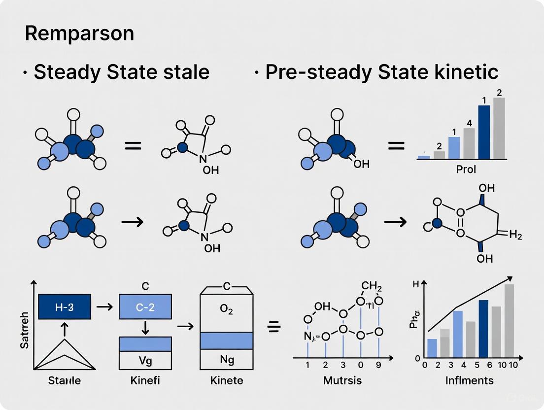
Enzyme kinetics is the study of the rates of enzyme-catalyzed reactions, crucial for understanding catalytic mechanisms, metabolic roles, and how drug molecules can alter reaction rates [1]. Within this field, the concept of steady-state kinetics, institutionalized by the Michaelis-Menten equation, forms the foundational framework for interpreting how enzymes interact with their substrates [2]. This model serves as the bedrock upon which much of modern enzymology is built, providing essential parameters to compare enzyme performance and efficiency.
However, a comprehensive understanding of enzyme action requires looking beyond this steady-state. A broader thesis is emerging that compares the established methods of steady-state analysis with the more detailed insights provided by pre-steady-state kinetic analysis. While steady-state kinetics offers a simplified and accessible view of the overall reaction, pre-steady-state kinetics delves into the transient phases at the reaction's start, revealing the individual steps and short-lived intermediates that are masked once a steady state is achieved [3] [4]. This guide will objectively compare these two approaches, outlining their principles, methodologies, applications, and how they complement each other in biochemical research and drug development.
The Michaelis-Menten Model: Principles and Assumptions
The modern definition of enzymology is synonymous with the Michaelis-Menten equation, introduced by Leonor Michaelis and Maud Menten in 1913 and later modified by Briggs and Haldane using the steady-state assumption [2]. This model describes the fundamental catalysis of enzymatic reactions through a series of steps. The enzyme (E) binds the substrate (S) to form an enzyme-substrate complex (ES), which then reacts to form the product (P) and release the free enzyme [5].
The entire reaction can be represented as: E + S ⇄ ES → E + P
The Michaelis-Menten equation is: V₀ = (Vₘₐₓ × [S]) / (Kₘ + [S])
Where:
- V₀ is the initial reaction velocity.
- Vₘₐₓ is the maximum reaction velocity, achieved when the enzyme is fully saturated with substrate.
- [S] is the substrate concentration.
- Kₘ is the Michaelis constant, defined as the substrate concentration at which the reaction velocity is half of Vₘₐₓ [5].
The derivation of this equation relies on several key assumptions [5]:
- Initial Velocity: The equation describes only the initial reaction velocity (V₀), when the substrate concentration is much greater than the product concentration.
- Steady-State Approximation: The concentration of the enzyme-substrate complex [ES] remains constant over time because the rate of its formation equals the rate of its consumption.
- Free Ligand Approximation: The total substrate concentration is approximately equal to the free substrate concentration, as the enzyme concentration is much lower than the substrate concentration ([S]>>>[E]).
- Single-Substrate Reaction: The model initially applies to reactions with a single substrate. The reverse reaction (product to substrate) is also considered negligible.
The relationship between substrate concentration and reaction velocity is graphically represented by a hyperbolic saturation curve [5]. As substrate concentration increases, the reaction velocity increases until it plateaus at Vₘₐₓ, indicating that all available enzyme molecules are saturated and operating at their maximum capacity.
Steady-State vs. Pre-Steady-State Kinetics: A Direct Comparison
The following table provides a structured, point-by-point comparison of these two fundamental approaches to kinetic analysis.
Comparison of Steady-State and Pre-Steady-State Kinetics
| Feature | Steady-State Kinetics | Pre-Steady-State Kinetics |
|---|---|---|
| Time Frame Analyzed | The period after initiation when [ES] is constant, typically seconds to minutes [1]. | The initial transient phase before steady-state is established, typically milliseconds to seconds [3] [4]. |
| Primary Kinetic Parameters Obtained | ( Km ) (Michaelis constant), ( V{max} ) (maximum velocity), and ( k_{cat} ) (turnover number) [3]. | Individual rate constants for binding, catalysis, and product release (e.g., ( k1 ), ( k2 ), ( k_3 )) [6] [3]. |
| Information Provided | Overall catalytic efficiency (( k{cat}/Km )) and substrate specificity. Provides a "black box" view of the overall reaction [3]. | Reveals the actual reaction mechanism, including short-lived intermediates and the rate-limiting step [3] [4]. |
| Typical Experiment Duration | Longer time scales (minutes). | Very short time scales (milliseconds to seconds) [3]. |
| Enzyme Concentration Required | Low, as the enzyme acts catalytically. | High, as the enzyme is treated as a stoichiometric reactant to observe its behavior directly [3]. |
| Key Assumptions | Steady-state of [ES], initial velocity conditions [5]. | No equilibrium or steady-state assumptions; observes the system as it evolves. |
| Example Experimental Data | FOR enzyme: ( Km ) (formaldehyde) = 21 µM; ( Km ) (ferredoxin) = 14 µM [6]. | FOR enzyme: Fast processes (( k{obs1} = 4.7 \, s^{-1} ), ( k{obs2} = 1.9 \, s^{-1} )) interpreted as substrate oxidation and active site rearrangement [6]. |
Experimental Protocols and Workflows
The choice between steady-state and pre-steady-state analysis dictates the experimental setup, instrumentation, and protocols required.
Steady-State Kinetics Protocol
Steady-state assays are typically performed by incubating an enzyme with varying concentrations of its substrate and monitoring the formation of product or disappearance of substrate over time.
A classic example is the study of Formaldehyde Ferredoxin Oxidoreductase (FOR) from Pyrococcus furiosus [6]:
- Objective: Determine steady-state parameters for FOR with formaldehyde as a substrate.
- Method: Activity was assayed at 80°C under anaerobic conditions.
- Substrate: Formaldehyde.
- Electron Acceptors: Benzyl viologen or ferredoxin at various concentrations.
- Measurement: The rate of electron acceptor reduction was monitored optically (using the extinction coefficients of the acceptors) in triplicate.
- Analysis: The ( K_m ) values for formaldehyde and the electron acceptors were determined from the activity measurements, fitting the data to the Michaelis-Menten model [6].
Pre-Steady-State Kinetics Protocol
Pre-steady-state experiments require specialized equipment to initiate a reaction and observe it on a millisecond timescale. Two common techniques are stopped-flow and chemical quench-flow.
Stopped-Flow Spectroscopy: Two reactant solutions are rapidly mixed and pushed into an observation cell. The flow is abruptly stopped, and an optical signal (e.g., absorbance or fluorescence) is monitored as a function of time as the reaction proceeds in the cell [3]. This was used in the FOR study to monitor the weak optical spectrum of the tungsten cofactor, revealing several fast processes in the first seconds of the reaction [6].
Chemical Quench-Flow: The reaction is initiated by rapid mixing of enzyme and substrate. After a precise, user-defined delay (milliseconds to seconds), the reaction mixture is combined with a quenching agent (e.g., acid or base) that denatures the enzyme and stops the reaction instantly. The quenched mixture is then analyzed offline, often using chromatography or mass spectrometry, to quantify reactants, intermediates, and products at that specific time point [7] [3].
The workflow for a pre-steady-state experiment, such as nucleotide incorporation by a DNA polymerase, can be visualized as follows:
The Scientist's Toolkit: Essential Reagents and Instruments
Successful kinetic analysis, particularly pre-steady-state, relies on specific reagents and sophisticated instrumentation.
Key Research Reagent Solutions
- Purified Enzyme: The enzyme of interest, often recombinant and highly purified (e.g., human DNA polymerase η or SARS-CoV-2 Mpro) [7] [4].
- Substrate: Natural or synthetic substrate, sometimes chromophoric or fluorescently labeled for detection (e.g., FAM-labeled DNA primer) [7].
- Cofactors: Essential ions or molecules, such as Mg²⁺ for DNA polymerases [7].
- Quenching Agent: A solution to stop reactions instantly (e.g., 500 mM EDTA to chelate essential metal ions, or strong acid/base) [7].
- Reaction Buffers: To maintain optimal pH and ionic strength (e.g., Tris-HCl, Epps) [6] [7].
Essential Instruments
- Spectrophotometer/Fluorometer: Standard for monitoring steady-state reactions by absorbance or fluorescence [6].
- Rapid Quench-Flow Instrument (e.g., RQF-3): A specialized apparatus that mixes, ages, and quashes reactions with millisecond precision [7].
- Stopped-Flow Spectrometer: Instrument for rapid mixing and subsequent continuous optical monitoring of reactions [3].
- Analytical Equipment: High-performance liquid chromatography (HPLC) or gel electrophoresis systems for analyzing quenched samples [7] [3].
Application in Action: Case Study on SARS-CoV-2 Main Protease
The power of pre-steady-state kinetics is exemplified in modern drug discovery, particularly in the development of antivirals against SARS-CoV-2.
The SARS-CoV-2 main protease (Mpro) is an essential viral enzyme, processing viral polyproteins and is a primary target for antiviral drugs [4]. Researchers used pre-steady-state kinetic analysis to investigate the mechanism of inhibition of Mpro by promising drug candidates like PF-00835231.
This approach allowed them to move beyond simple IC₅₀ values and uncover the detailed mechanism of action. The analysis revealed that PF-00835231 inhibition occurs via a two-step binding process, followed by the formation of a covalent complex with the catalytic cysteine of the enzyme [4]. Furthermore, by also studying a catalytically inactive mutant (C145A), they demonstrated that strong, non-covalent binding due to an excellent fit in the active site contributes significantly to the inhibitor's potency [4].
This deep mechanistic insight, provided by pre-steady-state kinetics, is invaluable for optimizing lead compounds and designing more effective drugs, showcasing a critical application that goes far beyond the capabilities of steady-state analysis alone.
This guide provides an objective comparison between steady-state and pre-steady-state kinetic analysis, two fundamental methodologies for studying enzyme mechanisms. While steady-state kinetics, underpinned by the assumption of a constant enzyme-substrate (ES) complex concentration, offers simplified parameters for routine characterization, pre-steady-state kinetics unveils the rapid, transient events of a single catalytic cycle. The selection between these methods is not a matter of superiority but of objective, as each provides distinct insights critical for different stages of research, from initial enzyme characterization to detailed mechanistic understanding and drug discovery.
Core Principles and Comparative Analysis
The steady-state assumption is a cornerstone of classical enzyme kinetics, positing that the concentration of the enzyme-substrate complex (ES) remains constant over time during the reaction. This occurs because the rate of ES formation is equal to the rate of its consumption (both dissociation back to E+S and formation of E+P) [5]. This assumption holds true provided the substrate concentration [S] is significantly greater than the enzyme concentration [E], ensuring that the free substrate concentration does not change appreciably [8] [5].
The following table contrasts the key features of steady-state and pre-steady-state kinetic analyses.
| Feature | Steady-State Kinetics | Pre-Steady-State Kinetics |
|---|---|---|
| Timeframe | Seconds to minutes; after ES complex stabilizes [9]. | Milliseconds to seconds; immediately after reaction initiation [9]. |
| Primary Measured Parameters | ( KM ) (Michaelis constant), ( V{max} ) (maximum velocity), ( k_{cat} ) (turnover number) [5]. | Individual rate constants (e.g., ( k1 ), ( k{-1} ), ( k_2 )), observation of transient intermediates [10] [9]. |
| Information Provided | Catalytic efficiency (( k{cat}/KM )), enzyme affinity for substrate (inversely related to ( K_M )) [5]. | Direct observation of binding, conformational changes, and chemical catalysis steps; identifies rate-limiting steps [11] [10]. |
| [ES] Complex Concentration | Assumed to be constant (steady-state assumption) [8] [5]. | Directly observed as it forms and decays; not constant [9]. |
| Data Interpretation | ( KM ) and ( k{cat} ) are composite constants, combinations of individual rate constants [9]. | Resolves individual rate constants for each step in the catalytic cycle [10]. |
| Typical Applications | Routine enzyme characterization, inhibitor screening, determining catalytic efficiency [5]. | Elucidating detailed reaction mechanisms, studying fast conformational changes, drug residence time [11] [12]. |
Experimental Protocols and Data Analysis
Steady-State Kinetics: The Michaelis-Menten Protocol
Objective: To determine the Michaelis constant (( KM )) and the maximum velocity (( V{max} )).
Methodology:
- A fixed, low concentration of enzyme is prepared.
- The initial velocity (( V_0 )) of the reaction is measured across a range of substrate concentrations ([S]).
- [S] must be in vast excess over [E] to satisfy the steady-state assumption [5].
- ( V_0 ) is plotted against [S], typically producing a hyperbolic saturation curve.
- The data are fitted to the Michaelis-Menten equation to extract ( KM ) and ( V{max} ): ( V0 = \frac{(V{max} [S])}{(K_M + [S])} ) [5].
Key Assumptions:
- The enzyme-substrate complex [ES] is in a steady state.
- The reaction is measured at initial velocity conditions, where product concentration is negligible and reverse reactions are minimal [5].
- The first step (E + S ⇌ ES) is often assumed to be faster than the catalytic step (ES → E + P) [8].
Pre-Steady-State Kinetics: Stopped-Flow Fluorescence Protocol
Objective: To observe the formation and decay of transient intermediates and measure individual rate constants.
Methodology:
- High Enzyme Concentration: Enzyme concentrations are typically much higher than in steady-state experiments to allow for stoichiometric detection of intermediates [9].
- Rapid Mixing: Enzyme and substrate solutions are rapidly mixed in a stopped-flow apparatus, initiating the reaction on a millisecond timescale.
- Signal Detection: A rapid detection method, such as fluorescence or absorbance spectroscopy, monitors the reaction in real-time. For instance, intrinsic tryptophan fluorescence in the Fpg protein was used to track conformational changes during lesion processing [11].
- Data Analysis: The resulting trace is fitted to exponential equations to extract observed rate constants (( k_{obs} )) for each phase. A study on Fpg protein identified up to five distinct conformational transitions in the pre-steady-state phase [11].
Key Insights from Pre-Steady-State Data:
- Burst Phase Kinetics: Evidence of a phosphoenzyme intermediate was found in a phosphotyrosyl protein phosphatase, where a rapid "burst" of product formation was observed, followed by a slower steady-state rate. This burst phase had a rate constant of 48 s⁻¹, while the steady-state turnover was 1.2 s⁻¹ [10].
- Conformational Changes: Research on formaldehyde ferredoxin oxidoreductase revealed multiple fast processes (( k{obs1} = 4.7 s^{-1}, k{obs2} = 1.9 s^{-1} )) interpreted as substrate oxidation and active site rearrangement [6].
Experimental Workflow and Data Interpretation
The diagram below illustrates the logical sequence of a kinetic analysis workflow, from initial setup to data interpretation for both steady-state and pre-steady-state methods.
The Scientist's Toolkit: Key Reagents and Techniques
The following table details essential materials and methods used in kinetic studies.
| Tool / Reagent | Function in Kinetic Analysis |
|---|---|
| Stopped-Flow Spectrometer | Rapid mixing device for initiating reactions and monitoring changes in absorbance or fluorescence on millisecond timescales [11] [6]. |
| Chemical Quench-Flow Apparatus | Rapidly mixes reactants and, after a precise time interval, mixes again with a quenching agent (e.g., acid) to stop the reaction for offline analysis [9]. |
| Synthetic Oligonucleotide Substrates | Defined DNA substrates containing specific lesions (e.g., 8-oxoguanine); essential for studying DNA repair enzymes like Fpg [11]. |
| Chromophoric Substrate Analogs | Artificial substrates (e.g., p-nitrophenyl phosphate) that produce a color change upon reaction, facilitating optical detection in steady-state studies [10] [9]. |
| Electrospray Ionization Mass Spectrometry (ESI-MS) | A powerful technique for directly detecting and identifying reactive intermediates and products in an enzymatic reaction, providing direct chemical insight [9]. |
The steady-state assumption provides a powerful simplification that makes enzyme characterization tractable, yielding the foundational parameters ( KM ) and ( V{max} ). However, this simplification necessarily obscures the rich, transient dynamics of the catalytic cycle. Pre-steady-state kinetics strips away this assumption, directly observing the formation of the ES complex and subsequent intermediates to dissect the mechanism at the level of individual rate constants. For researchers, the choice is contextual: steady-state for initial characterization and efficiency metrics, pre-steady-state for mechanistic elucidation and detailed drug interaction studies. A comprehensive kinetic strategy often leverages both to build a complete picture of enzyme function.
Enzyme kinetics, the study of reaction rates catalyzed by enzymes, provides fundamental insights into catalytic mechanisms, metabolic roles, cellular control, and inhibition by drugs or toxins [13]. Within this field, two complementary experimental approaches have emerged: steady-state kinetics and pre-steady-state kinetics. While steady-state kinetics has been the traditional workhorse for characterizing enzyme activity, pre-steady-state kinetics offers a window into the early, transient phases of enzymatic reactions that are critical for understanding mechanistic details [14] [15]. This guide provides an objective comparison of these methodologies, highlighting their respective capabilities, limitations, and appropriate applications in modern biochemical research and drug development.
The fundamental difference between these approaches lies in their observational timeframes. Steady-state kinetics observes enzyme behavior after reaction intermediates have reached constant concentrations, typically measuring substrate depletion or product formation over seconds to minutes [14] [13]. In contrast, pre-steady-state kinetics captures events during the initial burst phase before intermediates stabilize, requiring time resolution from milliseconds to seconds [14] [15]. This temporal distinction enables each method to illuminate different aspects of enzyme function, making them valuable for different research objectives.
Theoretical Foundations
Steady-State Kinetics Principles
Steady-state kinetics describes enzymatic behavior once the concentrations of reaction intermediates (such as enzyme-substrate complexes) have reached a constant level [14]. This occurs when the rates of formation and decay of these intermediates are equal, resulting in a period where the reaction rate remains relatively constant until substrate depletion becomes significant [14] [13]. The Michaelis-Menten equation, derived from steady-state assumptions, relates reaction velocity (v) to substrate concentration ([S]) through two key parameters: Vmax (maximum velocity) and Km (Michaelis constant) [13] [16]:
[v = \frac{V{\text{max}}[S]}{Km + [S]}]
Where (V{\text{max}}) represents the maximum rate achieved at saturating substrate concentrations, and (Km) is the substrate concentration at which the reaction rate is half of (V{\text{max}}) [14] [13]. Traditionally, (Km) has been interpreted as an inverse measure of substrate affinity, though this interpretation is only valid under specific mechanistic conditions [16].
Pre-Steady-State Kinetics Principles
Pre-steady-state kinetics describes the behavior of an enzymatic reaction during the initial phase, before the concentrations of reaction intermediates have reached a steady state [14]. In this regime, intermediate concentrations change rapidly over time as they are formed and consumed during the early reaction stages [14]. The approach typically involves changing conditions (e.g., rapidly mixing reactants) and observing how the system evolves over time as it approaches a new equilibrium [15].
The time course of this "relaxation" to equilibrium generally follows a single exponential or sum of exponentials, described by equations of the form [15]:
[\text{Signal}(t) = A(1 - e^{-k_{\text{obs}}t}) + C]
Where the signal (e.g., fluorescence, absorbance) tracks concentration changes, (k_{\text{obs}}) is the observed rate constant, and A and C are constants. These measurements provide direct access to individual rate constants for elementary steps in the reaction mechanism, offering insights not available from steady-state analysis alone [14] [15].
Methodological Comparison
Experimental Techniques and Time Resolution
The fundamental difference between steady-state and pre-steady-state kinetics lies in their temporal resolution and the corresponding experimental approaches required to capture reactions at these timescales.
Table 1: Experimental Approaches for Kinetic Analysis
| Methodological Aspect | Steady-State Kinetics | Pre-Steady-State Kinetics |
|---|---|---|
| Time Resolution | Seconds to minutes | Milliseconds to seconds |
| Typical Assay Format | Continuous monitoring or discrete sampling | Rapid mixing with continuous monitoring |
| Key Techniques | Traditional spectrophotometry, fluorimetry, radiometric assays | Stopped-flow, rapid quench-flow, temperature-jump |
| Data Collection | Linear initial rate period measured over minutes | Transient phase captured in first few seconds |
| Instrumentation Requirements | Standard laboratory equipment (spectrophotometers) | Specialized rapid-mixing equipment |
Information Content and Kinetic Parameters
Each kinetic approach provides distinct insights into enzyme function, with differing abilities to resolve individual steps in the catalytic cycle.
Table 2: Information Content of Kinetic Approaches
| Parameter/Insight | Steady-State Kinetics | Pre-Steady-State Kinetics |
|---|---|---|
| Primary Parameters | kcat, Km, kcat/Km | Individual rate constants for elementary steps |
| Intermediate Detection | Indirect inference | Direct observation of formation and decay |
| Rate-Determining Step | Identified but not characterized | Direct characterization of individual steps |
| Conformational Changes | Only if rate-limiting | Potentially observable even if not rate-limiting |
| Binding Constants | Apparent (Km) | Direct measurement of association/dissociation rates |
The specificity constant (kcat/Km) provides a measure of enzyme efficiency and is proportional to the apparent second-order rate constant for substrate binding [16]. However, as Johnson (2019) emphasizes, "kcat and kcat/Km should be considered as the two primary steady state kinetic parameters, rather than kcat and Km" because kcat/Km quantifies enzyme specificity, efficiency, and proficiency, while Km alone is often uninterpretable without additional information [16].
Experimental Protocols
Steady-State Kinetics Protocol
Objective: Determine kcat and Km for an enzymatic reaction under substrate saturation conditions.
Procedure:
- Prepare a constant concentration of enzyme (typically 0.0002 mM) [17] in appropriate buffer
- Create a series of substrate concentrations spanning below and above the expected Km (e.g., 0.1, 0.2, 0.5, 5, 10, 100 mM) [17]
- Initiate reactions by adding enzyme to substrate solutions
- Measure initial velocity by tracking product formation or substrate depletion
- Ensure measurements occur during the linear phase of reaction progress [13]
- Fit data to the Michaelis-Menten equation using nonlinear regression or linear transformations (Lineweaver-Burk, Eadie-Hofstee) [13] [18]
Data Analysis:
- Plot reaction velocity (v) versus substrate concentration ([S])
- Fit data to: (v = \frac{k{\text{cat}}[E]{\text{tot}}[S]}{K_m + [S]})
- kcat = Vmax/[E]tot
- Alternatively, use the form: (v = \frac{k{SP}[S]}{1 + k{SP}[S]/k_{\text{cat}}}) where kSP = kcat/Km to directly obtain the specificity constant [16]
Pre-Steady-State Kinetics Protocol
Objective: Measure individual rate constants for elementary steps in an enzymatic mechanism by capturing transient phases.
Procedure:
- Stopped-Flow Method [14]:
- Load syringes with enzyme and substrate solutions
- Rapidly mix solutions (mixing time ~1 ms)
- Monitor signal change (absorbance, fluorescence) immediately after mixing
- Collect data points at high temporal resolution (microsecond to millisecond intervals)
- Repeat at multiple substrate concentrations
- Rapid Quench-Flow Method [14]:
- Rapidly mix enzyme and substrate solutions
- Allow reaction to proceed for precisely controlled time intervals
- Quench reaction with acid, base, or denaturant at specific times
- Analyze quenched samples for product formation using separation methods (HPLC, electrophoresis) or spectroscopic techniques
Data Analysis:
- Fit time courses to single or multiple exponential functions
- Determine observed rate constants (kobs) at each substrate concentration
- Analyze dependence of kobs on substrate concentration to extract individual rate constants
- Use kinetic simulation software for complex mechanisms
Comparative Analysis of DNA Polymerase Fidelity
A comprehensive study comparing steady-state and pre-steady-state kinetic approaches for measuring DNA polymerase fidelity provides an excellent case study for methodological comparison [19]. This research directly evaluated the fidelity of the proofreading-deficient Klenow fragment (KF(-)) DNA polymerase using both traditional steady-state methods and pre-steady-state kinetics, comparing these results with direct competition measurements.
Table 3: DNA Polymerase Fidelity Measurement Comparison
| Methodological Approach | Measured Fidelity (R:W Ratio) | Key Advantages | Technical Challenges |
|---|---|---|---|
| Direct Competition | Variable across 12 natural base mispairs (9 measurable) | Direct physiological relevance; measures actual partitioning | Detection of minor W products masked by major R products |
| Steady-State Kinetics | Quantitative agreement with direct competition | Broadly accessible methodology; well-established protocols | Measures kcat/Km indirectly under substrate saturation |
| Pre-Steady-State Kinetics | Quantitative agreement with both other methods | Direct measurement of elementary steps; identifies rate-limiting steps | Requires specialized equipment; more complex data analysis |
The study found that "all the data are in quantitative agreement" across the three methods, validating the kinetic approaches for fidelity measurements [19]. This demonstrates that while pre-steady-state kinetics provides more detailed mechanistic information, steady-state methods can yield equivalent functional parameters when appropriately applied.
Research Toolkit: Essential Reagents and Methods
Successful implementation of kinetic studies requires specific reagents and methodologies tailored to each approach.
Table 4: Research Reagent Solutions for Kinetic Studies
| Reagent/Method | Function in Kinetic Studies | Applications |
|---|---|---|
| Stopped-Flow Spectrometer | Rapid mixing (∼1 ms) with continuous monitoring | Pre-steady-state kinetics of fast reactions |
| Rapid Quench-Flow Instrument | Rapid mixing followed by chemical quenching | Pre-steady-state analysis of reactions requiring product separation |
| Fluorescent Nucleotide Analogs | Reporting on binding and conformational changes | Pre-steady-state studies of nucleotide-dependent enzymes |
| Isotopically Labeled Substrates | Tracing chemical fate; kinetic isotope effects | Identifying rate-determining steps in both kinetic approaches |
| Synthetic Substrate Analogs | Monitoring specific bond cleavages | Steady-state activity assays (e.g., USP lactase units) [18] |
| Enzyme-Coupled Assay Systems | Amplifying signal for low-activity enzymes | Both approaches, particularly steady-state |
Advantages and Limitations in Research Applications
Strengths of Each Approach
Steady-State Kinetics Advantages:
- Technical Accessibility: Requires standard laboratory equipment available in most biochemical laboratories [18]
- Established Protocols: Well-characterized methodology with extensive literature support
- Parameter Efficiency: Directly provides kcat/Km, the key parameter for enzyme specificity and efficiency [16]
- Therapeutic Applications: Suitable for enzyme characterization required by regulatory agencies [18]
Pre-Steady-State Kinetics Advantages:
- Mechanistic Resolution: Direct observation of intermediate formation and decay [14]
- Elementary Step Characterization: Measurement of individual rate constants for distinct reaction steps [15]
- Dynamic Information: Captures conformational changes and transient species [14]
- Comprehensive Understanding: Reveals features not apparent from steady-state analysis alone [14]
Practical Limitations
Steady-State Kinetics Limitations:
- Averaged Parameters: Provides population averages that may mask individual steps
- Indirect Information: Intermediate steps must be inferred rather than directly observed
- Limited Temporal Resolution: Misses early transient phases rich in mechanistic information
Pre-Steady-State Kinetics Limitations:
- Specialized Equipment: Requires expensive instrumentation not universally available [15]
- Technical Expertise: Demands significant experience in experimental design and data interpretation
- Sample Consumption: Often requires substantial amounts of purified enzyme
- Time-Intensive Analysis: Complex data fitting and modeling requirements
Conceptual and Experimental Workflows
The relationship between steady-state and pre-steady-state kinetics, along with their experimental implementations, can be visualized through the following conceptual and technical workflows:
Diagram Title: Kinetic Approaches Relationship
Steady-state and pre-steady-state kinetics offer complementary rather than competing perspectives on enzyme function. Steady-state kinetics provides efficient characterization of overall enzyme efficiency through kcat/Km and is more accessible for routine characterization [16] [18]. Pre-steady-state kinetics delivers unparalleled mechanistic resolution of individual steps but requires specialized resources and expertise [14] [15].
The choice between these approaches depends entirely on research objectives: steady-state kinetics for functional characterization and screening applications, pre-steady-state kinetics for detailed mechanistic studies. As demonstrated in the DNA polymerase fidelity study [19], these methods can yield quantitatively equivalent parameters for functional comparisons while providing different levels of mechanistic insight. For comprehensive enzyme characterization, the most powerful strategy often combines both approaches, using steady-state analysis for initial functional assessment followed by pre-steady-state investigations to elucidate underlying mechanisms.
Pre-steady-state and steady-state kinetic analyses represent complementary approaches for elucidating the intricate mechanisms of enzyme catalysis and biological processes. While steady-state kinetics provides a macroscopic view of overall enzyme efficiency through parameters like kcat and KM, pre-steady-state kinetics captures the transient molecular events that occur before the system reaches equilibrium—the infamous "burst" phase that often reveals rate-limiting steps [10] [20]. This distinction is not merely technical but fundamental to understanding how enzymes truly operate at the molecular level.
The analysis of burst kinetics has become increasingly relevant across multiple fields, from enzymology to gene regulation. Recent research has demonstrated that these transient kinetic phases often conceal crucial mechanistic information about rate-limiting steps, conformational changes, and allosteric regulation [21] [20]. For researchers and drug development professionals, understanding this distinction provides critical insights for designing better inhibitors, engineering improved enzymes, and developing targeted therapeutic strategies. This guide objectively compares these complementary analytical approaches, their instrumentation requirements, and their respective capabilities for revealing the inner workings of biological systems.
Theoretical Foundation: Burst Phase and Steady-State Kinetics
Defining the Kinetic Regimes
The journey of enzyme-catalyzed reactions begins with a pre-steady-state phase (burst phase) where reaction intermediates accumulate, followed by a steady-state phase where intermediate concentrations remain relatively constant [10]. The burst phase represents the initial transient period when enzyme-substrate complexes form and the first turnover occurs, typically lasting milliseconds to seconds. The steady-state phase follows, characterized by a constant rate of product formation until substrate depletion becomes significant.
Mathematically, the burst phase often follows an exponential approach to the steady-state rate, described by the equation: [P = vss \cdot t + (vi - vss)(1 - e^{-kobs \cdot t})/k_obs] where P is product concentration, vi is initial velocity, vss is steady-state velocity, and kobs is the observed first-order rate constant for the transition [20].
The Significance of Burst Kinetics
The presence of a burst phase provides critical mechanistic information. A pronounced burst indicates that the catalytic step is faster than at least one subsequent step in the pathway, causing accumulation of a reaction intermediate [10]. For instance, in the hydrolysis of p-nitrophenyl phosphate catalyzed by bovine heart phosphotyrosyl protein phosphatase, a stoichiometric burst of p-nitrophenol formation revealed a phosphoenzyme intermediate with rapid formation followed by rate-limiting hydrolysis (k2 = 540 s-1, k3 = 36.5 s-1) [10].
Comparative Analysis: Methodologies and Applications
Experimental Designs and Workflows
Figure 1: Experimental workflows for pre-steady-state and steady-state kinetic analyses.
Technical Specifications and Data Output
Table 1: Comparative specifications of pre-steady-state versus steady-state kinetic approaches
| Parameter | Pre-Steady-State Kinetics | Steady-State Kinetics |
|---|---|---|
| Time Resolution | Microseconds to seconds | Seconds to hours |
| Key Measured Parameters | Burst amplitude (λ), individual rate constants (k1, k2, k3), transient intermediates | kcat, KM, kcat/KM |
| Instrumentation Requirements | Stopped-flow, quenched-flow, rapid-mixing devices with fast detection | Spectrophotometers, plate readers with temperature control |
| Information Obtained | Direct observation of reaction intermediates, elemental steps, conformational changes | Overall catalytic efficiency, substrate specificity, inhibitor potency |
| Sample Consumption | Typically higher (μmol range) | Typically lower (nmol-pmol range) |
| Throughput | Lower (single reactions monitored) | Higher (multiple conditions parallel) |
| Key Applications | Elucidating catalytic mechanisms, identifying rate-limiting steps, characterizing transient intermediates | Screening enzyme variants, determining inhibitor constants, comparative enzymology |
Case Studies: Revealing Mechanistic Insights
β-Lactamase Evolution and Epistasis
Directed evolution of the β-lactamase OXA-48 revealed striking epistatic interactions that dramatically enhanced ceftazidime resistance [21]. While wild-type enzyme showed minimal burst kinetics (1.5-fold higher burst rate than steady-state), evolved variants exhibited pronounced burst phases—F72L showed a 4.6-fold burst and the quadruple mutant Q4 displayed a dramatic 48-fold burst amplitude [21]. Pre-steady-state analysis revealed that epistasis emerged from mutations that sequentially changed the rate-limiting step, with early mutations accelerating substrate binding and later mutations enhancing chemical steps, culminating in 470-800 fold improvement in burst-phase kcat/KM that strongly correlated with in vivo resistance (R2 = 0.97) [21].
Butyrylcholinesterase-Catalyzed Hydrolysis
The hydrolysis of mirabegron by butyrylcholinesterase exhibits classic hysteretic behavior with an extensive pre-steady-state burst phase lasting up to 18 minutes at maximum velocity [20]. This burst results from a slow equilibrium between two active enzyme forms (E and E'), with the initial E form displaying higher activity (kcat = 7.3 min-1, Km = 23.5 μM) than the final E' form (kcat = 1.6 min-1, Km = 3.9 μM) [20]. This mechanistic insight, obtainable only through pre-steady-state analysis, explains the complex catalytic behavior and predicts minimal impact on pharmacological activity despite slow degradation in blood.
Transcriptional Bursting in Gene Regulation
In gene regulation, transcriptional bursting represents a biological analog to enzymatic burst kinetics, with stochastic switching between active (ON) and inactive (OFF) states [22]. Recent live-cell imaging reveals that burst frequency—rather than duration or amplitude—serves as the primary regulatory parameter, with transcription factor binding dynamics (occurring on timescales of seconds) somehow generating transcriptional bursts lasting minutes to hours [22]. This temporal mismatch suggests complex multi-step mechanisms where short-lived molecular events initiate prolonged output states, analogous to hysteretic enzyme behavior.
Table 2: Kinetic parameters from case studies demonstrating burst-phase phenomena
| Biological System | Observed Burst Amplitude | Burst Duration | Molecular Interpretation | Biological Impact |
|---|---|---|---|---|
| β-lactamase OXA-48 (Evolved Q4) | 48-fold higher than steady-state | Not specified | Change in rate-limiting step from substrate binding to chemical step | 40-fold increase in antibiotic resistance [21] |
| Butyrylcholinesterase with Mirabegron | vi > vss | τ ≈ 18 min at Vmax | Hysteresis between enzyme forms E and E' | Minimal pharmacological impact despite slow degradation [20] |
| Phosphotyrosyl Protein Phosphatase | Stoichiometric burst | Transient phase (k = 48 s-1) | Rate-limiting breakdown of phosphoenzyme intermediate (k3 = 1.2 s-1) | Identification of catalytic pathway with intermediate [10] |
| Transcriptional Activation | ON/OFF switching | Minutes to hours | Stochastic TF binding (seconds) vs. prolonged output | Regulation of burst frequency controls RNA output [22] |
Essential Research Tools and Reagents
Table 3: Key research reagent solutions for kinetic analysis
| Reagent/Instrument | Specifications | Application Context |
|---|---|---|
| Stopped-Flow Spectrometer | Millisecond time resolution, temperature control, multiple detection modes (absorbance, fluorescence) | Pre-steady-state burst phase measurement of rapid enzymatic reactions [21] |
| Rapid Quench-Flow Instrument | Millisecond mixing and quenching capabilities, chemical or freeze-quenching | Trapping enzymatic intermediates for structural analysis or quantification |
| p-Nitrophenyl Phosphate | Chromogenic substrate, λmax = 405 nm upon hydrolysis (pKa = 7.2) | Continuous assay for phosphatases and phosphohydrolases [10] |
| MS2/PP7 RNA Labeling System | Fluorescent RNA stem-loops for live-cell imaging of nascent transcripts | Visualization of transcriptional bursting kinetics in living cells [22] |
| Site-Directed Mutagenesis Kits | High-fidelity polymerases, efficient bacterial transformation | Creating point mutants to test mechanistic hypotheses (e.g., β-lactamase variants) [21] |
| HPLC with Rapid Sampling | Second-to-minute time resolution, quantitative detection | Monitoring substrate depletion and product formation in hysteretic enzymes [20] |
Integrated Analysis: Connecting Kinetic Signatures to Biological Mechanisms
Figure 2: Relationship between kinetic signatures and their underlying molecular mechanisms.
The connection between kinetic observations and molecular mechanisms becomes particularly evident when comparing burst phenomena across biological systems. In enzymatic systems, burst kinetics often reveal covalent intermediates or rate-limiting conformational changes [10] [20], while in gene regulation, transcriptional bursting reflects the stochastic nature of molecular interactions driving ON-OFF switching [22]. The emerging pattern across these diverse systems is that burst kinetics frequently signal rate-limiting steps with profound functional consequences—whether in antibiotic resistance evolution or drug metabolism.
The critical distinction between burst phase and steady-state analyses provides complementary insights into biological mechanisms. Pre-steady-state kinetics excels at identifying transient intermediates, elemental rate constants, and the molecular basis of rate-limiting steps, while steady-state kinetics offers efficient characterization of overall catalytic performance and inhibitor effects. The most powerful approaches strategically integrate both methodologies, using steady-state analysis to identify interesting phenomenological behavior and pre-steady-state methods to elucidate underlying mechanisms.
For researchers and drug development professionals, this distinction has practical implications. Enzyme engineers can exploit burst kinetics to identify mutations that change rate-limiting steps and create epistatic improvements [21]. Pharmacologists can understand how hysteretic enzyme behavior affects drug metabolism timelines [20]. Molecular biologists can connect transcription factor binding dynamics to transcriptional output control [22]. In each case, moving beyond steady-state analysis to examine transient kinetics provides the critical mechanistic insights needed to advance both basic understanding and applied research.
Kinetic analysis provides the foundation for understanding reaction mechanisms across chemical and biological sciences. This guide offers a comparative analysis of two fundamental kinetic approaches: the steady-state approximation and pre-steady-state burst phase analysis. While steady-state kinetics delivers macroscopic parameters for overall reactions, pre-steady-state kinetics reveals transient intermediates and individual steps within catalytic cycles. We objectively evaluate their complementary strengths, methodological requirements, and applications through experimental case studies spanning enzymology, drug delivery systems, and biotherapeutic development, providing researchers with a framework for selecting appropriate analytical strategies for their specific investigations.
Kinetic analysis serves as the cornerstone for elucidating reaction mechanisms across diverse scientific domains, from chemical catalysis to biological processes. The steady-state approximation represents one of the most fundamental principles in chemical kinetics, enabling the simplification of complex multi-step reactions by assuming that reactive intermediates maintain constant concentrations during the majority of the reaction timeline [23] [24]. This approach has proven invaluable for determining overall kinetic parameters and deriving rate laws for complex reaction mechanisms.
In contrast, pre-steady-state kinetics captures the transient early phases of reactions, often revealing mechanistic details obscured under steady-state conditions. This approach is particularly valuable for detecting and characterizing reaction intermediates and identifying rate-limiting steps [10] [20]. Burst phase kinetics, a specific manifestation of pre-steady-state behavior, often signifies the accumulation and subsequent turnover of intermediates, providing critical insights into catalytic mechanisms [10] [20].
This guide examines the theoretical foundations, experimental methodologies, and practical applications of these complementary kinetic frameworks, providing researchers with a comprehensive resource for mechanistic investigation across chemical and biological systems.
Theoretical Foundations
Steady-State Approximation
The steady-state approximation (SSA) applies to reaction mechanisms involving reactive intermediates that are generated and consumed in subsequent steps. This method assumes that the concentration of these intermediates remains constant throughout much of the reaction duration, meaning their rate of formation equals their rate of disappearance [23] [25] [24]. Mathematically, this is expressed as d[Int]/dt = 0, where [Int] represents the concentration of the intermediate species.
The SSA is particularly applicable to consecutive reactions where the first step is rate-limiting (k₁ << k₂), preventing significant accumulation of the intermediate product [24]. For a mechanism where A → I → P (where I is an intermediate and P is product), the SSA yields a relatively simple expression for product formation: [P] = [A]₀(1 - e^(-k₁t)) [24]. This stands in stark contrast to the complex exact solution, demonstrating the utility of SSA for simplifying kinetic analysis of multi-step mechanisms.
Pre-Steady-State and Burst Kinetics
Pre-steady-state kinetics describes the initial phase of a reaction before intermediates reach their steady-state concentrations. During this phase, reaction rates change rapidly as intermediates accumulate, providing a window into individual steps of the reaction mechanism [10] [20].
Burst kinetics refers to a specific pre-steady-state phenomenon characterized by a rapid initial release of product (the "burst") followed by a slower, linear steady-state phase. This behavior typically indicates that the first catalytic step (such as enzyme acylation or intermediate formation) is faster than subsequent steps (such as deacylation or intermediate breakdown) [10] [20]. The burst amplitude often correlates stoichiometrically with the concentration of active sites, while the steady-state rate reflects the rate-limiting step of the catalytic cycle.
Figure 1: Reaction mechanism underlying burst kinetics in a two-step catalytic process. The rapid formation and accumulation of the EP intermediate (k₃ > k₅) leads to an initial burst of P1 product, followed by slower steady-state turnover.
Experimental Methodologies
Steady-State Kinetic Analysis
Steady-state kinetic analysis typically involves measuring reaction velocities under conditions where substrate concentrations remain essentially constant (typically consumption of less than 5-10% of initial substrate) while varying substrate concentrations. For enzymatic reactions, this approach yields classic Michaelis-Menten parameters Km and Vmax, which provide macroscopic descriptions of enzyme-substrate interactions and catalytic efficiency [20].
Protocol: Standard Steady-State Kinetic Analysis
- Prepare a series of reactions with varying substrate concentrations while maintaining constant enzyme concentration
- Ensure initial linear rate conditions (typically monitoring ≤10% substrate depletion)
- Measure initial velocities for each substrate concentration
- Fit data to appropriate kinetic model (e.g., Michaelis-Menten equation for single-substrate reactions)
- Determine kinetic parameters (Km, Vmax, kcat) through nonlinear regression or linear transformations
For the hydrolysis of mirabegron by butyrylcholinesterase, steady-state analysis revealed Michaelian behavior with kcat = 1.6 min⁻¹ and Km = 3.9 μM at pH 7.0 and 25°C [20].
Pre-Steady-State Kinetic Analysis
Pre-steady-state kinetics requires specialized techniques capable of monitoring reactions on short timescales, often milliseconds to seconds, before the steady state is established.
Protocol: Pre-Steady-State Burst Kinetics Analysis
- Utilize rapid mixing/detection methods (stopped-flow, quenched-flow, or laser photolysis of caged compounds)
- Initiate reaction synchronously across all enzyme molecules
- Monitor product formation with high temporal resolution (millisecond timescale)
- Fit progress curves to integrated rate equations accounting for burst and steady-state phases
For BChE-catalyzed hydrolysis of mirabegron, progress curves were analyzed using the integrated rate equation: [ [P] = v{ss}t + \frac{(vi - v{ss})(1 - e^{-k{obs}t})}{k{obs}} ] where (vi) and (v{ss}) represent initial and steady-state velocities, respectively, and (k{obs}) is the observed first-order rate constant for the transition between these phases [20].
Laser pulse photolysis of caged alanine has been employed to study SLC6A14 amino acid transporter kinetics with sub-millisecond resolution, revealing rapid transient currents decaying with a time constant of <1 ms [26].
Figure 2: Comparative experimental workflows for steady-state and pre-steady-state kinetic analyses, highlighting fundamental methodological differences.
Comparative Analysis of Kinetic Approaches
Direct Comparison of Methodologies
Table 1: Comparative analysis of steady-state versus pre-steady-state kinetic approaches
| Parameter | Steady-State Kinetics | Pre-Steady-State Kinetics |
|---|---|---|
| Time Scale | Seconds to hours | Milliseconds to seconds |
| Key Measured Parameters | Km, Vmax, kcat | Burst amplitude, kobs, transient rate constants |
| Information Obtained | Overall catalytic efficiency, substrate specificity | Individual rate constants, catalytic intermediates, rate-limiting steps |
| Experimental Complexity | Relatively simple | Technically demanding |
| Instrumentation | Standard spectrophotometers, HPLC | Stopped-flow, quenched-flow, laser photolysis |
| Data Analysis | Michaelis-Menten, linear transformations | Integrated rate equations, exponential fitting |
| Detection Limit | Nanomolar to micromolar | Often requires higher enzyme concentrations |
Case Study Comparisons
Table 2: Experimental kinetic parameters from case studies
| System | Method | Key Parameters | Mechanistic Insight |
|---|---|---|---|
| BChE-catalyzed hydrolysis of mirabegron [20] | Pre-steady-state | Burst phase: kcat = 7.3 min⁻¹, Km = 23.5 μMSteady-state: kcat = 1.6 min⁻¹, Km = 3.9 μM | Hysteretic behavior with slow equilibrium between enzyme forms E and E′ |
| Bovine heart phosphotyrosyl protein phosphatase [10] | Pre-steady-state | Burst rate: 48 s⁻¹Turnover rate: 1.2 s⁻¹ | Phosphoenzyme intermediate with breakdown as rate-limiting step |
| SLC6A14 Amino Acid Transporter [26] | Pre-steady-state | Turnover rate: <1 ms transient current | Rapid substrate translocation not rate-limiting; Na+ binding likely rate-limiting |
| PLGA-based nanoparticle drug release [27] | Steady-state modeling | Weibull model: AIC = -36.37, OE = 7.24 | Best fit for multi-mechanistic drug release profiles |
Applications in Drug Development and Research
Biotherapeutic Stability Assessment
Kinetic modeling plays a crucial role in predicting biotherapeutic stability. First-order kinetic models combined with Arrhenius equations enable long-term stability predictions for various protein modalities, including IgG1, IgG2, bispecific IgG, Fc fusion proteins, scFv, nanobodies, and DARPins [28]. This approach reduces the number of parameters and samples required while enhancing prediction reliability. For aggregate formation in biotherapeutics, the reaction rate can be described by:
[ \frac{d\alpha}{dt} = v × A1 × \exp\left(-\frac{Ea1}{RT}\right) × (1-\alpha1)^{n1} × \alpha1^{m1} × C^{p1} + (1-v) × A2 × \exp\left(-\frac{Ea2}{RT}\right) × (1-\alpha2)^{n2} × \alpha2^{m2} × C^{p2} ]
where α represents the fraction of degradation products, A is the pre-exponential factor, Ea is activation energy, and n, m are reaction orders [28].
Drug Release Kinetics
Mathematical modeling of drug release kinetics from delivery systems like PLGA nanoparticles employs both steady-state and dynamic approaches. Comparative analyses of release models have demonstrated that the Weibull model shows superior fit for multi-mechanistic release from PLGA-based systems, with mean Akaike Information Criterion (AIC) = -36.37 and mean overall error (OE) = 7.24 [27]. Alternative models including zero-order, first-order, Higuchi, Hixson-Crowell, and Korsmeyer-Peppas provide varying degrees of accuracy depending on the specific drug delivery system and release mechanisms [27] [29].
The Scientist's Toolkit: Essential Research Reagents and Materials
Table 3: Key research reagents and materials for kinetic analysis
| Reagent/Material | Application | Function | Example Usage |
|---|---|---|---|
| p-Nitrophenyl phosphate | Enzyme kinetics | Chromogenic substrate | Burst titration kinetics with phosphotyrosyl protein phosphatase [10] |
| Mirabegron | Enzyme kinetics | Arylacylamide substrate | BChE-catalyzed hydrolysis studies [20] |
| PLGA polymers | Drug delivery | Nanoparticle matrix | Controlled release kinetics studies [27] [29] |
| Caged amino acids | Pre-steady-state kinetics | Photolabile precursors | Laser pulse photolysis studies of transporter kinetics [26] |
| Size exclusion chromatography columns | Biotherapeutic analysis | Aggregate quantification | Stability studies for various protein modalities [28] |
Steady-state and pre-steady-state kinetic analyses represent complementary approaches to mechanistic investigation, each with distinct strengths and applications. The steady-state approximation provides simplified, practical parameters for overall reaction characterization, while pre-steady-state methods, particularly burst phase analysis, reveal intricate mechanistic details often masked under steady-state conditions. The selection between these approaches depends on the specific research questions, available instrumentation, and time scale of the process under investigation. As kinetic modeling continues to evolve, these frameworks remain essential tools for advancing our understanding of reaction mechanisms across chemical, biological, and pharmaceutical sciences.
Practical Techniques: Methods and Applications in Kinetic Analysis
Kinetic analysis provides the foundation for understanding reaction mechanisms and rates in biochemical and chemical processes. Within this field, the steady-state approximation represents a cornerstone methodology, offering a powerful framework for determining kinetic parameters such as Vmax and Km. This approach assumes that the concentration of enzyme-substrate complexes remains constant over time, as their rate of formation equals their rate of breakdown. The resulting mathematical treatment enables researchers to extract meaningful kinetic data from velocity measurements plotted against substrate concentration, typically yielding characteristic hyperbolic plots that can be linearized through various transformations.
This guide objectively compares steady-state kinetic analysis with its more time-resolved counterpart—pre-steady-state kinetics—by examining their fundamental principles, experimental requirements, data interpretation frameworks, and respective applications in basic research and drug development. While steady-state methods provide essential macroscopic parameters for enzyme characterization, pre-steady-state techniques reveal the transient microscopic events that underlie catalytic mechanisms. The complementary nature of these approaches provides a more complete picture of enzyme function when applied to research problems ranging from fundamental mechanistic studies to pharmaceutical development.
Core Principles and Data Features
Fundamental Characteristics of Steady-State Kinetics
Steady-state kinetics operates under the fundamental assumption that the concentration of enzyme-substrate intermediates remains constant during the measurement period. This occurs because the rate of ES complex formation equals its rate of decomposition to product and free enzyme. The methodology focuses on the initial rate of reaction (v0) measured during this steady-state phase, where product accumulation remains linear with time. These velocity measurements are then plotted against substrate concentration to generate a rectangular hyperbola, described mathematically by the Michaelis-Menten equation: v0 = (Vmax × [S])/(Km + [S]).
To extract the key kinetic parameters Vmax (maximum velocity) and Km (substrate concentration at half Vmax), researchers typically employ linear transformations of the Michaelis-Menten equation. The most common linear plots include Lineweaver-Burk (double-reciprocal), Eadie-Hofstee, and Hanes-Woolf plots. Each transformation offers distinct advantages and sensitivities to experimental error, with the Lineweaver-Burk plot (1/v versus 1/[S]) being the most widely recognized despite its susceptibility to error propagation at low substrate concentrations.
Key Experimental Data from Steady-State Analysis
The phosphotyrosyl protein phosphatase study provides exemplary steady-state kinetic data, demonstrating how these methods yield quantitative parameters for enzyme characterization [10]. In this analysis, researchers determined an apparent Km of 0.38 mM for p-nitrophenyl phosphate hydrolysis alongside a Vmax that remained constant across multiple substrates. The study also established precise rate constants for the catalytic cycle, with phosphorylation proceeding at k2 = 540 s-1 and dephosphorylation at k3 = 36.5 s-1 under optimal conditions (pH 5.0, 37°C) [10].
Beyond these basic parameters, the investigation revealed fundamental thermodynamic properties through temperature-dependent measurements. The energy of activation for the enzyme-catalyzed hydrolysis was determined to be 13.6 kcal/mol at pH 5.0 and 14.1 kcal/mol at pH 7.0 [10]. These values, combined with the observed constant product ratio across substrates and the enzyme-catalyzed 18O exchange between inorganic phosphate and water (kcat = 4.47 × 10⁻³ s-1 at pH 5.0, 37°C), provided compelling evidence for a phosphoenzyme intermediate with breakdown as the rate-limiting step [10].
Table 1: Key Kinetic Parameters from Phosphotyrosyl Protein Phosphatase Study
| Parameter | Value | Conditions | Significance |
|---|---|---|---|
| Apparent Km | 0.38 mM | pH 5.0, 37°C | Michaelis constant for p-nitrophenyl phosphate |
| True Ks | 6.0 mM | pH 5.0, 37°C | Actual enzyme-substrate dissociation constant |
| Phosphorylation rate (k2) | 540 s-1 | pH 5.0, 37°C with phosphate acceptors | Rate constant for phosphoenzyme formation |
| Dephosphorylation rate (k3) | 36.5 s-1 | pH 5.0, 37°C with phosphate acceptors | Rate constant for phosphoenzyme breakdown |
| Activation energy (pH 5.0) | 13.6 kcal/mol | - | Temperature dependence of hydrolysis |
| Activation energy (pH 7.0) | 14.1 kcal/mol | - | Temperature dependence at physiological pH |
| 18O exchange rate | 4.47 × 10⁻³ s-1 | pH 5.0, 37°C | Exchange between phosphate and water |
Experimental Protocols and Methodologies
Standard Steady-State Kinetic Protocol
The implementation of steady-state kinetic analysis follows a systematic experimental workflow designed to ensure accurate velocity measurements and reliable parameter estimation:
Reaction Mixture Preparation: Prepare a master mix containing buffer, cofactors, and any essential salts required for enzyme activity. Maintain this mixture at the desired assay temperature using a precision water bath or thermostatted block.
Substrate Dilution Series: Create a series of substrate concentrations typically spanning values below and above the anticipated Km. A recommended range is 0.2Km to 5Km to adequately define the hyperbolic curve.
Reaction Initiation: Start reactions by adding a small volume of enzyme solution to each substrate concentration. Use techniques that ensure rapid mixing while maintaining constant temperature throughout.
Initial Velocity Measurement: Monitor product formation or substrate depletion using appropriate detection methods (spectrophotometric, fluorometric, radiometric) during the linear phase of the reaction. The observation period should capture less than 5-10% of total substrate conversion to maintain initial rate conditions.
Data Collection: Record time-course data for each substrate concentration, ensuring sufficient data points to establish a linear fit with high confidence (typically R2 > 0.98).
Parameter Calculation: Plot initial velocity versus substrate concentration and fit the data to the Michaelis-Menten equation using nonlinear regression. Alternatively, employ linear transformations for preliminary analysis and outlier identification.
This protocol emphasizes temperature control, precise timing, and verification of linearity throughout the measurement period—factors critical for obtaining reliable kinetic parameters.
Advanced Measurement Techniques
Beyond conventional spectrophotometric methods, sophisticated measurement technologies enable kinetic analysis in challenging systems. Magnetic Resonance Imaging (MRI) has been adapted for velocity and temperature measurements in porous media convection studies, providing non-invasive quantification of flow dynamics [30]. This approach couples velocity vector measurements with temperature field mapping to analyze heat transfer in fluid-saturated systems, demonstrating how traditional kinetic principles extend to complex physical systems [30].
Similarly, X-ray Particle Tracking Velocimetry (XPTV) has emerged as a powerful technique for quantifying velocity fields in opaque systems where conventional optical methods fail [31]. By embedding tracer particles within materials and tracking their motion using X-ray imaging, researchers can resolve complex flow patterns and extract rheological information from polymer melts and other challenging media [31]. These advanced methodologies share the fundamental principle of steady-state analysis—measuring system properties under conditions where key variables remain constant over the observation period.
Table 2: Comparison of Velocity Measurement Techniques in Steady-State Analysis
| Technique | Application Scope | Spatial Resolution | Temporal Resolution | Key Limitations |
|---|---|---|---|---|
| Conventional spectrophotometry | Enzyme kinetics, solution chemistry | N/A (bulk measurement) | Seconds to minutes | Limited to optically transparent systems |
| Magnetic Resonance Imaging (MRI) | Porous media, biological tissues | 10-100 μm | Seconds to minutes | Expensive instrumentation, requires specialized expertise |
| X-ray Particle Tracking Velocimetry (XPTV) | Polymer melts, opaque fluids | 1-10 μm | Millisecond to second | Requires tracer particles, radiation source |
| X-ray Rheography | Granular flows, amorphous materials | Single grain level | Sub-second | Complex setup, limited penetration depth |
Comparative Analysis: Steady-State vs. Pre-Steady-State Kinetics
Temporal Resolution and Information Content
The fundamental distinction between steady-state and pre-steady-state kinetics lies in their temporal resolution and the corresponding information obtained about the catalytic mechanism. Steady-state methods observe the repetitive turnover of enzyme molecules over multiple catalytic cycles, providing an average view of the process. In contrast, pre-steady-state kinetics examines the first turnover events after rapid mixing of enzyme and substrate, typically occurring on millisecond timescales [10].
This temporal distinction directly determines the mechanistic information accessible through each approach. The phosphotyrosyl protein phosphatase study exemplifies this difference, where pre-steady-state analysis revealed a rapid "burst" of p-nitrophenol formation (k = 48 s-1 at pH 7.0, 4.5°C) corresponding to the first enzyme turnover, followed by slower steady-state turnover (k = 1.2 s-1) representing the rate-limiting step in the catalytic cycle [10]. This burst phase provided direct evidence for a phosphoenzyme intermediate that would be invisible in conventional steady-state analysis alone.
Experimental Requirements and Technical Considerations
The implementation of pre-steady-state kinetics demands specialized equipment and more complex experimental designs compared to steady-state approaches. Rapid mixing techniques such as stopped-flow or quenched-flow instrumentation are essential to initiate reactions homogeneously and observe events on millisecond timescales. These systems require sophisticated detection methods including rapid spectrophotometry, fluorescence, or chemical quenching followed by product analysis.
Conversely, steady-state kinetics can typically be performed with standard laboratory equipment—spectrophotometers, pipettes, and temperature control systems—available in most biochemical laboratories. The minimal equipment requirements and straightforward data interpretation make steady-state methods more accessible for routine enzyme characterization, initial inhibitor screening, and comparative activity assays across enzyme variants or conditions.
Data Interpretation and Parameter Extraction
The mathematical treatment of steady-state kinetic data focuses primarily on two fundamental parameters—Km and Vmax—which provide essential information about substrate affinity and catalytic capacity. These parameters serve as valuable indicators for comparing enzyme variants, assessing inhibitor potency, and predicting metabolic flux in biochemical pathways. The linear transformations of Michaelis-Menten kinetics further facilitate the identification of inhibition patterns (competitive, noncompetitive, uncompetitive) through characteristic changes in the plots.
Pre-steady-state analysis yields more detailed information about individual rate constants within the catalytic cycle, including substrate binding (kon), chemical transformation (kchem), and product release (koff). The phosphotyrosyl protein phosphatase study demonstrated how this approach can distinguish between phosphorylation (k2 = 540 s-1) and dephosphorylation (k3 = 36.5 s-1) rates, identifying the latter as the rate-limiting step in the catalytic cycle [10]. This level of mechanistic detail proves invaluable for understanding enzyme specificity, designing transition-state analogs, and developing targeted inhibitors.
Table 3: Comprehensive Comparison of Steady-State and Pre-Steady-State Kinetic Methods
| Characteristic | Steady-State Kinetics | Pre-Steady-State Kinetics |
|---|---|---|
| Time resolution | Seconds to minutes | Milliseconds to seconds |
| Observation window | Multiple catalytic cycles | First turnover and transient phases |
| Primary parameters obtained | Km, Vmax, kcat | Individual rate constants for each catalytic step |
| Key mechanistic information | Overall catalytic efficiency, inhibition patterns | Reaction intermediates, rate-limiting steps |
| Equipment requirements | Standard spectrophotometer, temperature controller | Stopped-flow apparatus, rapid detection systems |
| Sample consumption | Moderate to low | Typically higher due to rapid mixing requirements |
| Data interpretation complexity | Low to moderate | High, requires sophisticated modeling |
| Primary applications | Enzyme characterization, inhibitor screening, functional comparisons | Mechanistic studies, elucidation of catalytic pathways |
The Scientist's Toolkit: Essential Research Reagents and Materials
Successful implementation of kinetic analysis requires specific reagents and materials tailored to each methodology. The following table details essential components for both steady-state and pre-steady-state approaches, along with their critical functions in kinetic experiments.
Table 4: Essential Research Reagents and Materials for Kinetic Analysis
| Reagent/Material | Function | Steady-State Applications | Pre-Steady-State Applications |
|---|---|---|---|
| High-purity enzyme preparations | Catalytic component free from contaminants | Essential for accurate kcat determination | Critical for observing unimpeded transient kinetics |
| Defined substrate solutions | Reactant at precise concentrations | Km determination, velocity measurements | Rapid mixing at known concentrations |
| Stopped-flow apparatus | Rapid mixing and observation | Limited use | Essential for millisecond time resolution |
| Spectrophotometric detection systems | Monitoring reaction progress | Standard cuvette-based systems | Rapid detection capabilities required |
| Temperature control systems | Maintaining constant assay temperature | Water baths, thermostatted cells | Jacketed flow systems, rapid temperature equilibration |
| Chemical quench agents | Stopping reactions at precise times | Limited use in standard protocols | Essential for quenched-flow experiments |
| Tracer particles | Flow visualization in complex systems | Not typically used | Required for XPTV measurements in opaque fluids [31] |
| Specialized buffers | pH maintenance, cofactor provision | Standard biochemical buffers | Often requires degassing for flow systems |
Visualization of Methodologies
Steady-State Kinetic Analysis Workflow
Comparative Experimental Setups
Steady-state kinetic analysis, with its focus on velocity measurements and linear transformations, remains an indispensable methodology in enzyme characterization and inhibitor screening. The approach provides robust parameters (Km, Vmax) that enable quantitative comparisons across enzyme variants, conditions, and potential therapeutic agents. The methodology's accessibility, relatively simple implementation, and well-established theoretical framework ensure its continued relevance in biochemical research and drug development.
However, the comprehensive understanding of enzymatic mechanisms often requires integration of both steady-state and pre-steady-state approaches. As demonstrated in the phosphotyrosyl protein phosphatase study [10], pre-steady-state kinetics reveals transient intermediates and individual rate constants that remain obscured in conventional steady-state analysis. The complementary application of these methodologies, enhanced by advanced measurement technologies like MRI [30] and XPTV [31], provides a more complete picture of catalytic mechanisms—from initial substrate binding to final product release.
This synergistic approach proves particularly valuable in pharmaceutical development, where detailed mechanistic understanding guides the design of specific, potent enzyme inhibitors. By combining the macroscopic perspective of steady-state analysis with the microscopic resolution of pre-steady-state techniques, researchers can accelerate the development of therapeutic agents while deepening fundamental knowledge of biochemical catalysis.
Enzymology, the branch of biochemistry dedicated to understanding enzyme function, relies heavily on kinetic analysis to unravel catalytic mechanisms. Traditional steady-state kinetics, which studies enzymes under conditions of repeated turnover, provides essential parameters like ( k{cat} ) and ( Km ) but offers limited insight into the individual steps comprising a catalytic cycle [32]. This approach measures the slowest step in the pathway but obscures faster intermediate steps that often hold the key to understanding enzymatic efficiency and specificity [32]. To address this limitation, pre-steady-state kinetics emerged as a powerful methodology for resolving the transient phases of enzyme reactions, typically occurring within milliseconds to seconds after mixing enzyme with substrate [32].
Pre-steady-state kinetic studies aim to resolve each individual step in a catalytic reaction sequence by rapidly mixing enzyme and substrate and monitoring changes in the system over time [32]. Among the most valuable tools for these investigations are rapid quench-flow and stopped-flow instruments, which enable researchers to capture reaction intermediates and measure fast rate constants that are inaccessible through manual mixing methods [33]. These techniques have become indispensable for elucidating complex enzymatic mechanisms, including the formation and breakdown of phosphoenzyme intermediates [10] and the catalytic strategies of ribozymes [33]. This guide provides a comprehensive comparison of these essential instruments, their applications, and their performance relative to alternative approaches in the context of modern kinetic analysis.
Understanding Pre-Steady-State Kinetics: Fundamental Concepts
Theoretical Foundation: Beyond Michaelis-Menten
While steady-state kinetics follows the Michaelis-Menten equation for the simplest enzyme mechanism (( E + S \leftrightarrow ES \rightarrow E + P )), actual enzyme catalytic cycles typically involve multiple intermediate steps (( E + S \leftrightarrow ES1 \rightarrow ... \rightarrow ESn \rightarrow I1 \rightarrow ... \rightarrow In \rightarrow EP1 \rightarrow ... \rightarrow EPn \rightarrow E + P )) [32]. The individual elementary steps in these mechanisms often proceed with first-order rate constants between ( 10^1 ) and ( 10^6 ) s⁻¹, while bimolecular rate constants range between ( 10^4 ) and ( 10^{10} ) M⁻¹s⁻¹ [32]. These rapid transitions cannot be captured by conventional steady-state approaches, necessitating specialized techniques that can operate on millisecond timescales.
The pre-steady-state phase represents the period before the enzyme-substrate complex reaches steady-state concentration, typically characterized by a "burst" of product formation that corresponds to the first turnover cycle [10]. For instance, in studies of bovine heart phosphotyrosyl protein phosphatase, a transient pre-steady-state burst of p-nitrophenol was observed with a rate constant of 48 s⁻¹, followed by slower steady-state turnover with a rate constant of 1.2 s⁻¹ [10]. Such burst kinetics provide direct evidence for the formation of covalent enzyme intermediates and allow researchers to isolate and characterize these transient species.
Advantages Over Steady-State Approaches
Pre-steady-state kinetics offers several distinct advantages for mechanistic enzymology:
- Identification of Intermediates: Allows direct observation and characterization of catalytic intermediates that are invisible in steady-state measurements [32].
- Elementary Rate Constants: Enables determination of individual rate constants for specific steps in the catalytic cycle rather than composite parameters [10].
- Chemical Mechanism Elucidation: Provides insights into chemical steps such as phosphotransfer, proton transfer, and conformational changes that often occur faster than overall turnover [32].
- Minimized Artifacts: Studying reactions under single-turnover conditions avoids complications from product inhibition, substrate depletion, and enzyme instability during multiple turnovers [33].
Diagram 1: Enzyme catalytic pathway showing transient intermediates detectable only in pre-steady-state phase.
Rapid Quench-Flow Instruments
Rapid quench-flow (RQF) instruments operate by rapidly mixing enzyme and substrate solutions, allowing the reaction to proceed for a precisely defined time period (milliseconds to seconds), then forcibly quenching the reaction with a chemical agent such as acid, base, or denaturant [33]. The quenched samples are collected and analyzed offline, typically using separation techniques like polyacrylamide gel electrophoresis (PAGE) or high-performance liquid chromatography (HPLC) coupled with appropriate detection methods [33].
Key Components and Operation:
- Drive System: Stepping motor-controlled drive plate that forces solutions through the instrument with high precision [33].
- Mixing Chamber: Where enzyme and substrate solutions initially combine to initiate the reaction.
- Reaction Loops: Tubes of varying lengths through which the mixed solution flows, determining the reaction time for short time points (≤100 ms) [33].
- Quench Mixer: Where quenching solution is introduced to stop the reaction at precise time points.
- Collection System: Expels quenched samples into collection tubes for subsequent analysis [33].
Modern RQF instruments like the KinTek RQF-3, TgK Scientific RQF-63, and BioLogic QFM-4000 offer multiple operation modes, including continuous flow for very short time points (as low as 2 ms) and push-pause-push mode for longer incubations (up to minutes or more) [33]. The BioLogic SFM series instruments can be converted between stopped-flow, freeze-quench, and chemical quench-flow configurations, enhancing their versatility [34].
Stopped-Flow Instruments
Stopped-flow instruments rapidly mix solutions and then immediately stop the flow, monitoring the reaction progress in real-time using spectroscopic methods such as UV-Vis absorption, fluorescence, or circular dichroism [32]. This approach enables hundreds of time points to be collected rapidly within a single experiment, but requires that the reaction produces a detectable spectroscopic signal [33].
Key Components and Operation:
- Syringe Drive System: High-speed actuators that force reagents from syringes into the mixing chamber.
- Mixing Chamber: Efficient mixer that ensures complete homogenization within milliseconds.
- Observation Cell: A flow cell positioned immediately after the mixer where the reaction is monitored spectroscopically after flow cessation.
- Detection System: Photomultiplier tubes or diode array detectors coupled to monochromators for signal detection.
- Data Acquisition System: High-speed electronics for capturing rapid spectral changes.
Advanced stopped-flow systems like the BioLogic SFM-3000/4000 series offer double or triple mixing capabilities, temperature control from 4°C to 70°C, and compatibility with various detection methods including fluorescence and circular dichroism [34]. These systems achieve dead times as short as 1-2 milliseconds, enabling observation of extremely fast biochemical processes.
Diagram 2: Comparison of rapid quench-flow and stopped-flow instrument workflows.
Performance Comparison and Experimental Data
Technical Specifications Comparison
Table 1: Technical comparison of commercial pre-steady-state instrumentation
| Parameter | Rapid Quench-Flow | Stopped-Flow | Freeze-Quench | Manual Mixing |
|---|---|---|---|---|
| Time Resolution | 2-100 ms (flow mode), up to minutes (pause mode) [33] | 1-2 ms dead time [34] | 5-10 ms [35] | 5-20 seconds [36] |
| Sample Volume | 10-50 μL per time point [34] [33] | 50-200 μL per shot | 50-100 μL [35] | 500-2000 μL |
| Detection Method | Offline (PAGE, HPLC, MS) [33] | Real-time spectroscopic (UV-Vis, Fluorescence, CD) [32] | Low-temperature spectroscopy (EPR, ENDOR, EXAFS) [32] | Spectroscopic or offline |
| Key Applications | Chemical mechanism studies, covalent intermediate detection, radiolabeled experiments [10] [33] | Conformational changes, binding events, fast folding studies [32] | Radical intermediates, metal center studies, EPR-silent species [32] | Slow reactions, educational demonstrations, method development |
| Data Collection | Discrete time points, labor-intensive | Continuous, high-time-resolution | Discrete time points, specialized analysis | Limited time resolution |
| Typical Error | 2-5% [36] | 1-3% | 5-10% | 5-20% [36] |
Experimental Performance Data
Table 2: Experimental performance data from published studies using pre-steady-state techniques
| Enzyme/System | Technique | Observed Rate Constants | Key Findings | Reference |
|---|---|---|---|---|
| Bovine heart phosphotyrosyl protein phosphatase | Chemical quench-flow | Burst: 48 s⁻¹ at 4.5°CTurnover: 1.2 s⁻¹ | Phosphoenzyme intermediate with stoichiometric burst phase | [10] |
| glmS ribozyme | Rapid quench-flow | ~0.2 s half-life (under biological conditions) | Fast processing under physiological conditions | [33] |
| Twister ribozyme | Rapid quench-flow | ~1.4 s half-life (under biological conditions) | Biological relevance of fast kinetics | [33] |
| Low-cost freeze-quench device | Freeze-quench | 5-10 ms time resolution | Accessible sequential mixing for solid-state spectroscopy | [35] |
| Flow-based analysis systems | FIA/SIA | 5-20x faster than manual methods | High precision with minimal sample consumption | [36] |
Detailed Experimental Protocols
Rapid Quench-Flow Protocol for Ribozyme Kinetics
Based on methodologies described for studying fast-reacting ribozymes under biological conditions [33]:
Sample Preparation:
- Prepare ribozyme RNA by in vitro transcription and purify via denaturing PAGE
- 5'-end label substrate strand with [γ-³²P]ATP using T4 polynucleotide kinase
- Purify labeled RNA using spin columns or ethanol precipitation
- Fold ribozyme in appropriate buffer by heating to 65°C for 2 minutes, then incubating at 37°C for 10-20 minutes
Instrument Setup:
- Equilibrate KinTek RQF-3 or similar instrument at desired temperature (e.g., 25°C) using external water bath
- Load Drive Syringes A and B with high-purity water
- Load Drive Syringe C with quench solution (appropriate for reaction: e.g., 100 mM EDTA, 3 M NaOAc, or formamide-containing denaturing buffer)
- Pre-load sample lines with reaction solutions:
- Sample A: Ribozyme in reaction buffer
- Sample B: Radiolabeled substrate in reaction buffer
Time Course Collection:
- For times ≤100 ms: Use continuous flow mode with appropriate reaction loop
- For times >100 ms: Use push-pause-push mode with calculated delay times
- Collect quenched samples into Eppendorf tubes pre-positioned in collection port
- Immediately place samples on dry ice or freeze at -20°C until analysis
Sample Analysis:
- Fractionate reaction products by denaturing PAGE (8-10% polyacrylamide, 7 M urea)
- Visualize and quantify bands using phosphorimager or autoradiography
- Plot fraction cleaved versus time and fit to appropriate kinetic model
Stopped-Flow Protocol for Burst Kinetic Analysis
Adapted from pre-steady-state analysis of enzymatic bursts [10] [32]:
Sample Preparation:
- Purify enzyme to homogeneity using appropriate chromatography methods
- Prepare substrate solution in reaction buffer, ensuring concentration is well above expected ( K_m )
- Degas buffers if necessary to avoid bubble formation during rapid mixing
Instrument Calibration:
- Calibrate detection wavelength based on spectral properties of substrate/product (e.g., 400-420 nm for p-nitrophenol detection)
- Perform dead time calibration using standard reactions with known kinetics
- Equilibrate sample handling unit at desired temperature
Data Collection:
- Load enzyme and substrate solutions into separate syringes
- Program instrument for sequential shots with varying observation times
- Perform multiple shots (typically 3-5) at each condition and average traces
- Include control shots with enzyme or substrate alone for background subtraction
Data Analysis:
- Fit the initial rapid phase (burst) to single or double exponential
- Fit the subsequent linear phase (steady-state turnover) to straight line
- Calculate burst amplitude and rate from exponential phase
- Determine turnover rate from linear phase slope
The Scientist's Toolkit: Essential Research Reagents and Materials
Table 3: Key research reagent solutions for pre-steady-state kinetic studies
| Reagent/Material | Function/Application | Examples/Specifications |
|---|---|---|
| High-Purity Enzymes | Catalytic subject of study | Recombinantly expressed, purified to >95% homogeneity, accurately quantified |
| Radiolabeled Substrates | Sensitive detection of reaction products | [γ-³²P]ATP for phosphorylation, [³H]- or [¹⁴C]-labeled substrates |
| Chemical Quench Solutions | Rapid reaction termination | Acids (HCl, TCA), bases (NaOH), denaturants (urea, guanidine), EDTA for metalloenzymes |
| Cryogenic Quench Media | Freezing reactions for analysis | Liquid isopentane (-160°C), liquid nitrogen, specialized freeze-quench apparatus [35] |
| Spectroscopic Probes | Real-time reaction monitoring | p-Nitrophenyl phosphate (400-420 nm), fluorescent tags (dansyl, fluorescein) |
| Stopped-Flow Buffers | Maintain pH and ionic strength during rapid mixing | HEPES, Tris, phosphate buffers; degassed to prevent bubble formation |
| HPLC Standards | Product identification and quantification | Authentic samples of suspected intermediates and products |
| Specialized Software | Data acquisition and analysis | KinTek Explorer, BioKine, MATLAB-based analysis tools [37] |
Applications and Case Studies
Phosphoenzyme Intermediate Characterization
The power of rapid quench-flow methodology is exemplified in studies of bovine heart phosphotyrosyl protein phosphatase [10]. Through careful pre-steady-state kinetic analysis, researchers demonstrated burst titration kinetics consistent with a phosphoenzyme intermediate. At pH 7.0 and 4.5°C, a transient burst of p-nitrophenol formation occurred with a rate constant of 48 s⁻¹, effectively stoichiometric with enzyme active sites. This burst phase was followed by slower steady-state turnover (1.2 s⁻¹), indicating that breakdown of the phosphoenzyme intermediate represented the rate-limiting step under these conditions [10]. This study highlights how rapid kinetic techniques can distinguish between chemical and physical steps in enzymatic mechanisms.
Ribozyme Kinetics Under Biological Conditions
Rapid quench-flow has proven particularly valuable for studying ribozyme kinetics, as many self-cleaving ribozymes react with half-lives too fast for manual mixing [33]. The glmS and twister ribozymes, for instance, display half-lives of approximately 0.2 and 1.4 seconds, respectively, under biological conditions [33]. Studying these systems under physiological conditions without artificially slowing reactions is crucial, as altered conditions may change the rate-limiting step and obscure key mechanistic features. The ability of RQF instruments to collect time points as short as 2 milliseconds enables direct observation of the chemical step under biologically relevant conditions [33].
Low-Cost Instrumentation Development
Recent innovations have focused on developing more accessible instrumentation for pre-steady-state studies. Bujnowicz et al. (2023) described a low-cost stopped-flow and freeze-quench device that enables double mixing while combining both methodologies in a single instrument [35]. This system addresses the limitation of most commercial instruments, which typically do not combine stopped-flow and freeze-quench capabilities despite their high cost. The device enables sequential double mixing, which is particularly valuable for studying reactions that require addition of multiple reagents at specific time intervals [35]. Such developments make pre-steady-state techniques more accessible to research groups with limited budgets.
The choice between rapid quench-flow, stopped-flow, and related techniques depends primarily on the scientific question, the nature of the reaction under investigation, and the available detection methods. Stopped-flow instruments offer superior time resolution and rapid data collection for reactions that produce detectable spectroscopic changes, making them ideal for studying conformational changes, binding events, and reactions with convenient chromophores or fluorophores [32]. In contrast, rapid quench-flow systems provide unparalleled flexibility in detection methodology, allowing researchers to use separation techniques, radioactivity measurements, or other offline analysis methods that can distinguish between multiple reaction products [33].
For the modern enzymology laboratory, both techniques offer complementary approaches to unraveling complex catalytic mechanisms. As instrumentation becomes more accessible and methodologies continue to advance [35], pre-steady-state kinetic analysis will remain an essential component of the mechanistic enzymologist's toolkit, providing insights into enzymatic mechanisms that simply cannot be obtained through steady-state approaches alone. The integration of these techniques with structural biology and computational approaches promises to further enhance our understanding of enzyme function at the atomic level.
The choice of detection method is pivotal in biochemical research, directly influencing the quality and interpretation of kinetic data. Within the framework of kinetic analysis, the distinction between steady-state and pre-steady-state phases offers a fundamental paradigm for studying enzyme mechanisms. Steady-state kinetics examines the system when the enzyme-substrate complex remains approximately constant over time, providing parameters like ( Km ) and ( V{max} ) that describe overall catalytic efficiency [10] [38]. In contrast, pre-steady-state kinetics investigates the early, transient phases of the reaction—typically the first few milliseconds to seconds—allowing researchers to isolate and observe individual catalytic steps, such as substrate binding, chemical transformation, and product release [38]. The selection of spectrophotometry, fluorescence, or mass spectrometry is therefore not merely technical but strategic, as each technique offers distinct advantages and limitations for resolving specific aspects of enzyme behavior across these different kinetic regimes. This guide provides an objective comparison of these three core detection technologies, framing their performance within the context of kinetic research to inform method selection for researchers and drug development professionals.
Technology Comparison and Performance Data
Fundamental Principles and Key Applications
Spectrophotometry: This technique measures the absorption of light by molecules. When a molecule absorbs light in the ultraviolet (UV) or visible range, electrons are promoted to higher energy states. The absorbance value, governed by the Beer-Lambert law, is used for quantification. It is a workhorse for quantifying biomolecules like nucleic acids and proteins and for monitoring reactions that involve a change in chromophores, such as NADH/NAD+ interconversion in dehydrogenase assays [39].
Fluorescence Spectroscopy: Fluorometry measures the light emitted by molecules after they have absorbed photons. A molecule is excited by light at a specific wavelength, and then emits light of a longer, lower-energy wavelength. This technique provides extremely high sensitivity for detecting low-abundance analytes. Its applications are diverse, ranging from DNA quantification using intercalating dyes [39] to drug quantification in pharmaceutical formulations and monitoring protein folding and molecular interactions [40].
Mass Spectrometry (MS): MS separates and detects ions based on their mass-to-charge ratio (( m/z )). Its principle involves ionizing chemical compounds, then using electric and magnetic fields to sort the resulting ions. The Orbitrap mass spectrometer, a high-resolution MS, uses electrostatic fields to trap and measure ions with exceptional accuracy and mass resolution [41]. MS is indispensable in proteomics for identifying and quantifying thousands of proteins [41], characterizing biotherapeutics [41], and conducting high-throughput screening.
Comparative Performance Metrics
The following table summarizes the quantitative performance characteristics of each technique, which are critical for experimental design and selection.
Table 1: Performance Comparison of Spectrophotometry, Fluorescence, and Mass Spectrometry
| Parameter | Spectrophotometry | Fluorescence Spectroscopy | Mass Spectrometry |
|---|---|---|---|
| Typical Sensitivity | ng-µg range [39] | pg-ng range; LODs as low as 0.007 µg/mL [40] | Sub-femtomole for proteins [41] |
| Dynamic Range | ~2 orders of magnitude | 4-6 orders of magnitude [42] | >4 orders of magnitude [41] |
| Key Strengths | Simple, fast, low-cost; provides sample purity ratios (A260/A280) [39] | Exceptional sensitivity and specificity; adaptable to high-throughput formats [40] | Unmatched specificity and compound identification; high-resolution and multiplexing capability [41] |
| Major Limitations | Low sensitivity; interfered by contaminants; cannot discriminate dsDNA/ssDNA/RNA [39] | Susceptible to quenching and photobleaching; may require derivatization [40] | High instrument cost and complexity; requires expert operation [43] |
| Suitability for Pre-steady-state | Moderate (requires a significant absorbance change) | High (excellent for rapid, sensitive detection of transient intermediates) | Moderate to High (with specialized rapid-introduction systems) |
| Throughput | High | Very High | Medium (increasing with new instrumentation [41]) |
Experimental Data in Kinetic Analysis
Objective performance data from direct comparative studies and specific applications highlight the practical trade-offs.
DNA Quantification Analysis: A direct comparison between spectrophotometry (NanoDrop) and fluorometry (Qubit, AccuGreen) revealed that spectrophotometry tends to overestimate DNA concentration compared to dsDNA-specific fluorometric methods. This is because UV absorbance cannot distinguish between dsDNA, ssDNA, and RNA, whereas fluorescent dyes like PicoGreen selectively bind dsDNA. For accurate results, a combination of both methods is recommended—spectrophotometry for assessing sample purity and fluorometry for determining the true dsDNA concentration [39].
Pre-steady-state Kinetic Studies: In a study on DNA polymerase δ (Pol δ), pre-steady-state kinetics were essential for elucidating its nucleotide incorporation mechanism. The research utilized rapid chemical quench-flow apparatus to measure reactions on a timescale of milliseconds to seconds. This approach allowed for the direct determination of the maximal rate constant of incorporation (( k{pol} )) and the apparent dissociation constant for the nucleotide (( Kd )), revealing that Pol δ has a surprisingly slow ( k_{pol} ) of ~1 s⁻¹ for correct nucleotides, which is significantly enhanced by accessory factors like PCNA [38].
Advanced MS in Proteomics: The introduction of next-generation Orbitrap mass spectrometers, such as the Orbitrap Astral Zoom, has dramatically improved the capabilities of MS in kinetic and high-throughput studies. This instrument demonstrates 35% faster scan speeds and 40% higher throughput, enabling researchers to capture deeper proteomic coverage and identify more biomarker candidates in clinical research. Such performance makes MS increasingly powerful for complex kinetic analyses in systems biology [41].
Experimental Protocols
Protocol: Fluorometric Quantitation of a Pharmaceutical Compound
This protocol, adapted from methodologies detailed by Kotadiya (2025), outlines the steps for the highly sensitive quantification of an active pharmaceutical ingredient (API) using fluorescence spectroscopy [40].
Sample Preparation:
- Dissolve the standard API and the sample (e.g., a ground tablet) in an appropriate solvent. Methanol and water are the most commonly used solvents in pharmaceutical fluorometry [40].
- If the API is not intrinsically fluorescent, a derivatization step is required. This involves reacting the compound with a fluorogenic reagent (e.g., fluorescamine or NBD-Cl) to produce a fluorescent derivative. This step must be optimized for time, temperature, and pH.
Instrument Calibration:
- Prepare a series of standard solutions of the API across a concentration range (e.g., 0.05–600 ng/mL, typical for these methods [40]).
- Using a spectrofluorometer, measure the fluorescence intensity of each standard at the predetermined optimal excitation (λex) and emission (λem) wavelengths. These wavelengths are specific to the compound or its derivative and must be established prior to analysis.
- Generate a calibration curve by plotting fluorescence intensity against concentration.
Sample Measurement:
- Under the same instrumental conditions (slit widths, gain, etc.), measure the fluorescence intensity of the prepared sample solutions.
Data Analysis:
- Calculate the concentration of the API in the sample by interpolating its fluorescence intensity from the calibration curve.
- Report key validation parameters such as the Limit of Detection (LOD) and Limit of Quantification (LOQ), which for these methods can be as low as nanogram per milliliter levels [40].
Protocol: Pre-steady-state Kinetic Analysis of a DNA Polymerase
This protocol is based on the work of Chimeno et al. (2010) and describes a pre-steady-state kinetic experiment using a rapid quench-flow instrument to study a single nucleotide incorporation event [38].
Reagent Preparation:
- DNA Substrate: Prepare a 5'-end-radiolabeled (³²P) DNA primer/template duplex. The template should have a specific base at the position of interest.
- Enzyme: Purify the DNA polymerase (e.g., Pol δ). Active site concentration should be accurately determined.
- Nucleotide Solution: Prepare a solution of the incoming dNTP.
Rapid Quench-Flow Experiment:
- Load one syringe of the quench-flow instrument with the Enzyme-DNA binary complex.
- Load the second syringe with a dNTP solution prepared in reaction buffer.
- Initiate the reaction by rapidly mixing the contents of both syringes.
- After a precise, variable time (from milliseconds to seconds), quench the reaction by injecting a strong acid (e.g., HCl) or EDTA from a third syringe to stop catalysis.
Product Analysis:
- Separate the extended primer from the substrate using polyacrylamide gel electrophoresis under denaturing conditions.
- Visualize and quantify the ratio of product to substrate using a phosphorimager or similar device.
Data Fitting and Parameter Determination:
- For each dNTP concentration, plot the concentration of product formed versus time.
- Fit the data to a single exponential equation: ( [Product] = A(1 - e^{-k_{obs}t}) + C ).
- Plot the observed rate constants (( k{obs} )) against the dNTP concentration and fit to a hyperbolic equation: ( k{obs} = (k{pol} [dNTP]) / (Kd^{app} + [dNTP]) ) to determine the kinetic parameters ( k{pol} ) (maximum rate of incorporation) and ( Kd^{app} ) (apparent dissociation constant for the nucleotide).
Diagram 1: Pre-steady-state kinetic analysis workflow for nucleotide incorporation.
The Scientist's Toolkit: Essential Research Reagent Solutions
Successful experimentation relies on a foundation of high-quality, purpose-built reagents and materials. The following table details key solutions used in the experiments and fields discussed in this guide.
Table 2: Key Research Reagent Solutions and Their Functions
| Reagent / Material | Function / Application | Example Kits & Instruments |
|---|---|---|
| Fluorescent DNA Binding Dyes | Selective quantification of dsDNA by intercalating and emitting a enhanced fluorescent signal. | Qubit dsDNA HS Assay Kit, AccuGreen High Sensitivity Kit [39] |
| Spectrophotometric Nucleic Acid Analyzers | Rapid, micro-volume absorbance measurement for quantifying DNA/RNA and assessing sample purity. | NanoDrop Spectrophotometers [39] |
| High-Resolution Mass Spectrometers | Precise separation and detection of ions for identifying and quantifying proteins, metabolites, and other biomolecules. | Thermo Scientific Orbitrap Astral Zoom and Excedion Pro MS [41] |
| Derivatization Reagents | Chemically tag non-fluorescent analytes to create fluorescent derivatives for highly sensitive detection. | Fluorescamine, NBD-Cl [40] |
| Rapid Kinetics Instruments | Mechanically mix and quench reactions on millisecond timescales to study transient enzyme intermediates. | KinTek Rapid Chemical Quench-Flow Apparatus [38] |
The objective comparison of spectrophotometry, fluorescence, and mass spectrometry reveals a clear trade-off between sensitivity, specificity, and analytical depth versus accessibility, cost, and operational simplicity.
Choose Spectrophotometry for initial, high-throughput quality control checks where high analyte concentrations are available and the primary need is for speed and cost-effectiveness, such as verifying nucleic acid purity or protein concentration [39].
Choose Fluorescence Spectroscopy when the experimental demands require high sensitivity for low-abundance analytes, or when conducting pre-steady-state kinetic studies that rely on detecting rapid changes in signal. It is the preferred method for accurate dsDNA quantification and is widely used in pharmaceutical analysis and molecular interaction studies [39] [40].
Choose Mass Spectrometry when the highest level of analytical specificity and compound identification is required. It is unmatched for complex mixture analysis, proteomic profiling, and the detailed characterization of biomolecules like biotherapeutics, especially when supported by modern, high-throughput instruments like the Orbitrap Astral [41].
The overarching thesis of steady-state versus pre-steady-state kinetic analysis provides a critical lens for this selection. While steady-state parameters can often be obtained with simpler techniques, unraveling the rapid, transient steps of a catalytic mechanism during the pre-steady-state phase often demands the superior sensitivity of fluorescence or the molecular specificity of mass spectrometry. The ongoing integration of machine learning and AI with these spectroscopic techniques is poised to further revolutionize data analysis, enabling more precise interpretations and predictive modeling in kinetic research [44] [40].
Thesis Context: This guide objectively compares the performance of steady-state and pre-steady-state kinetic analysis in enzymology research, focusing on their respective roles in characterizing inhibitors and therapeutic enzymes for drug discovery.
The pursuit of novel therapeutics relies heavily on a deep understanding of enzyme function and inhibition. Enzymes, as essential physiological catalysts, are among the most druggable targets due to their discrete substrate-binding pockets [45]. Kinetic analysis provides the framework to understand how enzymes interact with substrates and inhibitors, offering insights into their mechanism of action, regulation, and the mode of action of pharmaceutical enzyme modifiers [45] [46]. While classical steady-state kinetics has long been the workhorse of enzymology, pre-steady-state kinetics is increasingly recognized for its ability to capture transient phases of enzymatic reactions, revealing complexities often masked in steady-state measurements [47]. This guide compares these two foundational approaches, highlighting their complementary strengths in the drug discovery pipeline.
Direct Comparison: Steady-State vs. Pre-Steady-State Kinetics
The following table summarizes the core characteristics, advantages, and limitations of steady-state and pre-steady-state kinetic analysis.
Table 1: Comparative overview of steady-state and pre-steady-state kinetic analysis
| Feature | Steady-State Kinetics | Pre-Steady-State Kinetics |
|---|---|---|
| Time Scale | Seconds to minutes; measures the linear phase of product formation [48]. | Microseconds to milliseconds; measures the burst phase before the steady state is established [47] [49]. |
| Primary Measured Parameters | ( Km ), ( V{\text{max}} ), ( k{\text{cat}} ), IC({50}), inhibition constants ((K{ic}), (K{iu})) [50] [51] [48]. | Rates of individual catalytic steps (e.g., substrate binding, chemical transformation, product release), amplitude of burst/lag phases [20] [49]. |
| Key Information Revealed | Overall catalytic efficiency, enzyme specificity, potency and mechanism of inhibitors [45]. | Transient intermediates, order of substrate binding/product release, detailed catalytic mechanism [47] [49]. |
| Typical Experimental Setup | Continuous or discontinuous assays monitoring a single, late time-point or the initial linear rate; uses spectrophotometers, plate readers, or glucometers [46] [48]. | Rapid-mixing techniques (e.g., stopped-flow); requires specialized instrumentation for fast data acquisition [47] [49]. |
| Data Analysis Complexity | Lower; often uses linearized plots (Lineweaver-Burk) or direct nonlinear fitting of the Michaelis-Menten equation [46]. | Higher; requires fitting full progress curves to complex, multi-parameter mechanistic models [20] [47]. |
| Application in Drug Discovery | High-throughput inhibitor screening, ranking compound potency, determining inhibition mechanisms (competitive, uncompetitive, mixed) [50] [45]. | Characterizing time-dependent inhibition, elucidating the molecular basis of hysteresis, identifying and quantifying transient enzyme-inhibitor complexes [20] [52]. |
Experimental Data and Protocols
This section details specific experimental approaches, providing methodologies and data from both kinetic regimes.
Steady-State Kinetics: Protocol and Data
Steady-state analysis forms the basis for most routine enzyme characterization and inhibitor screening.
Protocol: Determining IC({50}) and Inhibition Constants A contemporary method for precise estimation of inhibition constants uses a reduced experimental design known as the 50-BOA (IC({50})-Based Optimal Approach) [50].
- Estimate IC({50}): Prior to the main experiment, determine the half-maximal inhibitory concentration (IC({50})) by measuring the enzyme's percentage control activity across a range of inhibitor concentrations at a single substrate concentration (typically near ( K_m )) [50].
- Establish Experimental Design: Use a minimum of three substrate concentrations (e.g., ( 0.2Km ), ( Km ), and ( 5Km )) and a single inhibitor concentration that is greater than the estimated IC({50}) [50].
- Measure Initial Velocity: For each substrate-inhibitor combination, measure the initial reaction velocity ((v_0)).
- Fit Data and Determine Constants: Fit the initial velocity data to the general mixed inhibition model (Equation 1) incorporating the relationship between IC({50}) and the inhibition constants to estimate (K{ic}) and (K_{iu}) [50].
[v0 = \frac{V{\max} [S]}{Km (1 + \frac{[I]}{K{ic}}) + [S] (1 + \frac{[I]}{K_{iu}})}]
Supporting Data: This optimized approach demonstrates that using a single, well-chosen inhibitor concentration can yield precise estimates of inhibition constants, substantially reducing the number of required experiments by over 75% while maintaining accuracy and precision compared to traditional multi-concentration designs [50].
Pre-Steady-State Kinetics: Protocol and Data
Pre-steady-state kinetics is crucial for uncovering time-dependent phenomena and detailed mechanistic steps.
Protocol: Analyzing Hysteretic Behavior in Enzymes Hysteresis is an atypical kinetic behavior where an enzyme shows a slow response to a sudden change in substrate concentration, characterized by a burst or lag phase before reaching a steady state [20] [47].
- Monitor Full Progress Curves: Use a continuous assay (e.g., spectrophotometry) to monitor product formation from the moment of reaction initiation over a sufficient time course to capture both the initial transient phase and the subsequent linear steady-state phase [20].
- Collect Data at Multiple Substrate Concentrations: Perform the experiment across a wide range of substrate concentrations. The duration of the transient phase (induction time, (\tau)) often changes with substrate concentration [20].
- Fit the Progress Curve: For a hysteretic enzyme showing a burst (initial velocity (vi) > steady-state velocity (v{ss})), fit the full progress curve data to Equation 2 to extract the initial velocity, steady-state velocity, and the observed first-order rate constant ((k{obs})) for the transition [20] [47]. [ [P] = v{ss}t + (vi - v{ss})(1 - e^{-k{obs}t})/k{obs} ]
- Analyze (k{obs}) Dependence: Analyze how (k{obs}) varies with substrate concentration ([S]) using a model, such as Frieden's for hysteretic enzymes, which accounts for a slow equilibrium between two active enzyme forms (E and E'), to determine the individual catalytic parameters for each form [20].
Supporting Data: A pre-steady-state study of Butyrylcholinesterase (BChE)-catalyzed hydrolysis of the drug Mirabegron revealed a pronounced burst phase. Kinetic analysis showed the reaction was consistent with hysteretic behavior. The initial burst phase corresponded to a high-activity enzyme form (E) with (k{cat} = 7.3 \text{ min}^{-1}) and (Km = 23.5 \mu M), while the final steady state was governed by a different form (E') with higher affinity but lower activity ((k{cat} = 1.6 \text{ min}^{-1}), (Km = 3.9 \mu M)) [20]. This mechanistic insight, only accessible via pre-steady-state analysis, has implications for understanding the drug's metabolic profile.
Table 2: Kinetic parameters for the hysteretic enzyme BChE with Mirabegron, resolved via pre-steady-state analysis [20]
| Enzyme Form | (k_{cat}) (min⁻¹) | (K_m) ((\mu M)) | Phase |
|---|---|---|---|
| Initial Form (E) | 7.3 | 23.5 | Burst |
| Final Form (E') | 1.6 | 3.9 | Steady-State |
Workflow and Mechanism Visualization
The following diagrams illustrate the logical workflow for a kinetic study and the mechanism of a complex, time-dependent inhibition.
A successful kinetic study requires carefully selected enzymes, substrates, and analytical tools.
Table 3: Essential research reagents and solutions for kinetic analysis
| Item | Function/Description | Example in Context |
|---|---|---|
| Therapeutic Enzyme | The protein target of interest; often recombinantly expressed and purified to high homogeneity. | Human butyrylcholinesterase (BChE) for metabolizer studies [20]; Cystathionine β-synthase (CBS) for H₂S signaling research [49]. |
| Chemical Inhibitor | A small molecule that reduces enzyme activity; used to probe active site and develop drugs. | Mirabegron (substrate and probe for hysteresis) [20]; Saxagliptin (reversible covalent DPPIV inhibitor) [52]. |
| Validated Substrate | A molecule transformed by the enzyme; choice dictates assay detectability (e.g., chromogenic). | Synthetic chromogenic/fluorogenic substrates; natural substrates like lactose for lactase [46]. |
| Continuous Assay System | Allows real-time monitoring of reaction progress without stopping the reaction. | Spectrophotometers, fluorometers, or rapid-mixing stopped-flow instruments [49] [51]. |
| Analysis Software | Tools for fitting kinetic data to models to extract parameters. | ICEKAT (for steady-state) [51], DYNAFIT, KinTek Explorer (for pre-steady-state and complex mechanisms) [51]. |
| Cost-Effective Assay Kits | Accessible solutions for educational labs or high-throughput screening. | Commercially available lactase pills and milk, monitored with a glucometer [46]. |
Butyrylcholinesterase (BChE) is a serine hydrolase enzyme with significant pharmacological and toxicological importance, particularly in detoxifying organophosphorus compounds and ester-containing drugs [53] [54]. While its catalytic mechanism shares similarities with acetylcholinesterase (AChE), BChE exhibits unique kinetic complexities, including hysteretic behavior observed during substrate hydrolysis [53] [55]. This phenomenon manifests as a slow transient phase preceding the steady-state reaction, indicating a time-dependent conformational change in the enzyme [55]. Understanding hysteretic behavior is crucial for accurate kinetic characterization, as it significantly influences enzyme activity under pre-steady-state conditions and challenges traditional Michaelis-Menten kinetic assumptions [55]. This case study provides a comparative analysis of experimental approaches for investigating hysteretic behavior in BChE-catalyzed hydrolysis, specifically examining the neutral substrate N-methylindoxyl acetate (NMIA).
Comparative Experimental Platforms for Kinetic Analysis
Pre-Steady-State Kinetic Approach
Pre-steady-state kinetics captures the early reaction phases where enzyme-substrate complexes form and undergo conformational changes. For hysteretic BChE behavior, this method is essential for observing the initial lag phase indicative of slow conformational transitions [55].
Key Experimental Protocol:
- Technique: Stopped-flow spectrometry with pressure dependence analysis
- Substrate: N-methylindoxyl acetate (NMIA) as a neutral ester substrate
- Enzyme Preparation: Wild-type human BChE and peripheral site mutants (D70G, Y332A, D70G/Y332A)
- Assay Conditions: Multiple fixed substrate concentrations (0.05-2.0 mM) with hydrostatic pressure variation
- Detection Method: Real-time monitoring of product formation (N-methylindoxyl) via fluorescence or absorbance
- Key Measurements: Induction time quantification, volume change calculations during binding and activation [55]
Data Interpretation Framework: The observed lag phase is interpreted as hysteresis without kinetic cooperativity, resulting from a slow conformational equilibrium between two enzyme states (E and E′). NMIA binds preferentially to the E′ conformation. The equilibrium is influenced by solution conditions, with kosmotropic salts and hydrostatic pressure shifting it toward the compact E′ form [55].
Steady-State Kinetic Approach
Steady-state kinetics measures enzyme activity when substrate and product concentrations remain relatively constant over time, typically after the initial transient phase has passed.
Key Experimental Protocol:
- Technique: Traditional spectrophotometric assay (Ellman's method for thiocholine esters)
- Substrate Variety: Butyrylthiocholine (BTC), benzoylcholine (BzCh), or NMIA
- Enzyme Source: Purified BChE from human plasma or recombinant expression systems
- Assay Conditions: Substrate concentration series (0.05-2.0 mM) in appropriate buffer
- Detection Method: Continuous monitoring of thionitrobenzoate formation at 412 nm for thiocholine esters, or direct product detection for NMIA
- Inhibition Studies: Potential use of specific inhibitors like ethopropazine (20 μM) for BChE selectivity [56] [57]
Data Interpretation Framework: Standard Michaelis-Menten parameters (Km, Vmax, kcat) are derived, though these values may mask the underlying hysteretic behavior. For BChE with neutral substrates like NMIA, the kinetics typically follow Michaelis-Menten patterns despite the hysteretic transient [55].
Comparative Experimental Data and Findings
Quantitative Kinetic Parameters
Table 1: Comparative Kinetic Parameters for BChE-Catalyzed NMIA Hydrolysis
| Enzyme Variant | Km (mM) | kcat (min⁻¹) | Induction Time | Pressure Sensitivity |
|---|---|---|---|---|
| Wild-type BChE | 0.14 | 12,000-18,000 | Several minutes | High |
| D70G Mutant | 0.16 | 15,000 | Reduced | Moderate |
| Y332A Mutant | 0.07 | 18,000 | Similar to WT | High |
| D70G/Y332A Double | 0.11 | 14,000 | Intermediate | Moderate |
Source: Adapted from Masson et al. [55]
Volume Change Analysis from Pressure Dependence
Table 2: Thermodynamic Parameters for BChE-NMIA Interaction
| Parameter | Wild-type BChE | D70G Mutant | Interpretation |
|---|---|---|---|
| Binding Volume (-ΔVKm) | Significant negative | Reduced | Water release upon substrate binding |
| Activation Volume (ΔVkcat‡) | Positive | Faintly positive | Water re-entry during catalysis |
| E→E' Volume (ΔV0‡) | -45 ± 10 ml/mol | Not reported | E' form more compact than E |
Source: Adapted from Masson et al. [55]
Methodological Advantages and Limitations
Pre-Steady-State Kinetic Analysis
Advantages:
- Direct observation of hysteretic behavior through lag phase quantification
- Ability to measure volume changes associated with conformational transitions
- Detailed mechanistic insights into the E⇌E' equilibrium
- Pressure dependence studies provide unique thermodynamic information
Limitations:
- Requires specialized equipment (stopped-flow, high-pressure apparatus)
- Complex data interpretation requiring advanced kinetic modeling
- Limited throughput compared to steady-state methods
- Potential interference from experimental artifacts in rapid mixing
Steady-State Kinetic Analysis
Advantages:
- Technically accessible with standard laboratory equipment
- Higher throughput for screening multiple conditions
- Direct determination of catalytic efficiency parameters
- Established protocols (Ellman's method) for routine characterization
Limitations:
- Masks hysteretic behavior occurring in pre-steady-state phase
- Provides limited insight into conformational dynamics
- May yield apparent parameters that don't reflect true mechanism
- Cannot detect the E⇌E' equilibrium directly
Molecular Mechanism of Hysteretic Behavior
Mechanistic Interpretation of Hysteresis: The hysteretic behavior in BChE arises from a slow conformational equilibrium between two distinct enzyme states (E and E′) that exists prior to substrate binding [55]. The transition between these states involves significant hydration changes within the active site gorge, with the E′ state exhibiting more structured water molecules. Residue D70 plays a critical role in controlling gorge hydration and stabilizing the conformational states [55]. This hysteretic mechanism has important implications for understanding BChE's biological function, particularly its ability to adapt to different substrates and environmental conditions.
The Scientist's Toolkit: Essential Research Reagents
Table 3: Key Research Reagents for BChE Hysteresis Studies
| Reagent/Category | Specific Examples | Function/Application |
|---|---|---|
| Enzyme Sources | Human plasma BChE, Recombinant BChE, Site-directed mutants (D70G, Y332A) | Provide enzyme material with defined characteristics and specific functional properties [55] |
| Substrates | N-methylindoxyl acetate (NMIA), Butyrylthiocholine, Benzoylcholine | Probe neutral and charged ester hydrolysis with different detection methods [55] |
| Inhibitors | Ethopropazine, Huperzine A, Organophosphates (chlorpyrifos-oxon) | Characterize active site specificity and study inhibition kinetics [56] [58] |
| Detection Reagents | DTNB (Ellman's reagent), Fluorescent detection systems | Enable spectrophotometric or fluorometric activity monitoring [55] [57] |
| Solubilizing Agents | Propylene glycol, Acetonitrile, Tween 20, Brij detergents | Improve aqueous solubility of hydrophobic compounds without significant enzyme inhibition [57] |
| Specialized Equipment | Stopped-flow spectrometer, High-pressure cells, Temperature controllers | Enable pre-steady-state kinetics and thermodynamic parameter determination [55] |
The comparative analysis of steady-state versus pre-steady-state kinetic approaches reveals complementary insights into BChE hysteretic behavior. While steady-state methods provide practical catalytic efficiency parameters for routine characterization, pre-steady-state kinetics is essential for uncovering the fundamental conformational dynamics that underlie BChE function [55]. The hysteretic behavior, characterized by slow E⇌E′ transitions and associated hydration changes, represents an important regulatory mechanism for BChE's promiscuous substrate specificity [53] [55]. These findings have significant implications for drug development targeting BChE, particularly for Alzheimer's disease therapy and organophosphate detoxification strategies, where understanding the full kinetic profile is essential for designing effective inhibitors or reactivators [59] [54]. Future research should focus on integrating both kinetic approaches to fully characterize BChE variants and their interactions with therapeutic compounds.
The accurate replication of genetic material by DNA polymerases is a fundamental process in biology, with direct implications for genetic fidelity, disease research, and biotechnological application. A critical step in understanding these enzymes is quantifying their catalytic activity and specificity at the most fundamental level: the incorporation of a single nucleotide. This case study focuses on the methodologies employed to measure this single-nucleotide incorporation event, objectively comparing two primary kinetic approaches: pre-steady-state and steady-state kinetic analysis. The thesis central to this discussion is that while steady-state analysis provides a quantitative measure of overall catalytic efficiency, pre-steady-state kinetics is indispensable for isolating and characterizing the individual chemical and conformational steps that constitute a single catalytic cycle. This guide provides a detailed comparison of these methodologies, complete with experimental protocols and data, to inform the work of researchers, scientists, and drug development professionals.
Kinetic analysis of DNA polymerases allows researchers to determine key parameters that define an enzyme's efficiency and accuracy. The two main methodologies offer complementary insights.
Pre-steady-state kinetics examines the enzyme's behavior during the first few catalytic cycles before the concentration of enzyme-substrate complexes becomes constant. This approach is capable of directly observing the isolated single-nucleotide incorporation event, typically by using rapid mixing and quenching techniques like stopped-flow or quench-flow instruments [60]. The high enzyme concentrations used relative to the DNA substrate ensure that every substrate molecule is bound to the enzyme at time zero, allowing for the direct observation of a single catalytic turnover [60] [61].
Steady-state kinetics, in contrast, analyzes the enzyme's activity under conditions where the concentration of enzyme-substrate complexes remains constant over time. This is achieved by using a DNA substrate concentration that greatly exceeds the enzyme concentration. The reaction is monitored over multiple turnover events, and the parameters obtained represent the average activity of the enzyme across many cycles [62].
Table: Core Characteristics of Pre-Steady-State and Steady-State Kinetic Analysis
| Feature | Pre-Steady-State Kinetics | Steady-State Kinetics |
|---|---|---|
| Temporal Resolution | Millisecond to second timescale [60] | Second to minute timescale [62] |
| Enzyme Concentration | Higher than or equal to substrate concentration [60] | Significantly lower than substrate concentration [62] |
| Key Measured Parameters | Maximal rate of incorporation ((k{pol})), dissociation constant for nucleotide ((Kd)), burst amplitude [60] [61] | Michaelis constant ((KM)), turnover number ((k{cat})), catalytic efficiency ((k{cat}/KM)) [62] |
| Information Gained | Individual steps in the catalytic cycle (e.g., nucleotide binding, conformational change, chemistry) [60] [63] | Overall catalytic efficiency and processivity over multiple cycles [62] |
| Primary Application | Mechanistic studies of nucleotide selection, fidelity, and inhibition [60] [61] | Comparative analysis of enzyme activity, optimization of industrial/clinical assay conditions [62] |
Experimental Protocols for Single-Nucleotide Incorporation
The following sections provide detailed methodologies for conducting both pre-steady-state and steady-state single-nucleotide incorporation assays.
Pre-Steady-State Single-Turnover Protocol
This protocol, adapted from Renders et al., is designed to measure the kinetics of a single nucleotide incorporation event [60].
Strategic Planning:
- Instrumentation: A quench-flow instrument is required for rapid mixing and quenching on millisecond timescales.
- DNA Substrate: A synthetic DNA primer/template duplex where the primer is 5'-end-labeled with a fluorophore (e.g., 6-carboxyfluorescein, FAM) or a radioisotope.
- Enzyme: Purified DNA polymerase. The active concentration must be accurately determined via active site titration.
Procedure:
- Preparation of Reactants:
- Solution A: Mix DNA polymerase at a concentration higher than the DNA substrate (typically 50-200 nM) with the labeled primer/template DNA (e.g., 20-50 nM) in an appropriate reaction buffer [60] [64].
- Solution B: Prepare a solution of a single dNTP substrate at varying concentrations (e.g., 1-500 µM) in the same buffer. Mg²⁺ or Mn²⁺ is included as the catalytic metal ion.
Rapid Mixing and Quenching:
- Load Solutions A and B into the syringes of the quench-flow instrument.
- Rapidly mix equal volumes of the two solutions for a predetermined time (e.g., 5 ms to 10 s) [60].
- Quench the reaction by injecting a strong acid (e.g., 0.5 M EDTA) or base to denature the enzyme and stop the reaction.
Product Analysis:
Data Analysis:
- The fraction of extended primer is plotted as a function of time for each dNTP concentration.
- These time courses are fit to a single-exponential equation to obtain the observed rate of incorporation ((k_{obs})).
- A hyperbola is then fit to a plot of (k{obs}) vs. dNTP concentration to determine the maximal rate of phosphorylation ((k{pol})) and the apparent equilibrium dissociation constant for the nucleotide ((K_d)) [60].
Steady-State Single-Nucleotide Incorporation Protocol
This protocol, based on established steady-state methods, is used to measure the average catalytic efficiency of nucleotide incorporation, including across DNA lesions [62].
Strategic Planning:
- DNA Substrate: A primer/template duplex where the primer is 5'-end-labeled with FAM. The template can be undamaged or contain a specific lesion (e.g., O4-methylthymidine) for translesion synthesis studies [62].
- Design: The primer should be short enough (e.g., 13-25 nucleotides) to allow for clear resolution of the substrate and the product extended by a single nucleotide via PAGE.
Procedure:
- Reaction Setup:
- Prepare a master mix containing reaction buffer, the FAM-labeled primer/template duplex (concentration >> enzyme concentration), and MgCl₂.
- Dispense the master mix into separate tubes and initiate the reaction by adding DNA polymerase and a single dNTP at varying concentrations.
Incubation and Quenching:
- Incubate the reactions at a constant temperature (e.g., 37°C) for a fixed time, ensuring that less than 20% of the substrate is converted to product to maintain steady-state conditions [62].
- Quench the reactions by adding a large volume of quench solution (e.g., 95% formamide, 20 mM EDTA, and tracking dyes) [62].
Product Analysis:
- Separate the reaction products using denaturing PAGE.
- Visualize and quantify the fluorescent bands using a scanner and image analysis software (e.g., ImageJ).
Data Analysis:
- Plot the initial velocity of the reaction (nM/s) against the dNTP concentration.
- Fit the data to the Michaelis-Menten equation to obtain the parameters (KM) (Michaelis constant) and (k{cat}) (catalytic rate constant, calculated as (V_{max}/[E]), where [E] is the total enzyme concentration).
- The specificity constant, (k{cat}/KM), is then calculated as the measure of catalytic efficiency.
Comparative Kinetic Data from Diverse DNA Polymerases
The application of these kinetic methods across different DNA polymerase families reveals distinct functional profiles, informing our understanding of their biological roles and utility.
Table: Pre-Steady-State Kinetic Parameters for Single-Nucleotide Incorporation
| DNA Polymerase (Family & Source) | Correct dNTP | (k_{pol}) (s⁻¹) | (K_d) (µM) | (k{pol}/Kd) (µM⁻¹s⁻¹) | Reference & Key Finding |
|---|---|---|---|---|---|
| polD (Family D)Thermococcus sp. 9°N | dGTP | ~2.5 | ~1.7 | ~1.47 | [61] [64]Slow polymerization rate but very tight nucleotide binding. |
| Dbh (Y-Family)Sulfolobus solfataricus | dCTP | 5.6 | ~600 | ~0.009 | [63]Extremely weak nucleotide binding and slow incorporation. |
| KOD (B-Family)Thermococcus kodakarensis | dATP | Not Reported | Not Reported | Not Reported | [65]Engineered variant (Mut_E10) showed >20-fold activity improvement for modified dATP. |
Table: Fidelity and Error Rate Comparisons Across Polymerase Families
| DNA Polymerase Family | Representative Enzyme | Key Fidelity Mechanism | Reported Error Rate/Fidelity Insight |
|---|---|---|---|
| A | E. coli Klenow Fragment | Polymerase active site selection, DnaQ-like exonuclease proofreading [66] | High-fidelity replication [66]. |
| B | P. abyssi PolB | Polymerase active site selection, DnaQ-like exonuclease proofreading [66] | High-fidelity replication [66]. |
| C | E. coli Pol III | Polymerase active site selection, DnaQ-like exonuclease proofreading [66] | High-fidelity replication [66]. |
| D | P. abyssi PolD | Polymerase active site selection, PDE-like exonuclease proofreading [66] | Lower intrinsic fidelity compared to other replicative polymerases [61] [66]. |
| Y | Dbh | Specialized active site for bypassing DNA lesions, often lacks proofreading [63] | Low-fidelity synthesis, error-prone [63]. |
Essential Research Reagent Solutions
The following reagents and instruments are critical for successfully executing the kinetic experiments described in this guide.
Table: Essential Reagents and Tools for Polymerase Kinetic Studies
| Research Reagent / Tool | Function / Role in Experiment | Example & Notes |
|---|---|---|
| Quench-Flow Instrument | Enables rapid mixing and quenching of reactions on millisecond timescales for pre-steady-state kinetics [60]. | KinTek RQF-3 Rapid Quench-Flow. Essential for capturing single-turnover events. |
| Fluorophore-Labeled Primers | Allows for sensitive detection and quantification of primer extension products without radioactivity [62]. | 5'-6-Carboxyfluorescein (FAM)-labeled primer. |
| High-Fidelity/Engineered Polymerases | Provides enzymes with desired properties such as high thermostability, reverse transcriptase activity, or modified nucleotide incorporation. | KOD DNA polymerase (B-Family) [65]; Engineered Taq variants for RT-PCR [67]. |
| Denaturing Polyacrylamide Gels | High-resolution separation of extended and unextended DNA primers for product quantification [60] [62]. | Urea-PAGE gels. Standard for analyzing primer extension assays. |
| Nucleotide Analogs | Used to study substrate specificity, enzyme engineering, and for applications like sequencing. | 3’-O-azidomethyl-dATP (used to engineer KOD pol variants) [65]. |
The comparative data presented in this guide underscores the complementary nature of steady-state and pre-steady-state kinetic analyses. Pre-steady-state kinetics provides an unparalleled, high-resolution view into the mechanistic steps of nucleotide incorporation, revealing how differences in enzyme architecture translate into functional diversity. For instance, the slow (k{pol}) but tight (Kd) of polD [61] [64] contrasts sharply with the weak nucleotide binding of the Y-family Dbh polymerase [63], highlighting distinct evolutionary strategies for managing genomic replication versus damage tolerance.
In contrast, steady-state kinetics offers a practical and quantitative measure of an enzyme's overall catalytic performance, which is highly valuable for screening enzyme variants [65], comparing different polymerases, and optimizing conditions for applied uses in diagnostics and sequencing [67] [62]. The thesis that pre-steady-state analysis is required to deconstruct the catalytic cycle is strongly supported by the data; it is the only method that can determine whether a mutation or external factor affects nucleotide binding ((Kd)), the rate of the chemical step ((k{pol})), or both.
The ongoing engineering of DNA polymerases for biotechnology, such as the development of novel Taq variants for quantitative multiplex RT-PCR [67] or KOD mutants for incorporating modified nucleotides in sequencing [65], relies heavily on the insights from both these kinetic approaches. Understanding the fundamental kinetics of single-nucleotide incorporation is therefore not merely an academic exercise but a critical endeavor for advancing molecular diagnostics, drug development, and genomic technologies.
Overcoming Challenges: Troubleshooting and Optimizing Kinetic Assays
Steady-state kinetic analysis is a foundational technique in enzymology and drug metabolism studies, providing critical parameters such as kcat and Km to quantify enzyme efficiency and substrate affinity. This approach, typically characterized by reactions where enzyme concentration is significantly lower than substrate concentration ([E] << [S]) to maintain multiple turnover conditions, allows researchers to extrapolate fundamental constants governing catalytic efficiency [68]. However, the integrity of this paradigm is critically dependent on maintaining specific experimental conditions. When these conditions are compromised, the resulting data can be fundamentally flawed, leading to inaccurate predictions of in vivo metabolic clearance and incorrect mechanistic interpretations. This guide objectively compares the performance and pitfalls of steady-state analysis against pre-steady-state methods, with particular focus on two prevalent yet often overlooked vulnerabilities: substrate depletion and enzyme instability. Through experimental data and methodological comparisons, we demonstrate how these pitfalls significantly impact data reliability in pharmaceutical research and development, and provide validated protocols for their identification and mitigation.
Theoretical Framework: Steady-State vs. Pre-Steady-State Kinetic Analysis
The distinction between steady-state and pre-steady-state kinetic analysis extends beyond mere experimental design; it represents a fundamental difference in the temporal resolution and mechanistic information obtainable from enzymatic studies. Steady-state kinetics observes enzyme activity over timescales where the enzyme-substrate complex concentration remains approximately constant, typically using enzyme concentrations much lower than substrate ([E] << [S]) to observe multiple catalytic turnovers [68]. This approach yields the classic Michaelis-Menten parameters kcat and Km, which are composite constants reflecting the overall catalytic cycle. In contrast, pre-steady-state kinetics examines the early transient phase of reactions, typically at high enzyme concentrations ([E] ≈ [S] or [E] > [S]), capturing events during the first catalytic turnover before the steady-state condition is established [68]. This method provides direct observation of individual steps in the catalytic cycle, such as substrate binding, chemical conversion, and product release.
For enzymes that exhibit burst-phase kinetics, where a rapid exponential phase of product formation is followed by a slower linear steady-state phase, the limitations of steady-state analysis become particularly apparent [68]. In such cases, the steady-state rate (koff) often reflects the rate-limiting product release step rather than the chemical catalytic step, which can only be accurately measured using pre-steady-state approaches [68]. The following diagram illustrates the fundamental relationship between these approaches and the critical vulnerability points:
Diagram 1: Relationship between kinetic approaches and key pitfalls. Substrate depletion and enzyme instability specifically undermine steady-state analysis.
Pitfall 1: Substrate Depletion - Mechanisms and Consequences
Fundamental Principles and Experimental Evidence
Substrate depletion occurs when the initial substrate concentration diminishes significantly during the assay timeframe, violating the fundamental steady-state assumption that [S] remains approximately constant. This phenomenon is particularly problematic in drug metabolism studies employing the substrate depletion method for determining intrinsic clearance (CLint), where the test compound itself serves as the substrate [69]. The consequences are not merely mathematical deviations but fundamentally altered kinetic profiles that generate misleading results.
Research on benzodiazepine metabolism demonstrates that substrate depletion profiles are strongly influenced by enzyme source and concentration [69]. In hepatocyte incubations, the metabolism of compounds like triazolam, clonazepam, and diazepam typically followed monoexponential depletion across various cell densities. However, in microsomal systems, particularly at higher protein concentrations (2-5 mg/ml), distinctly biphasic depletion profiles emerged [69]. This biphasic pattern signifies a departure from ideal single-phase kinetics and indicates that the measured clearance rates do not represent the true intrinsic enzyme activity. The transition from linear to nonlinear depletion kinetics systematically underestimates the actual metabolic capacity, potentially leading to significant errors in predicting in vivo hepatic clearance, which for benzodiazepines ranges from 0.3 to 15 ml/min [69].
Detection and Diagnostic Methodologies
Identifying substrate depletion requires monitoring substrate concentrations throughout the assay duration, not merely at endpoint measurements. For continuous monitoring systems, such as automated chemistry analyzers, substrate depletion can be detected in real-time through deviations from linear reaction progress curves [70]. In the Mindray BS-2000M2 system, for instance, the substrate depletion limit parameter triggers an alarm when the number of absorbance points with linear change (N) falls to 1 or 0 during the default reaction time, prompting linearity extension protocols [70].
For discontinuous assays, including many radiometric and chromatographic methods, the diagnosis requires constructing complete substrate depletion time courses rather than single-timepoint measurements. The following experimental workflow outlines a robust approach for detecting and addressing substrate depletion:
Diagram 2: Experimental workflow for detecting and addressing substrate depletion.
Comparative Stability Across Experimental Systems
Enzyme instability during assay conditions represents a second critical vulnerability in steady-state analysis, potentially leading to significant underestimation of enzymatic activity. Systematic investigations reveal substantial differences in stability between enzyme sources. In comparative metabolic studies, hepatocyte systems demonstrated remarkable stability, maintaining approximately 100% of initial activity after 1-hour incubations [69]. In contrast, microsomal preparations showed substantial activity loss, ranging from 8% to 65% degradation after the same incubation period [69]. This instability directly contributes to the observed biphasic depletion profiles for low-clearance substrates like clonazepam, where the declining phase correlates with diminishing enzyme activity rather than true kinetic characteristics [69].
The mechanistic basis for this instability varies by system. In microsomal preparations, factors include thermal denaturation of membrane-bound cytochrome P450 enzymes, proteolytic degradation, and lipid peroxidation. Recombinant enzyme systems may suffer from aggregation or surface adsorption effects. The consequence is a time-dependent change in the actual active enzyme concentration, violating the steady-state assumption of constant [E] throughout the assay duration.
Experimental Protocols for Stability Assessment
Protocol: Time-Dependent Enzyme Activity Assessment
Preparation: Prepare identical enzyme incubations (microsomal, cytosolic, or recombinant) at the standard protein concentration used for assays.
Time-Course Sampling: Remove aliquots at specific time intervals (e.g., 0, 5, 15, 30, 45, 60 minutes) from the incubation mixture maintained at the standard assay temperature (typically 37°C).
Activity Measurement: Immediately assay each aliquot for enzyme activity using saturating substrate concentrations to measure Vmax.
Data Analysis: Plot remaining activity (%) versus pre-incubation time. Fit data to first-order decay model: Activity = A₀e^(-kdecay×t), where kdecay is the first-order decay constant.
This protocol directly quantifies the instability that compromises steady-state measurements, particularly for extended incubations. For the benzodiazepine metabolism studies, this approach revealed that enzyme loss was more pronounced in microsomal systems, explaining the biphasic patterns observed particularly at higher microsomal protein concentrations (2-5 mg/ml) [69].
Comparative Experimental Data: Quantitative Impact of Pitfalls
Systematic Comparison of Kinetic Approaches
The quantitative impact of these pitfalls becomes evident when comparing kinetic parameters derived from different methodological approaches. The following table summarizes key comparative data from multiple experimental systems:
Table 1: Comparative Kinetic Data Across Methodologies and Systems
| Experimental System | Key Comparative Metric | Numerical Finding | Experimental Context |
|---|---|---|---|
| Microsomal vs. Hepatocyte Systems | Enzyme activity retention after 1-hour incubation | Hepatocytes: ~100% activityMicrosomes: 35-92% activity (8-65% loss) | Benzodiazepine metabolism studies [69] |
| Continuous-flow Column (CM) vs. Batch Bottle (BM) Microcosms | Observed first-order rate constants (kOBS) | CM rate constants: 6.1 ± 1.1 times higher than BM [71] | Groundwater treatability studies for chlorinated solvents [71] |
| Continuous-flow Column (CM) vs. Batch Bottle (BM) Microcosms | PCE transformation rate constants | CM: 1.23 ± 0.87 d⁻¹BM: 0.16 ± 0.05 d⁻¹CM rates 8.0 ± 4.8 times faster [72] | Experimental investigation with PCE-impacted groundwater [72] |
| Continuous-flow vs. Batch Systems | Mass transformation capacity | CMs transformed 16.1 ± 8.0 times more mass than BMs [72] | Equivalent microcosm pairs with PCE [72] |
Methodological Recommendations Based on Comparative Performance
The consistent superiority of continuous systems in both metabolic and environmental applications provides compelling evidence for the limitations of batch-based steady-state approaches. Continuous-flow systems maintain more stable enzyme activity and substrate concentrations, minimizing the two key pitfalls addressed in this guide. Based on this comparative analysis, the following methodological recommendations emerge:
For metabolic clearance studies: Use lower enzyme concentrations and shorter incubation times to minimize enzyme instability effects [69]. Validate linear range for both time and protein concentration.
For robust kinetic parameter estimation: When possible, employ continuous-flow methodologies or confirm batch-derived kinetics with alternative approaches.
For analytical systems: Implement automated substrate depletion monitoring, such as the linearity extension function in Mindray BS-2000M2, which extends reportable range for enzymes like ALT to 3339 U/L, AST to 7411 U/L, and CK to 14106 U/L [70].
The Scientist's Toolkit: Essential Research Reagents and Solutions
Table 2: Research Reagent Solutions for Robust Kinetic Analysis
| Reagent/System | Function in Kinetic Analysis | Specific Application Context |
|---|---|---|
| Hepatocyte Systems | Physiologically relevant metabolic enzymes with superior stability | In vitro drug metabolism studies; maintains ~100% activity after 1-hr incubation [69] |
| Microsomal Preparations | Membrane-associated enzyme source (CYPs, UGTs) | Metabolic clearance studies; monitor instability (8-65% activity loss possible) [69] |
| Controlled-Release Carbon Source (CRCS) | Sustained substrate delivery maintains concentration | Column microcosm studies; prevents substrate depletion [71] |
| Proprietary Dechlorinating Culture (KB-1) | Biological transformation system for chlorinated compounds | Groundwater remediation studies; used in comparative kinetic investigations [72] |
| 5'-6-carboxyfluorescein (6-FAM) Labeled Oligonucleotides | Fluorescent substrate for pre-steady-state kinetics | OGG1 glycosylase activity studies; enables burst phase kinetic measurements [68] |
| Rapid Quench-Flow Instrumentation | Millisecond-time resolution for pre-steady-state kinetics | Direct measurement of chemical step in enzyme catalysis (kchem) [68] |
Integrated Experimental Protocols for Pitfall Mitigation
Comprehensive Substrate Depletion Assay Protocol
Objective: Determine in vitro metabolic clearance while avoiding substrate depletion artifacts.
Materials Preparation:
- Enzyme source: Hepatocytes (recommended for stability) or microsomes (with instability awareness)
- Substrate: Test compound at concentrations spanning anticipated Km
- Incubation buffer: Appropriate physiological buffer (e.g., HEPES, pH 7.4-7.5)
- Termination method: Acid, organic solvent, or specific inhibitor
Methodology:
- Initial Rate Determination: Conduct time course experiments at multiple enzyme concentrations (e.g., 0.1-1.0 mg/mL microsomal protein) to identify linear range.
- Substrate Depletion Phase: Using conditions within linear range, incubate substrate (≤ 1 μM recommended for low-clearance compounds) with enzyme system.
- High-Frequency Sampling: Collect timepoints at short intervals (e.g., 0, 1, 2, 5, 10, 15, 20, 30, 45, 60 min) to define depletion profile accurately.
- Analysis Method: Quantify substrate concentration using LC-MS/MS or radiometric detection.
- Data Processing: Plot natural logarithm of substrate concentration versus time. Use initial linear portion for clearance calculation.
Critical Validation Steps:
- Confirm monoexponential depletion profile; biphasic curves indicate methodological artifacts [69]
- Verify that clearance values are independent of enzyme concentration within tested range
- For microsomal systems, include enzyme activity controls at beginning and end of incubation period
Pre-Steady-State Burst Phase Assay Protocol
Objective: Measure the true chemical catalytic rate (kchem) independent of product release limitations.
Materials Preparation:
- Purified enzyme at high concentration (≥ 10 μM)
- Fluorescent or radiolabeled substrate
- Rapid quench-flow instrumentation
- Quenching solution (e.g., 0.5 M NaOH for OGG1 studies)
Methodology [68]:
- Single-Turnover Conditions: Saturate substrate with enzyme ([E] >> [S]) to prevent catalytic cycling.
- Rapid Mixing and Quenching: Use rapid quench-flow apparatus to mix enzyme and substrate, then quench at millisecond intervals.
- Short Time Intervals: Focus on early time points (< 1 second) before steady-state phase establishes.
- Product Quantification: Separate and quantify product formation (e.g., via denaturing PAGE for DNA glycosylases).
- Data Analysis: Fit time course to single-exponential equation: [Product] = A(1 - e^(-kobst)), where kobs represents the intrinsic chemical rate.
Interpretation Guidelines:
- Burst amplitude provides active enzyme concentration
- Exponential rate constant (kobs) measures chemistry step, independent of product release
- Comparison with steady-state kcat reveals which step is rate-limiting
This comparative analysis demonstrates that traditional steady-state kinetic approaches remain vulnerable to significant artifacts from substrate depletion and enzyme instability, particularly in batch-based systems. The experimental evidence consistently shows that continuous-flow systems and pre-steady-state methods provide more reliable kinetic estimates across diverse applications, from drug metabolism to environmental biotechnology. The implementation of the methodological safeguards and validation protocols outlined in this guide—including substrate depletion monitoring, enzyme stability assessments, and when appropriate, pre-steady-state analyses—enables researchers to generate more predictive and physiologically relevant kinetic data. As kinetic analysis continues to inform critical decisions in drug development and environmental remediation, acknowledging and addressing these fundamental vulnerabilities becomes essential for scientific and translational progress.
Enzyme kinetics is a cornerstone of mechanistic enzymology, drug discovery, and understanding metabolic pathways. The analysis is typically divided into two temporal regimes: steady-state and pre-steady-state. Steady-state kinetics, described by the classic Michaelis-Menten model, studies the enzyme's behavior after the reaction rate has stabilized, providing crucial parameters like ( Km ) and ( V{max} ) [73] [74]. In contrast, pre-steady-state kinetics investigates the transient phases of the reaction—the first turnover and the formation of enzyme-bound intermediates—which occur before the steady state is established [6] [10]. This initial phase, often lasting from milliseconds to minutes, reveals the intimate details of the catalytic mechanism, including individual rate constants for substrate binding, chemical conversion, and product release [10] [20]. Optimizing these experiments, particularly the enzyme-substrate ratio and the experimental time range, is critical for capturing these fleeting yet information-rich events.
Core Principles and Direct Comparative Analysis
The fundamental difference between the two approaches lies in their temporal focus and the concentration of enzyme used. The table below summarizes the key distinctions.
Table 1: A direct comparison of steady-state and pre-steady-state kinetic analysis.
| Feature | Steady-State Kinetics | Pre-Steady-State Kinetics |
|---|---|---|
| Temporal Scope | Studies the reaction after a stable rate is achieved (seconds to minutes). | Studies the transient phase before the steady state, often the first enzyme turnover (milliseconds to seconds). |
| Enzyme Concentration | [E] << [S]. The enzyme concentration is negligible compared to the substrate. | [E] ≈ [S] or [E] > [S]. Enzyme concentration is often high to visualize the first turnover. |
| Primary Kinetic Parameters | ( Km ), ( V{max} ), ( k_{cat} ) [73] [74]. | Individual rate constants (e.g., ( k_{obs} )) for elemental steps like substrate binding and catalysis [6] [10]. |
| Typical Output | A hyperbolic rate vs. [S] plot. | A time-dependent trajectory (e.g., a "burst" phase) showing product formation or spectroscopic changes [10] [20]. |
| Key Technical Requirement | Standard spectrophotometer. | Rapid-mixing equipment like a stopped-flow instrument [6]. |
| Information Gained | Overall catalytic efficiency and substrate affinity. | Direct observation of reaction intermediates and the chemical mechanism. |
A critical optimization in pre-steady-state kinetics is the enzyme-substrate relationship. The governing principle of Michaelis-Menten kinetics is that the reaction rate ((v)) depends on both enzyme [E] and substrate [S] concentrations: ( v = \frac{k{cat}[E][S]}{Km + [S]} ) [75] [74]. To observe the transient formation of the enzyme-substrate complex (ES), the experimental conditions must shift from the steady-state paradigm. This often requires using enzyme concentrations comparable to or even exceeding the substrate concentration to ensure a sufficient signal from the ES complex and to monitor the first catalytic turnover [6].
The following diagram illustrates the logical workflow for designing a pre-steady-state experiment, emphasizing the critical decision points for time range and enzyme-substrate ratio.
Experimental Protocols and Data Presentation
The optimization of pre-steady-state experiments is best understood through real-world examples. The following case studies and summary table provide specific methodologies and quantitative data.
Case Study 1: Formaldehyde Ferredoxin Oxidoreductase (FOR) [6]
- Objective: To elucidate the catalytic cycle of the tungsten-containing FOR enzyme from Pyrococcus furiosus.
- Protocol:
- Enzyme-Substrate Ratio: The pre-steady-state analysis was performed using a stopped-flow apparatus at 50°C.
- Time Range: The reaction was monitored over multiple time scales (16 s, 65 s, and 250 s), recording 400 spectra to capture both fast and slow processes.
- Detection Method: The relatively weak optical spectrum of the tungsten cofactor (ε ≈ 2 mM⁻¹ cm⁻¹) was monitored directly using diode-array rapid-scan detection.
- Key Findings: The analysis revealed four distinct kinetic processes:
- Two fast processes ((k{obs1} = 4.7 \, \text{s}^{-1}), (k{obs2} = 1.9 \, \text{s}^{-1})) interpreted as substrate oxidation and active site rearrangement.
- Two slower processes ((k{obs3} = 6.10 \times 10^{-2} \, \text{s}^{-1}), (k{obs4} = 2.18 \times 10^{-2} \, \text{s}^{-1})) attributed to product release and internal electron transfer.
Case Study 2: Butyrylcholinesterase (BChE) and Mirabegron [20]
- Objective: To characterize the hysteretic hydrolysis of the drug Mirabegron by BChE.
- Protocol:
- Enzyme-Substrate Ratio: Standard steady-state conditions were used, but the analysis focused on the long pre-steady-state phase.
- Time Range: The "burst" phase before reaching steady-state was found to be remarkably long, with an induction time (τ) that increased with substrate concentration, reaching a maximum of approximately 18 minutes.
- Data Analysis: Progress curves were fitted to the equation: ( [P1] = v{ss}t + (vi - v{ss})(1 - \exp(-k{obs}t))/k{obs} ), where (vi) is the initial burst velocity and (v{ss}) is the steady-state velocity.
- Key Findings: The observed kinetics were consistent with a slow equilibrium between two active enzyme forms (E and E'), with the burst phase representing the activity of the higher-activity form (E).
Table 2: Summary of optimized parameters from cited pre-steady-state kinetic studies.
| Enzyme | Substrate | Key Optimized Parameter | Observed Rate Constants ((k_{obs})) | Experimental Time Range | Primary Detection Method |
|---|---|---|---|---|---|
| Formaldehyde Ferredoxin Oxidoreductase (FOR) [6] | Formaldehyde | Direct monitoring of tungsten cofactor | (k{obs1} = 4.7 \, \text{s}^{-1}), (k{obs2} = 1.9 \, \text{s}^{-1}), (k{obs3} = 0.061 \, \text{s}^{-1}), (k{obs4} = 0.0218 \, \text{s}^{-1}) | Milliseconds to 250 seconds | Stopped-flow spectrophotometry |
| Butyrylcholinesterase (BChE) [20] | Mirabegron | Analysis of long burst phase | Induction time τ ≈ 18 min at high [S] | 0 to >60 minutes | Standard spectrophotometry |
| Bovine Heart Phosphotyrosyl Protein Phosphatase [10] | p-nitrophenyl phosphate | Burst titration kinetics | Burst phase: (k = 48 \, \text{s}^{-1}) at 4.5°C; Steady-state: (k = 1.2 \, \text{s}^{-1}) | Not specified (pre-steady-state) | Spectrophotometry (p-nitrophenol release) |
The Scientist's Toolkit: Essential Research Reagent Solutions
Successful pre-steady-state experiments rely on specialized reagents and instruments. The following table details key materials and their functions.
Table 3: Key research reagents and tools for pre-steady-state kinetic analysis.
| Tool / Reagent | Function in Pre-Steady-State Kinetics | Example from Literature |
|---|---|---|
| Stopped-Flow Spectrophotometer | Rapidly mixes enzyme and substrate and monitors spectroscopic changes (absorbance, fluorescence) on millisecond timescales. | Used to study the fast kinetics of FOR by monitoring its tungsten cofactor [6]. |
| Electron Paramagnetic Resonance (EPR) Spectroscopy | Detects and characterizes paramagnetic intermediates (e.g., metal centers in oxidation state V) formed during catalysis. | Employed in nitrogenase biohybrid studies to measure transient populations of catalytic intermediates [76]. |
| Competing/Invisible Substrates | An "invisible" substrate (B) competes with a "reporter" substrate (A), allowing determination of catalytic parameters for B from its effect on A's progress curve. | A theoretical approach for analyzing enzymes showing activation/inhibition by excess substrate [77]. |
| Diode-Array Rapid-Scan Detector | Captures full UV-Vis spectra at high speed during a reaction, allowing identification of multiple intermediates. | Integral part of the stopped-flow system used in the FOR study [6]. |
The conceptual relationship between the reagents, the experiment, and the data output can be visualized as a pathway from tool application to kinetic insight.
The strategic optimization of enzyme-substrate ratio and experimental time range is what unlocks the powerful mechanistic insights of pre-steady-state kinetics. Moving from traditional steady-state conditions, which use catalytic amounts of enzyme, to conditions where the enzyme concentration is significant allows researchers to directly observe the formation and decay of enzyme-substrate complexes and intermediates. Furthermore, selecting an appropriate and broad time range—from milliseconds to minutes, as dictated by the system under study—is crucial for capturing the full spectrum of kinetic events, from fast chemical steps to slow conformational changes. As evidenced by the studies on diverse enzymes, from hyperthermophilic oxidoreductases to human drug-metabolizing enzymes, this optimized approach provides an unparalleled window into the fundamental workings of biological catalysts, directly informing drug development and basic biochemical research.
In the fields of drug development and biochemical research, the selection of appropriate data analysis models is not merely a statistical exercise but a fundamental determinant of research validity. This is particularly true in enzyme kinetics, where the choice between steady-state and pre-steady-state analysis frameworks can reveal fundamentally different mechanistic insights. Model selection in machine learning and statistics is the process of choosing the most appropriate model for a given task, typically the one that generalizes best to unseen data while successfully meeting relevant performance metrics [78].
The consequences of improper model selection are not merely academic; they carry significant financial and research implications. As evidenced by one fintech case, selecting an inappropriate model for document verification led to $50,000 in unnecessary API usage costs over six weeks [79]. In pharmaceutical contexts, such missteps could potentially derail drug development programs or lead to incorrect conclusions about compound mechanisms.
Nonlinear regression refers to a broader category of regression models where the relationship between dependent and independent variables is not assumed to be linear [80]. These techniques are particularly valuable for modeling the complex, curvilinear relationships commonly encountered in biochemical systems, such as enzyme kinetics, dose-response curves, and growth models [81]. As we move into 2025, mastering these techniques has become essential for researchers working in data-driven fields [82].
Within this framework, this article examines the comparative applications of steady-state versus pre-steady-state kinetic analysis, using concrete examples from recent research to illustrate how proper model selection and nonlinear regression techniques can yield more accurate mechanistic interpretations and drive more informed decisions in drug development.
Theoretical Foundations: Nonlinear Regression and Model Selection
Fundamental Principles of Nonlinear Regression
Nonlinear regression provides a powerful statistical framework for modeling complex relationships between variables when such relationships cannot be appropriately described by a straight line [81]. Unlike linear regression, which assumes a constant rate of change, nonlinear regression allows for dynamic, curvilinear changes that more accurately reflect many natural phenomena in biochemistry and pharmacology [81].
The general form of a nonlinear regression model can be expressed as:
Y = f(X, β) + ε
Where:
- Y represents the dependent variable
- X is the vector of independent variables
- β denotes the parameters that the model aims to estimate
- ε signifies the error term [80] [81]
This formulation allows for considerably more flexibility in capturing complex biological relationships compared to linear models, which must follow the more restrictive form of Dependent variable = constant + parameter * IV + ... + parameter * IV, where all terms are either the constant or a parameter multiplied by an independent variable [83].
Comparison of Linear and Nonlinear Regression Approaches
Table 1: Key Differences Between Linear and Nonlinear Regression
| Feature | Linear Regression | Nonlinear Regression |
|---|---|---|
| Relationship | Linear | Curved, dynamic |
| Estimation Method | Ordinary Least Squares (OLS) | Iterative (e.g., Gauss-Newton) |
| Interpretation | Direct | Often complex |
| Error Structure | Additive | Can be additive or multiplicative |
| Model Flexibility | Restricted in curve shapes | Can fit many more curve types |
| R-squared | Valid | Invalid |
| Parameter p-values | Can be calculated | Impossible to calculate |
The choice between these approaches should follow a general guideline: use linear regression first to determine whether it can fit the particular type of curve in your data, and only resort to nonlinear regression when linear approaches prove inadequate [83]. This systematic approach ensures that researchers benefit from the simpler interpretation of linear models when possible, while recognizing when more complex nonlinear approaches are necessary.
Model Selection Frameworks and Techniques
The model selection process is designed to produce a model custom-fit to the target use case through several stages: establishing the machine learning challenge, choosing candidate models, determining evaluation metrics, and performing model training and evaluation [78].
Several established techniques facilitate robust model selection:
- Hyperparameter tuning: Optimizing a model's external settings that determine its structure and behavior, using methods like grid search, random search, or Bayesian optimization [78]
- Cross-validation: Dividing data into k sets where the model is validated on different folds iteratively to obtain a more holistic performance overview [78]
- Information criteria: Using metrics like Akaike Information Criterion (AIC) and Bayesian Information Criterion (BIC) to adopt models with the lowest possible complexity that adequately handle the dataset [78]
These techniques help balance the trade-off between model performance and generalizability with complexity and resource usage [78].
Experimental Comparison: Steady-State vs. Pre-Steady-State Kinetic Analysis
Case Study: Butyrylcholinesterase-Catalyzed Hydrolysis of Mirabegron
Recent research on the hydrolysis of Mirabegron, a β-adrenergic drug initially used for overactive bladder treatment but with potential new indications, provides an excellent case study comparing steady-state and pre-steady-state kinetic approaches [20]. Butyrylcholinesterase (BChE) catalyzes the hydrolysis of this arylacylamide drug, and kinetic analysis revealed complex behavior that simple steady-state approaches could not fully capture.
The experimental protocol involved:
- Enzyme preparation: Purified butyrylcholinesterase
- Substrate: Mirabegron at varying concentrations
- Conditions: Steady-state analysis at 25°C and pH 7.0
- Monitoring: Progress curves tracking product formation over time
- Analysis: Application of both steady-state and pre-steady-state kinetic models [20]
The research demonstrated that establishment of the steady state is prolonged, showing non-linear portions of progress curves preceding the steady-state hydrolysis phase [20]. For each substrate concentration, the initial velocity (vi) was faster than the steady-state velocity (vss), characteristic of "burst" kinetics where the duration of the burst increased with substrate concentration [20].
Comparative Kinetic Parameters from Dual-Phase Analysis
Table 2: Kinetic Parameters for BChE-Catalyzed Hydrolysis of Mirabegron
| Parameter | Initial (Burst) Form E | Final Form E′ |
|---|---|---|
| k_cat (min⁻¹) | 7.3 | 1.6 |
| K_m (μM) | 23.5 | 3.9 |
| Catalytic Efficiency (kcat/Km) | 0.31 | 0.41 |
| Phase Characteristics | High activity, lower affinity | Lower activity, higher affinity |
| Interpretation | Represents initial catalytic form | Represents equilibrium-shifted form |
Progress curves were analyzed using the general integrated rate equation that describes product release as a function of time from the mono-exponential pre-steady-state phase to the steady state [20]:
P₁ = vss × t + (vi - vss) × (1 - exp(-kobs × t))/k_obs
The reciprocal of the first-order rate constant (k_obs) associated with the pre-steady-state phase is the induction time τ, which increased with substrate concentration, reaching approximately 18 minutes at maximum velocity [20].
Hysteretic Behavior and Mechanistic Interpretation
The kinetic behavior was interpreted as hysteretic behavior, resulting from a slow equilibrium between two enzyme forms, E and E′ [20]. The pre-steady-state phase with the highest activity corresponds to the E form, while the steady state corresponds to the E′ form. The structural basis for this hysteresis may involve subtle conformational changes, such as a flip of the His438 ring (the catalytic histidine) within the catalytic triad, which determines the efficiency of proton transfer during the catalytic cycle [20].
This hysteretic behavior follows the general model proposed by Frieden, where two active enzyme forms in slow equilibrium differ in their binding properties (Ks and K's) and catalytic activity (kcat and k'cat) [20]. The higher affinity of E′ for Mirabegron triggers the slow enzyme state equilibrium toward a slow steady state, explaining the observed kinetic complexity [20].
Diagram 1: Hysteretic enzyme mechanism with two active forms
Methodological Protocols for Nonlinear Regression in Kinetic Analysis
Implementing Nonlinear Regression for Enzyme Kinetic Data
The application of nonlinear regression to enzyme kinetic data represents a statistically valid approach that avoids the distortions introduced by linearization methods. As demonstrated in the analysis of enzyme kinetic data with substrate contamination, nonlinear regression allows direct calculation of actual values for Km, Vmax, and contaminating substrate concentration, along with estimates of their standard errors [84]. In contrast, linear regression methods using rearranged data transformations give only apparent values and considerably distort error distribution, rendering simple unweighted linear regression inappropriate [84].
The experimental workflow for proper nonlinear regression analysis involves:
Diagram 2: Nonlinear regression analysis workflow
Algorithm Selection for Parameter Estimation
Unlike linear regression, nonlinear models rely on iterative numerical methods for parameter estimation [80] [81]:
- Gauss-Newton Algorithm: An iterative optimization method designed for minimizing the sum of squared differences between observed and predicted values in nonlinear least squares regression [80]
- Levenberg-Marquardt Algorithm: A modification of Gauss-Newton that introduces a damping parameter to enhance robustness by combining advantages of both Gauss-Newton and gradient descent methods [80]
- Gradient Descent: A widely used iterative optimization technique that updates parameter estimates by moving towards the direction of the steepest decrease in the objective function [80]
The Levenberg-Marquardt algorithm is particularly valuable for enzyme kinetic studies as it provides versatility for solving nonlinear least squares problems commonly encountered with complex kinetic mechanisms [80].
Research Reagent Solutions for Kinetic Studies
Table 3: Essential Research Reagents for Kinetic Analysis Experiments
| Reagent/Resource | Function/Purpose | Application Notes |
|---|---|---|
| Purified BChE | Catalyzes hydrolysis reaction | Source and purity affect specific activity |
| Mirabegron | Substrate for kinetic studies | Concentration range must span K_m |
| Buffer Systems | Maintain optimal pH | Typically phosphate buffer, pH 7.0 |
| Detection Reagents | Monitor product formation | Spectrophotometric or fluorometric assays |
| Nonlinear Regression Software | Parameter estimation | Implement algorithms like Levenberg-Marquardt |
Evaluation Metrics and Model Diagnostics
Assessing Model Performance and Quality
Evaluating the performance of nonlinear regression models is crucial to ensure they accurately represent the underlying relationship between variables [80]. Several key metrics facilitate this assessment:
- R-squared (R²): Measures the proportion of variance in the dependent variable explained by the independent variables, ranging from 0 to 1 [80]
- Adjusted R-squared: A modified version that accounts for the number of independent variables, penalizing models with more variables [80]
- Root Mean Squared Error (RMSE): The square root of MSE, providing an intuitive measure of average error in predictions in the same units as the dependent variable [80]
- Akaike Information Criterion (AIC) and Bayesian Information Criterion (BIC): Compare model complexity with chances of overfitting or underfitting [78]
It is important to note that R-squared is not valid for nonlinear regression, contrary to its common use in linear modeling contexts [83].
Diagnostic Procedures for Model Validation
To ensure reliability, several assumptions must be verified when working with nonlinear regression models:
- Residual Patterns: Should be random, not patterned [81]
- Homoscedasticity: Constant variance across fitted values [80]
- Normality of Residuals: Check using Q-Q plots [81]
- Parameter Identifiability: Avoid parameter redundancy [81]
- Convergence: Model must reach a solution [81]
Violations of these assumptions may necessitate model re-specification or variable transformation. Additionally, researchers should guard against common pitfalls including poor initial parameter guesses, overfitting, convergence to local minima, and multicollinearity among predictors [81].
The comparison between steady-state and pre-steady-state kinetic analysis of BChE-catalyzed Mirabegron hydrolysis demonstrates the critical importance of appropriate model selection in biochemical research. While steady-state analysis provides valuable insights, pre-steady-state analysis revealed hysteretic behavior with two active enzyme forms that would have remained undetected using conventional approaches [20].
Based on the case studies and methodologies reviewed, researchers should adopt the following best practices:
- Visualize Data First: Plot your data to guide model selection and identify curvilinear relationships [81]
- Start Simple: Begin with biologically plausible models before progressing to more complex formulations [81]
- Use Robust Software: Choose platforms with comprehensive diagnostics and graphical output capabilities [81]
- Report Thoroughly: Include parameter estimates, standard errors, convergence status, and model diagnostics in research publications [81]
Nonlinear regression provides an essential toolkit for understanding complex biological systems, particularly in enzyme kinetics, drug response modeling, and disease progression studies [81]. When implemented with proper diagnostics and validation procedures, these techniques yield critical insights that advance health research, drug development, and clinical decision-making [81]. As kinetic analysis methodologies continue to evolve, the integration of comprehensive nonlinear regression approaches with both steady-state and pre-steady-state frameworks will remain fundamental to extracting maximal mechanistic understanding from experimental data.
Addressing Hysteresis and Slow Conformational Changes
Kinetic analysis provides the foundation for understanding enzyme and transporter function, yet the presence of hysteresis and slow conformational changes presents significant methodological challenges. This comparison guide objectively evaluates steady-state versus pre-steady-state kinetic approaches for characterizing systems exhibiting these complex kinetic behaviors. Through systematic analysis of experimental methodologies, data interpretation frameworks, and technical requirements, we provide researchers with a practical framework for selecting appropriate kinetic strategies based on their specific research objectives, system characteristics, and available resources. The comparative data presented highlight how these complementary approaches yield divergent yet equally valuable insights into complex kinetic mechanisms, with particular relevance for drug target validation and mechanistic enzymology.
Hysteresis and slow conformational changes represent significant deviations from classical Michaelis-Menten kinetics, where enzymes or transporters exhibit slow transitions between different functional states in response to ligand binding. These phenomena are characterized by a gradual shift from an initial velocity (Vi) to a final steady-state velocity (Vss), resulting in distinctive lag or burst phases in progress curves that reflect underlying conformational rearrangements [47]. Understanding these complexities is not merely an academic exercise—it has direct implications for drug discovery, as many therapeutic targets exhibit such kinetic behaviors, and misinterpretation can lead to flawed conclusions about mechanism and inhibition.
The fundamental distinction between steady-state and pre-steady-state analysis lies in their respective temporal resolutions and the mechanistic information they provide. Steady-state kinetics examines reactions over seconds to minutes, focusing on substrate conversion and product formation after the system reaches a stable rate. In contrast, pre-steady-state kinetics operates in the millisecond to second timeframe, capturing transient intermediates and conformational changes that occur during the initial catalytic steps [47] [26]. For systems displaying hysteresis, this temporal distinction becomes critically important, as the initial velocity measured in pre-steady-state experiments may differ substantially from the steady-state velocity achieved later in the reaction progress [47].
Theoretical Foundations of Hysteretic Behavior
Hysteretic enzymes and transporters display atypical kinetic behavior characterized by slow transitions between different kinetic states. This behavior manifests as either a lag phase (where initial velocity increases to reach steady state) or a burst phase (where initial velocity decreases to reach steady state) [47]. These patterns arise from slow conformational changes that occur after substrate binding but before catalysis, often involving oligomerization, isomerization, or ligand-induced rearrangement events.
The mathematical framework describing hysteretic behavior incorporates these slow transitions through specific rate constants. For a simple hysteretic system, product formation over time can be described by the equation: [ [P] = V{ss}t - (V{ss} - Vi)(1 - e^{-kt})/k ] where (Vi) represents the initial velocity, (V_{ss}) the steady-state velocity, and (k) the rate constant for the slow transition [47]. The amplitude of the transition reflects either the excess (burst) or deficit (lag) in product formation relative to the steady-state rate.
From a biological perspective, hysteretic behavior often serves regulatory functions, allowing enzymes to respond gradually to substrate concentration changes or to integrate signals over time. In drug development, understanding these kinetic complexities is essential for designing effective inhibitors, as compounds may differentially affect the various conformational states of a target protein.
Methodological Comparison: Steady-State vs. Pre-Steady-State Analysis
Steady-State Kinetic Approaches
Steady-state kinetics represents the most common approach for enzymatic characterization, focusing on the period where intermediate complexes remain at constant concentration. For hysteretic systems, this approach specifically examines the velocity after the initial transition period ((V_{ss})), when the system has reached a stable catalytic rate [47].
Key Methodological Considerations:
- Timeframe: Measurements typically span seconds to minutes, focusing on the linear portion of progress curves after any initial lag or burst phase
- Data Collection: Continuous monitoring via spectrophotometry or discontinuous assays using HPLC, chromatography, or electrophoretic methods [85]
- Parameter Determination: (Km) and (V{max}) are derived from initial velocity measurements at varying substrate concentrations, though for hysteretic enzymes, true "initial" velocity may be challenging to capture
- Progress Curve Analysis: The integrated form of the Michaelis-Menten equation enables parameter estimation from single time-point measurements, potentially converting up to 70% of substrate while maintaining reasonable parameter accuracy [85]
Advantages for Hysteretic Systems:
- Provides information about the stable catalytic state most relevant to physiological conditions
- Technically accessible with standard laboratory equipment
- Enables characterization of the final functional state after conformational equilibration
Limitations:
- May miss transient species and conformational changes occurring before steady state
- Provides limited insight into the mechanistic basis of hysteretic behavior
- Risk of misinterpretation if hysteretic phases are not recognized and accounted for
Pre-Steady-State Kinetic Approaches
Pre-steady-state kinetics captures the transient phases of enzymatic reactions, making it particularly valuable for studying hysteretic behavior and slow conformational changes. This approach monitors the system from the moment reagents are mixed, typically using specialized equipment with high temporal resolution [26].
Key Methodological Considerations:
- Timeframe: Measurements focus on millisecond-to-second timescales, capturing events before steady state is established
- Rapid-Mixing Techniques: Stopped-flow and continuous-flow instruments enable measurements immediately after reagent mixing
- Specialized Probes: Caged compounds (e.g., caged alanine) released by laser photolysis allow synchronization of reaction initiation with sub-millisecond resolution [26]
- Voltage Jump Methods: For electrogenic transporters, rapid voltage changes probe charge movements associated with conformational transitions [26]
Advantages for Hysteretic Systems:
- Directly characterizes the kinetic parameters of slow conformational changes ((k), amplitude)
- Identifies transient intermediates and distinct conformational states
- Provides mechanistic insight into the structural basis of hysteretic behavior
- Enables determination of individual rate constants for complex mechanisms
Limitations:
- Requires specialized instrumentation not available in most laboratories
- Higher protein concentrations typically needed
- Data interpretation requires more sophisticated modeling and analysis
- Technically challenging experiments with steeper learning curves
Table 1: Comparative Analysis of Kinetic Approaches for Studying Hysteretic Systems
| Parameter | Steady-State Analysis | Pre-Steady-State Analysis |
|---|---|---|
| Time Resolution | Seconds to minutes | Milliseconds to seconds |
| Key Measured Parameters | (Km), (V{max}), (V_{ss}) | (V_i), (k) (transition rate constant), amplitude |
| Information Obtained | Catalytic efficiency after conformational equilibration | Rates of conformational changes, transient intermediates |
| Technical Requirements | Standard spectrophotometer, HPLC | Stopped-flow, rapid mixing, laser photolysis, specialized detectors |
| Data Interpretation | Michaelis-Menten analysis, integrated rate equations | Global analysis, multi-exponential fitting, kinetic modeling |
| Sample Consumption | Typically low | Often higher due to concentrated stocks |
| Throughput | Moderate to high | Generally lower due to instrument constraints |
Experimental Design and Protocols
Detecting and Characterizing Hysteretic Behavior
The first step in analyzing hysteretic systems is recognizing their characteristic kinetic signatures. Researchers should implement the following protocol to identify potential hysteretic behavior:
Progress Curve Analysis:
- Monitor reactions continuously from initiation, collecting data points at appropriate intervals (millisecond to second resolution initially, transitioning to longer intervals as the reaction progresses)
- Plot substrate depletion or product formation over time, examining the early phase for deviations from classical exponential progress curves
- Compare progress curves at different enzyme and substrate concentrations, as hysteretic behavior may be concentration-dependent [47]
Derivative Analysis:
- Calculate the first derivative of progress curves to visualize changes in reaction velocity over time
- Identify transitions where velocity significantly increases (lag phase) or decreases (burst phase) before stabilizing
- Determine (Vi) (initial velocity) and (V{ss}) (steady-state velocity) from the derivative plot [47]
Transition Kinetics:
- For confirmed hysteretic behavior, fit progress curves to the hysteretic enzyme equation to extract (k), the rate constant for the slow transition
- Determine the transition amplitude by extrapolating the steady-state velocity line to the y-axis [47]
- Investigate the concentration dependence of (k) and amplitude to distinguish between different hysteretic mechanisms
Pre-Steady-State Kinetic Analysis Protocol
For detailed mechanistic studies of hysteretic systems, pre-steady-state analysis provides unparalleled insight into transient kinetics:
Rapid Kinetics with Stopped-Flow:
- Prepare concentrated solutions of enzyme and substrate in appropriate buffers
- Load syringes with enzyme and substrate solutions, ensuring careful temperature control
- Rapidly mix solutions and monitor reaction progress using absorbance, fluorescence, or other suitable detection methods
- Collect multiple time traces at different wavelengths if possible to capture different aspects of the reaction
- Repeat experiments at varying concentrations to establish concentration dependence of observed rates
Laser Photolysis of Caged Compounds:
- Synthesize or purchase caged substrate molecules (e.g., caged alanine)
- Incubate enzyme with caged substrate in the observation chamber
- Apply a brief laser pulse to rapidly release the active substrate, synchronizing reaction initiation
- Monitor subsequent reaction steps with high temporal resolution [26]
- Vary laser intensity to control the amount of substrate released and probe concentration dependence
Voltage-Jump Methods for Electrogenic Transporters:
- Express transporters in appropriate expression systems (e.g., HEK293 cells)
- Establish whole-cell patch clamp configuration
- Apply rapid voltage changes (-100mV to +60mV) to perturb transporter equilibrium
- Monitor transient currents associated with charge movements during conformational changes [26]
- Perform experiments under different ionic conditions and substrate concentrations
Data Analysis:
- Perform global analysis of multiple datasets simultaneously using software such as Kintek Explorer [86]
- Fit data to appropriate kinetic models, testing different mechanisms
- Extract individual rate constants for elementary steps in the reaction mechanism
- Validate models through predictive simulations and additional experiments
Integrated Steady-State Analysis Protocol
For comprehensive characterization, steady-state analysis provides essential functional parameters:
Initial Rate Determination:
- For non-hysteretic systems: Measure initial velocities at different substrate concentrations, ensuring less than 5-10% substrate conversion to maintain constant substrate concentration
- For hysteretic systems: Determine both (Vi) and (V{ss}) from full progress curves, recognizing that these may differ significantly [47]
- Use continuous assays when possible, or discontinuous methods with sufficient time points to establish reaction progress
Progress Curve Analysis with Integrated Equations:
- Monitor reactions until significant substrate conversion occurs (up to 70% in some cases)
- Fit full progress curves to the integrated form of the Michaelis-Menten equation: [ t = \frac{[P]}{V} + \frac{Km}{V} \cdot \ln\left(\frac{[S]0}{[S]_0 - [P]}\right) ]
- Extract (Km) and (V{max}) directly from the fit, recognizing that these represent apparent parameters for hysteretic systems [85]
Selwyn's Test:
- Verify enzyme stability during assays by testing progress curves at different enzyme concentrations
- For stable enzymes, progress curves at different enzyme concentrations should overlay when plotted as product concentration versus enzyme concentration × time [85]
Table 2: Essential Research Reagents and Solutions for Kinetic Studies of Hysteretic Systems
| Reagent/Solution | Function/Application | Example from Literature |
|---|---|---|
| Caged Substrates | Rapid, synchronized reaction initiation via laser photolysis | Caged alanine for SLC6A14 transporter studies [26] |
| Isotopically Labeled Substrates | Tracing reaction pathways, measuring rates of individual steps | Deuterated glucose-6-phosphate for KIE studies in F420-dependent dehydrogenases [86] |
| Specialized Buffers | Maintaining specific ionic conditions for electrogenic transport systems | Various anion compositions (Cl⁻, I⁻, gluconate) for SLC6A14 characterization [26] |
| Recombinant Enzymes/Transporters | Controlled expression for mechanistic studies | His-tagged Cryar-FGD from Cryptosporangium arvum [86] |
| Inhibitors/Analogs | Probing active site properties, conformational equilibria | α-methyl-L-tryptophan for SLC6A14 inhibition studies [26] |
| Cofactor Analogs | Investigating cofactor specificity and role in catalysis | F420 cofactor for F420-dependent dehydrogenase studies [86] |
Data Interpretation and Kinetic Modeling
Distinguishing Hysteretic Mechanisms
Different hysteretic mechanisms produce distinctive kinetic signatures that can be distinguished through careful experimental design and data analysis:
Slow Transition Models:
- Monomer-Oligomer Equilibrium: Hysteresis dependent on protein concentration, with slow association/dissociation kinetics
- Ligand-Induced Isomerization: Substrate binding triggers slow conformational change (E E*), characterized by substrate concentration dependence of the transition rate
- Slow Ligand Binding: Direct slow binding of substrate to enzyme, often showing hyperbolic dependence of observed rate on substrate concentration
Diagnostic Experiments:
- Protein concentration dependence tests distinguish oligomerization mechanisms
- Substrate concentration dependence of transition rates identifies ligand-induced mechanisms
- Pre-incubation experiments with substrates or analogs can distinguish between different hysteretic models
- Temperature dependence studies reveal energy barriers associated with conformational changes
Global Analysis and Kinetic Modeling
For complex hysteretic systems, global analysis of multiple datasets provides the most robust mechanistic insights:
Global Analysis Approach:
- Simultaneously fit data from multiple experiments (different substrate concentrations, detection methods, pre-steady-state and steady-state data) to a single kinetic model
- Use software tools like Kintek Explorer [86] or similar packages for computational modeling
- Extract individual rate constants for each step in the proposed mechanism
- Validate models through statistical comparison and predictive accuracy
Mechanistic Insights from Global Analysis:
- Identify rate-limiting steps in catalytic cycles
- Detect and characterize kinetically significant intermediates
- Determine microscopic rate constants for elementary steps
- Establish consistency between pre-steady-state and steady-state data
Visualization of Kinetic Approaches and Workflows
Experimental Workflow for Hysteretic System Analysis
Hysteretic Enzyme Kinetic Mechanism
The comparative analysis of steady-state and pre-steady-state kinetic approaches reveals their complementary strengths in characterizing hysteretic systems and slow conformational changes. While steady-state kinetics provides essential functional parameters under physiologically relevant conditions, pre-steady-state kinetics offers unparalleled mechanistic insight into transient species and rate-limiting conformational changes.
For researchers studying complex kinetic behavior, an integrated approach that combines both methodologies delivers the most comprehensive understanding. Initial steady-state characterization identifies systems exhibiting hysteretic behavior, while subsequent pre-steady-state analysis elucidates the underlying mechanistic basis. This combined strategy proves particularly valuable in drug discovery, where understanding the full kinetic landscape of a target protein informs the design of effective therapeutic interventions.
The continuing development of rapid kinetic techniques, global analysis methods, and specialized research tools promises to further enhance our ability to decipher complex kinetic behavior, ultimately advancing both basic enzymology and targeted therapeutic development.
Kinetic characterization forms the cornerstone of enzymology and drug development, providing critical insights into reaction rates, substrate specificity, and inhibitor efficacy. For decades, researchers have relied on steady-state kinetics to determine fundamental parameters such as kcat and Km, which describe enzyme activity under conditions where enzyme-substrate complexes remain constant. However, the emergence of pre-steady-state kinetics has enabled scientists to capture transient intermediates and elucidate individual catalytic steps that occur within milliseconds to seconds before the steady state is established. While pre-steady-state analysis offers unparalleled mechanistic insight, its implementation has traditionally been limited by technical challenges and low throughput. The growing demand for rapid characterization in pharmaceutical development has driven innovations in high-throughput adaptation, particularly through advanced platforms like surface plasmon resonance (SPR) that now enable parallel analysis of hundreds of binding interactions simultaneously [87]. This evolution represents a paradigm shift in kinetic characterization, allowing researchers to bridge the informational gap between steady-state and pre-steady-state methodologies while dramatically increasing experimental efficiency.
Comparative Analysis: Steady-State vs. Pre-Steady-State Kinetics
Steady-state kinetics provides a macroscopic view of enzyme activity, measuring the overall reaction rate when the concentration of enzyme-substrate complexes remains constant over time. This approach yields the classic Michaelis-Menten parameters Km and Vmax, which are invaluable for understanding substrate affinity and catalytic efficiency under sustained reaction conditions [6]. The methodology typically involves monitoring product formation or substrate depletion over minutes to hours using continuous assays or fixed-time point measurements. For instance, studies of ornithine 4,5-aminomutase utilized a coupled spectrophotometric assay to determine steady-state parameters through NAD+ consumption monitoring [88]. Similarly, butyrylcholinesterase-catalyzed hydrolysis of Mirabegron exhibited Michaelian behavior with a long pre-steady-state phase characterized by a burst before reaching steady state [20].
In contrast, pre-steady-state kinetics captures the molecular events occurring during the first few enzyme turnovers, revealing transient intermediates and individual rate constants for each step in the catalytic cycle. This approach requires specialized equipment such as stopped-flow instruments or rapid chemical quench flow systems that can initiate and monitor reactions on millisecond timescales [89]. Pre-steady-state analysis often reveals burst kinetics, where a rapid initial product formation (burst phase) precedes the slower linear steady-state phase, indicating that a step after chemistry is rate-limiting [10]. For example, studies of DNA polymerases using pre-steady-state kinetics have identified distinct conformational changes preceding and following phosphodiester bond formation [89].
Table 1: Fundamental Differences Between Steady-State and Pre-Steady-State Kinetic Approaches
| Parameter | Steady-State Kinetics | Pre-Steady-State Kinetics |
|---|---|---|
| Timescale | Seconds to hours | Milliseconds to seconds |
| Information Obtained | Overall catalytic efficiency (kcat/Km), substrate affinity (Km) | Individual rate constants, transient intermediates, mechanistic details |
| Typical Assays | Continuous monitoring, fixed-time points | Stopped-flow, rapid quench, relaxation methods |
| Data Output | Michaelis-Menten parameters (kcat, Km) | Burst kinetics, single-turnover events, conformational changes |
| Throughput Potential | Moderate to high | Traditionally low, now improving with new technologies |
| Sample Requirement | Varies, typically moderate | Often high for signal detection |
The complementary nature of these approaches is exemplified in studies of formaldehyde ferredoxin oxidoreductase from Pyrococcus furiosus, where steady-state analysis revealed KM values for formaldehyde and ferredoxin, while pre-steady-state measurements using the tungsten cofactor spectrum identified four distinct processes with rates ranging from 4.7 s-1 to 2.18 × 10⁻² s-1 [6]. Similarly, pre-steady-state analysis of butyrylcholinesterase with Mirabegron as substrate revealed hysteretic behavior characterized by a slow equilibrium between two enzyme forms with different catalytic activities [20].
High-Throughput Kinetic Platforms: Technological Advances
Traditional kinetic characterization has been limited by low throughput, but recent technological innovations have dramatically accelerated data acquisition. Surface plasmon resonance (SPR) platforms now enable simultaneous kinetic analysis of hundreds of molecular interactions in parallel, addressing a critical bottleneck in drug discovery [87]. Modern SPR instruments can characterize binding kinetics (ka, kd) and affinity (KD) for up to 384 antibodies simultaneously using only minimal sample volumes (<200 μL per interactant) [87]. This high-throughput capacity allows researchers to kinetically screen and rank up to 1,152 monoclonal antibodies in a single automated run, providing comprehensive datasets that inform candidate selection during therapeutic development.
The methodological workflow for high-throughput kinetics begins with sample immobilization through direct coupling to sensor chips using proprietary flow printing technology. The captured molecules are then probed with multiple concentrations of analytes, with each concentration flowed over the entire array in parallel from a single 200 μL sample volume [87]. This parallel processing approach significantly reduces both time and material requirements compared to traditional sequential analysis. Data acquisition is followed by global kinetic analysis using powerful software tools that rapidly fit large datasets and automatically validate results against multiple quality parameters [87].
Table 2: Comparison of High-Throughput Kinetic Platforms
| Platform | Throughput Capacity | Sample Requirement | Key Applications | Data Output |
|---|---|---|---|---|
| High-Throughput SPR | Up to 1,152 clones per run | <200 μL per interactant | Therapeutic antibody screening | ka, kd, KD, epitope binning |
| Rapid Chemical Quench-Flow | Moderate (multiple samples per day) | ~50 nM enzyme, 200 nM DNA | Enzyme mechanism studies | Burst kinetics, single-turnover rates |
| Stopped-Flow Spectrophotometry | Moderate (rapid data acquisition) | Varies with extinction coefficients | Rapid kinetic measurements | Transient state kinetics, conformational changes |
| Microfluidic Systems | High (parallel processing) | Minimal volume requirements | Drug metabolism studies | Multiple kinetic parameters |
The experimental workflow for high-throughput kinetics involves several critical steps that ensure data quality and reproducibility. For antibody characterization, the process typically includes: (1) antibody immobilization onto sensor surfaces through direct coupling or capture methods; (2) multi-concentration analyte injection with simultaneous parallel processing; (3) regeneration of sensor surfaces between analysis cycles; and (4) data processing and quality validation using integrated software tools [87]. This streamlined approach enables researchers to obtain complete kinetic profiles for large molecular libraries with unprecedented efficiency.
Figure 1: High-Throughput Kinetic Screening Workflow
Experimental Protocols and Methodologies
Pre-Steady-State Kinetic Assays
Rapid chemical quench-flow methodology represents a cornerstone technique in pre-steady-state kinetics, particularly for studying DNA polymerases and other enzymes with rapid catalytic cycles. The standard protocol involves pre-incubating polymerase (approximately 50 nM concentration) with a slight excess of DNA substrate (typically 200 nM concentration) in one syringe of the quench flow instrument [89]. Reactions are initiated by mixing with a solution containing dNTPs (usually 50 μM concentration) from the second syringe, with quenching times ranging from milliseconds to seconds depending on the reaction rate. The products are separated from substrates by denaturing polyacrylamide gel electrophoresis, and product formation is quantified as a function of reaction time [89].
Data analysis typically reveals biphasic kinetics (burst kinetics) described by the equation: [P] = A(1 - e^{-k_{obs}t}) + vt ] where [P] is product concentration, A is the burst amplitude, kobs is the observed first-order rate constant for the burst phase, and v is the steady-state velocity [89]. The burst amplitude provides information about the concentration of active enzyme, while kobs relates to the rate of nucleotide incorporation. To determine the dissociation constant (Kd) for DNA binding, researchers perform active site titration by varying DNA concentration (10-200 nM) at fixed enzyme concentration (50 nM) and plotting burst amplitudes against DNA concentration [89].
For dNTP binding characterization, the observed rate constant (kobs) is measured at various dNTP concentrations and fitted to the hyperbolic equation: [ k{obs} = \frac{k{pol}[dNTP]}{K_d + [dNTP]} ] where kpol represents the maximum rate of nucleotide incorporation and Kd is the dissociation constant for the dNTP [89]. This approach has revealed fundamental mechanistic insights, such as the fact that yeast pol η discriminates between correct and incorrect nucleotides primarily at the incorporation step rather than the initial binding step [89].
Stopped-Flow Spectrophotometric Analysis
Stopped-flow spectrophotometry enables rapid kinetic measurements for reactions occurring on timescales as short as milliseconds, making it invaluable for studying transient reaction intermediates. For oxygen-sensitive enzymes like ornithine 4,5-aminomutase, this technique requires specialized anaerobic implementation within a glove box maintained with oxygen-free atmosphere [88]. The typical protocol involves loading enzyme and substrate solutions into separate syringes, with rapid mixing initiating the reaction and data collection through photodiode array detectors monitoring spectral changes [6] [88].
In studies of formaldehyde ferredoxin oxidoreductase, stopped-flow measurements at 50°C monitored the relatively weak optical spectrum of the tungsten cofactor (ε ≈ 2 mM⁻¹ cm⁻¹), revealing four distinct kinetic processes with observed rate constants of 4.7 s⁻¹, 1.9 s⁻¹, 6.10 × 10⁻² s⁻¹, and 2.18 × 10⁻² s⁻¹ [6]. These processes were interpreted as substrate oxidation, active site rearrangement, product release, and electron transfer in the absence of external electron acceptor, respectively [6].
Hysteretic Enzyme Behavior Analysis
Some enzymes exhibit hysteretic behavior, characterized by slow transitions between multiple active forms during catalysis. Butyrylcholinesterase-catalyzed hydrolysis of the arylacylamide drug Mirabegron demonstrates this phenomenon, with progress curves showing an initial burst phase followed by a slower steady-state phase [20]. The duration of this burst phase (induction time, τ) increases with substrate concentration, reaching approximately 18 minutes at maximum velocity [20].
The kinetic analysis of hysteretic behavior employs the Frieden equation to describe the dependence of the observed rate constant (kobs) on substrate concentration: [ k{obs} = \frac{k0 + k1[S]/Ks}{1 + [S]/Ks} + \frac{k{-0} + k{-1}[S]/K's}{1 + [S]/K'_s} ] where E and E' represent two interconvertible enzyme forms with different catalytic activities and substrate affinities [20]. For butyrylcholinesterase with Mirabegron, this analysis revealed two active forms with distinct catalytic parameters: the initial burst form E (kcat = 7.3 min⁻¹, Km = 23.5 μM) and the final steady-state form E' (kcat = 1.6 min⁻¹, Km = 3.9 μM) [20].
The Scientist's Toolkit: Essential Research Reagents and Materials
Successful kinetic characterization requires carefully selected reagents and specialized materials that ensure data quality and reproducibility. The following table details essential solutions and their specific functions in kinetic studies:
Table 3: Essential Research Reagent Solutions for Kinetic Characterization
| Reagent/Material | Function | Application Examples | Technical Considerations |
|---|---|---|---|
| Stopped-Flow Instrument | Rapid mixing and data collection | Pre-steady-state kinetics of enzyme reactions | Anaerobic models available for oxygen-sensitive enzymes [88] |
| Rapid Chemical Quench-Flow | Reaction quenching at millisecond timescales | DNA polymerase kinetics, transient state analysis | Requires denaturing gel electrophoresis for product separation [89] |
| SPR Sensor Chips | Molecular immobilization for binding studies | High-throughput antibody screening, protein-protein interactions | Various surface chemistries available for different capture strategies [87] |
| Adenosylcobalamin (AdoCbl) | Cofactor for radical-mediated enzymes | Ornithine 4,5-aminomutase studies | Light-sensitive, requires anaerobic handling [88] |
| Pyridoxal 5′-phosphate (PLP) | Cofactor for aminotransferases | Ornithine 4,5-aminomutase, various aminotransferases | Stable when protected from light and moisture |
| Deuterated Substrates | Kinetic isotope effect studies | Reaction mechanism elucidation | Synthetic access required, often through custom synthesis [88] |
| Specialized Buffers | pH maintenance and cofactor stabilization | All kinetic studies | Often require anaerobic conditions for oxygen-sensitive enzymes [88] |
Additional specialized materials include enzyme-specific substrates such as p-nitrophenyl phosphate for phosphotyrosyl protein phosphatase studies [10], inhibitors for mechanism elucidation, and stable isotope-labeled compounds for kinetic isotope effect measurements. For AdoCbl-dependent enzymes like ornithine 4,5-aminomutase, the preparation of deuterated d,l-ornithine-3,3,4,4,5,5-d6 enables determination of intrinsic kinetic isotope effects on kcat and kcat/KM parameters, providing insights into the chemical step of the radical rearrangement mechanism [88].
Data Analysis and Interpretation Strategies
Transient-State Kinetic Analysis
The analysis of transient-state kinetics focuses on measuring rate constants that characterize molecular interactions and catalytic events. For binding reactions, the association rate (vassociation) is described by: [ v{association} = k+[A][B] ] where k+ is the second-order association rate constant (M⁻¹s⁻¹), and [A] and [B] are the concentrations of free reactants [15]. Conversely, dissociation follows first-order kinetics: [ v{dissociation} = k-[AB] ] where k- is the dissociation rate constant (s⁻¹) and [AB] is the concentration of the complex [15]. The ratio of these rate constants yields the equilibrium constant: [ Kd = \frac{k-}{k+} ] providing a connection between kinetic parameters and thermodynamic properties [15].
The time course of approach to a new equilibrium typically follows a single exponential or sum of exponentials, described by: [ Signal(t) = A(1 - e^{-k_{obs}t}) + C ] where Signal(t) represents the concentration-dependent signal, A is the amplitude, kobs is the observed rate constant, and C is the baseline signal [15]. This mathematical framework enables researchers to extract meaningful kinetic parameters from experimental data, providing insights into reaction mechanisms that are inaccessible through equilibrium measurements alone.
Steady-State Parameter Determination
Steady-state kinetics relies on the Michaelis-Menten equation to describe enzyme activity: [ v = \frac{k{cat}[E][S]}{Km + [S]} ] where v is the reaction velocity, [E] is the enzyme concentration, [S] is the substrate concentration, kcat is the catalytic constant, and Km is the Michaelis constant [20]. For reactions following a two-step mechanism involving an acyl-enzyme intermediate (ES → EA → E + P), the kinetic parameters relate to individual rate constants as: [ k{cat} = \frac{k2k3}{k2 + k3} ] and [ Km = \frac{Ksk3}{k2 + k3} ] where Ks is the dissociation constant for the enzyme-substrate complex, k2 is the acylation rate constant, and k3 is the deacylation rate constant [20]. When acylation is rate-limiting (k2 << k3), kcat ≈ k2 and Km ≈ Ks, simplifying parameter interpretation.
Figure 2: Integration of Kinetic Approaches in Modern Research
The strategic integration of steady-state kinetics, pre-steady-state kinetics, and high-throughput adaptation represents the current gold standard for comprehensive kinetic characterization in modern research and drug development. Each approach offers distinct advantages: steady-state kinetics provides efficient screening of enzyme activity under physiological conditions, pre-steady-state kinetics reveals detailed mechanistic information, and high-throughput platforms enable rapid analysis of large molecular libraries. The emerging trend toward hybrid methodologies that combine the information richness of pre-steady-state analysis with the efficiency of high-throughput implementation is particularly promising for accelerating drug discovery timelines.
For research programs aiming to optimize kinetic characterization efficiency, we recommend a tiered approach beginning with high-throughput steady-state screening to identify promising candidates, followed by detailed pre-steady-state analysis of selected hits to elucidate mechanistic details. This strategy leverages the strengths of each methodology while maximizing resource utilization. As technological advances continue to enhance the throughput and accessibility of pre-steady-state techniques, the distinction between these approaches is likely to diminish, leading to integrated platforms that provide comprehensive kinetic characterization with unprecedented speed and resolution.
Integrative Analysis: Validating Mechanisms Through Combined Kinetic Approaches
Why Use Both Methods? Complementary Insights from Steady-State and Pre-Steady-State Data
Enzyme kinetics provides a fundamental framework for understanding catalytic mechanisms, substrate specificity, and inhibitor interactions, which are crucial for drug discovery and development. Two complementary approaches—steady-state and pre-steady-state kinetic analysis—form the cornerstone of enzymatic investigation. Steady-state kinetics examines enzyme behavior under conditions where the enzyme-substrate complex remains approximately constant over time, typically providing macroscopic parameters such as kcat and Km that describe overall catalytic efficiency and substrate affinity. In contrast, pre-steady-state kinetics investigates the early, transient phases of enzymatic reactions, often occurring within milliseconds to seconds before the steady state is established. This approach allows researchers to isolate and characterize individual steps in the catalytic cycle, including substrate binding, chemical transformation, and product release, providing mechanistic details that are often obscured in steady-state measurements [20] [4].
The integration of both methodologies offers a more complete picture of enzyme function, revealing complexities such as burst kinetics, hysteretic behavior, and transient intermediate formation that inform both basic science and applied pharmaceutical development. This guide objectively compares the performance and applications of these complementary techniques, supported by experimental data from diverse enzyme systems.
Fundamental Principles and Comparative Framework
Core Theoretical Foundations
Steady-state kinetics operates under the Briggs-Haldane steady-state assumption, where the concentration of enzyme-substrate complexes remains constant during the measurement period. This approach yields the classic Michaelis-Menten parameters: Km (Michaelis constant, reflecting substrate affinity), kcat (catalytic turnover number), and kcat/Km (catalytic efficiency). These parameters are invaluable for comparing enzyme variants, assessing substrate preferences, and evaluating inhibitor potency under equilibrium-like conditions [20].
Pre-steady-state kinetics captures the formation and decay of transient intermediates during the initial reaction phase, requiring specialized rapid-mixing techniques such as stopped-flow and chemical quench methods. This approach provides individual rate constants for discrete catalytic steps, including substrate binding (k₁, k₋₁), chemical conversion (k₂), and product release (k₃). The ability to resolve these elementary steps is critical for identifying rate-limiting processes and characterizing short-lived catalytic intermediates [10] [90].
Direct Methodological Comparison
Table 1: Core Characteristics of Steady-State and Pre-Steady-State Kinetic Approaches
| Feature | Steady-State Kinetics | Pre-Steady-State Kinetics |
|---|---|---|
| Time Resolution | Seconds to minutes | Milliseconds to seconds |
| Key Measured Parameters | kcat, Km, kcat/Km | Individual rate constants for each catalytic step |
| Technical Requirements | Standard spectrophotometer | Stopped-flow apparatus, rapid quench instrument |
| Information Obtained | Overall catalytic efficiency and substrate affinity | Transient intermediate formation, chemical mechanism |
| Detection Capabilities | Conceals rapid kinetic phases | Reveals burst kinetics, hysteretic behavior |
| Typical Enzyme Concentration | Nanomolar range | Micromolar range (often ≥ substrate) |
| Data Interpretation | Linear transformations (Lineweaver-Burk) | Multi-exponential curve fitting, global analysis |
Experimental Evidence from Diverse Enzyme Systems
Case Study 1: MshC Ligase in Mycothiol Biosynthesis
Mycobacterium tuberculosis MshC catalyzes the ATP-dependent condensation of cysteine and GlcN-Ins, a crucial step in mycothiol biosynthesis. Steady-state analysis revealed a Bi Uni Uni Bi Ping Pong mechanism with kinetic parameters of kcat = 3.15 s⁻¹, Km(ATP) = 1.8 mM, Km(cysteine) = 0.1 mM, and Km(GlcN-Ins) = 0.16 mM [91].
Pre-steady-state analysis using rapid quench techniques provided deeper mechanistic insights, demonstrating that the cysteinyl adenylate serves as a kinetically competent intermediate. The measured rate constants for the first and second half-reactions were approximately 9.4 s⁻¹ and 5.2 s⁻¹, respectively. This detailed characterization identified the cysteinyl adenylate as a bona fide reaction intermediate and confirmed that the chemical step is not rate-limiting, with product release likely controlling overall turnover [91].
Experimental Protocols for MshC Analysis
Steady-State Protocol:
- Prepare reaction mixtures containing 50 mM HEPES buffer (pH 7.5), 10 mM MgCl₂, and varying concentrations of ATP (0.1-5 mM), cysteine (0.01-1 mM), and GlcN-Ins (0.02-2 mM)
- Initiate reactions by adding enzyme (10-100 nM final concentration)
- Monitor ADP production using coupled enzyme assay with pyruvate kinase and lactate dehydrogenase at 340 nm
- Fit initial velocity data to the Ping Pong equation to extract kinetic parameters
Pre-Steady-State Protocol:
- Prepare enzyme (5 μM) and substrate solutions in reaction buffer
- Use chemical quench-flow apparatus to mix enzyme with [α-³³P]-ATP and cysteine
- Quench reactions with formic acid (0.1-500 second time range)
- Separate reactants and products by TLC chromatography
- Quantify radiolabeled cysteinyl-adenylate intermediate using phosphorimager analysis
- Fit time courses to exponential equations to determine individual rate constants
Case Study 2: Butyrylcholinesterase-Catalyzed Hydrolysis of Mirabegron
Butyrylcholinesterase (BChE) hydrolyzes the arylacylamide drug mirabegron with distinctive hysteretic kinetics characterized by a prolonged pre-steady-state phase. Steady-state analysis showed Michaelis-Menten behavior with kcat = 1.6 min⁻¹ and Km = 3.9 μM for the final enzyme form (E′) [20].
Pre-steady-state analysis revealed a pronounced burst phase with significantly higher initial activity (kcat = 7.3 min⁻¹, Km = 23.5 μM) for the initial enzyme form (E). The observed kinetics were consistent with a slow equilibrium between two active enzyme conformers, E and E′, with substrate binding shifting the equilibrium toward the lower-activity E′ form. The induction time (τ) increased with substrate concentration, reaching approximately 18 minutes at maximum velocity, highlighting the critical importance of pre-steady-state analysis for detecting this hysteretic behavior [20].
Experimental Protocols for Hysteretic Enzyme Analysis
Steady-State Protocol:
- Prepare BChE (10-50 nM) in 0.1 M phosphate buffer, pH 7.0
- Add mirabegron (5-150 μM range) and monitor product formation at 247 nm for 60+ minutes
- Use linear portion of progress curves (after 30 minutes) to calculate steady-state velocities
- Fit velocity versus [S] data to Michaelis-Menten equation to obtain kcat and Km for E′ form
Pre-Steady-State Protocol:
- Load enzyme and substrate solutions into stopped-flow spectrophotometer
- Monitor rapid absorbance changes at 247 nm (1-2 second dead time)
- Collect data for 0-30 minute time range with high sampling frequency
- Fit progress curves to integrated rate equation: P₁ = vsst + (vi - vss)(1 - exp(-kobst))/kobs
- Determine dependence of kobs on substrate concentration and fit to hysteretic model
Case Study 3: SARS-CoV-2 Main Protease (Mpro) Inhibition
SARS-CoV-2 main protease (Mpro) represents a prime pharmaceutical target for COVID-19 treatment. Steady-state analysis of Mpro with peptide substrates established classic Michaelis-Menten parameters and enabled IC₅₀ determination for inhibitors such as boceprevir (1.82 μM), telaprevir (2.21 μM), and GC-376 (0.04 μM) [4].
Pre-steady-state analysis provided superior mechanistic resolution, revealing that the potent inhibitor PF-00835231 engages Mpro through a two-step binding mechanism followed by covalent complex formation. This approach quantified the individual association and dissociation rate constants, demonstrating that even the catalytically inactive C145A mutant retained significant binding affinity for PF-00835231 (approximately 100-fold reduced). This insight highlighted the importance of non-covalent recognition in inhibitor efficacy, guiding the selection of promising non-covalent inhibitor leads through structure-based virtual screening [4].
Table 2: Kinetic and Inhibition Parameters from Representative Enzyme Studies
| Enzyme System | Steady-State Parameters | Pre-Steady-State Parameters | Key Mechanistic Insight |
|---|---|---|---|
| MshC Ligase | kcat = 3.15 s⁻¹Km(ATP) = 1.8 mMKm(Cys) = 0.1 mM | k₁ = 9.4 s⁻¹k₂ = 5.2 s⁻¹ | Cysteinyl-adenylate intermediate confirmed; Product release rate-limiting |
| BChE with Mirabegron | kcat = 1.6 min⁻¹Km = 3.9 μM(E′ form) | kcat = 7.3 min⁻¹Km = 23.5 μM(E form) | Hysteretic behavior with slow E⇄E′ equilibrium; Burst kinetics observed |
| cis-CaaD Dehalogenase | kcat = 1.2 s⁻¹Km = 0.38 mM | Phosphorylation: k₂ = 540 s⁻¹Dephosphorylation: k₃ = 36.5 s⁻¹ | Phosphoenzyme intermediate; Product release rate-limiting (burst kinetics) |
| SARS-CoV-2 Mpro | IC₅₀(PF-00835231) = 0.007 μM | Two-step binding observedRapid covalent complex formation | Non-covalent binding sufficient for inhibition; C145A mutant studies informative |
Visualization of Kinetic Mechanisms and Workflows
Kinetic Mechanism of MshC Ligase
Diagram 1: Bi Uni Uni Bi Ping Pong mechanism of MshC. The mechanism involves sequential substrate binding, chemical transformation, and ordered product release, with a cysteinyl-adenylate intermediate (EQ) [91].
Hysteretic Enzyme Mechanism with Two Conformers
Diagram 2: Hysteretic enzyme mechanism with slow E⇄E′ equilibrium. Substrate binding to the high-activity E form produces an initial burst, followed by slow conversion to the low-activity E′ form, establishing the steady state [20].
Experimental Workflow for Integrated Kinetic Analysis
Diagram 3: Integrated workflow combining steady-state and pre-steady-state kinetic approaches. The complementary methodologies provide a comprehensive understanding of enzyme mechanism when analyzed together.
Essential Research Reagent Solutions
Table 3: Key Research Reagents for Kinetic Analysis
| Reagent/Category | Specific Examples | Function and Application |
|---|---|---|
| Specialized Substrates | [α-³³P]-ATP, p-Nitrophenyl phosphate, cis-3-Bromoacrylic acid | Radiolabeled and chromogenic substrates for tracking reaction progress and intermediate formation |
| Rapid Kinetics Instrumentation | Stopped-flow spectrophotometer, Chemical quench-flow apparatus | Millisecond time resolution for pre-steady-state phase analysis |
| Enzyme Expression Systems | E. coli BL21(DE3), Rosetta(DE3)pLysS, Baculovirus systems | Recombinant production of wild-type and mutant enzymes for mechanistic studies |
| Chromatographic Materials | PEI-F-TLC plates, Silica-TLC plates, Ion chromatography systems | Separation and quantification of reactants, products, and transient intermediates |
| Inhibitor Chemotypes | Boceprevir, Telaprevir, GC-376, PF-00835231 | Covalent and non-covalent inhibitors for characterizing active site properties and drug discovery |
| Key Buffer Components | Tris-SO₄, HEPES, Phosphate buffers at varying pH | Optimization of enzyme activity and stability during kinetic assays |
Steady-state and pre-steady-state kinetic analyses provide distinct yet complementary insights into enzyme function. Steady-state approaches efficiently characterize overall catalytic parameters and inhibition patterns under physiologically relevant conditions, making them ideal for initial enzyme characterization and inhibitor screening. Pre-steady-state methodologies resolve individual steps in the catalytic cycle, identifying rate-limiting processes, characterizing transient intermediates, and revealing complex kinetic behaviors such as hysteresis and burst phases.
The most comprehensive understanding emerges from strategic integration of both approaches, as demonstrated across diverse enzyme systems from MshC ligase to SARS-CoV-2 main protease. This combined methodology enables researchers to connect macroscopic kinetic parameters with microscopic mechanistic steps, providing a powerful framework for fundamental enzymology studies and rational drug design. For researchers designing kinetic studies, initial steady-state characterization should inform the design of more targeted pre-steady-state experiments, ultimately yielding a complete kinetic and mechanistic model that accounts for both overall catalytic efficiency and the individual steps that govern it.
In enzymatic research, validating a catalytic mechanism requires integrating multiple kinetic approaches. Steady-state kinetics measures the overall reaction cycle under conditions where enzyme-substrate complexes remain constant, providing parameters like kcat and Km. In contrast, pre-steady-state kinetics examines the early phases of the reaction, from milliseconds to seconds, allowing direct observation of transient intermediates and individual catalytic steps [92] [91]. This guide compares these approaches using Mycobacterium smegmatis cysteine ligase (MshC), a promising drug target involved in mycothiol biosynthesis, as a case study. MshC catalyzes the ATP-dependent condensation of L-cysteine and glucosamine-inositol (GlcN-Ins) to form Cys-GlcN-Ins [93] [94].
MshC Catalytic Mechanism: A Tale of Two Half-Reactions
The overall reaction follows a Bi Uni Uni Bi Ping Pong mechanism [92] [91]. This can be divided into two distinct half-reactions:
- Cysteine Activation: ATP and L-cysteine bind to form a cysteinyl-adenylate intermediate (Cys-AMP) and inorganic pyrophosphate (PP~i~).
- Ligation: The cysteinyl moiety is transferred to GlcN-Ins, forming Cys-GlcN-Ins and AMP [92] [91].
Single-turnover experiments using rapid-quench techniques confirmed the cysteinyl-adenylate as a kinetically competent intermediate, with rates of approximately 9.4 s⁻¹ for the first half-reaction and 5.2 s⁻¹ for the second [92] [91]. The table below summarizes the kinetic parameters obtained for MshC.
Table 1: Steady-State Kinetic Parameters for Wild-Type M. smegmatis MshC
| Parameter | Value | Experimental Conditions |
|---|---|---|
| kcat | 3.15 s⁻¹ | 25°C, pH 7.8 [92] [91] |
| Km (ATP) | 1.8 mM | 25°C, pH 7.8 [92] [91] |
| Km (Cysteine) | 0.1 mM | 25°C, pH 7.8 [92] [91] |
| Km (GlcN-Ins) | 0.16 mM | 25°C, pH 7.8 [92] [91] |
| Inhibition Constant (K~i~) for CSA | ~306 nM vs. ATP | Competitive inhibition versus ATP [92] [91] |
Figure 1: Workflow for the Kinetic Analysis of the MshC Catalytic Mechanism
Experimental Protocols for Kinetic Validation
Steady-State Kinetic Analysis Protocol
Objective: To determine the overall kinetic mechanism and parameters for the MshC-catalyzed reaction [92] [91].
Coupled Spectrophotometric Assay:
- Principle: Couple AMP production to the oxidation of NADH, monitored by a decrease in absorbance at 340 nm (ε₃₄₀ = 6,220 M⁻¹cm⁻¹).
- Reaction Mixture (1 mL): 100 mM HEPES (pH 7.8), 10 mM MgCl₂, 10 mM ATP, 1 mM L-cysteine, 2 mM DTT, 100 μM GlcN-Ins, 1 mM phosphoenolpyruvate (PEP), 300 μM NADH, 18 units each of myokinase, pyruvate kinase, and lactate dehydrogenase.
- Initiation: Start the reaction by adding MshC (final concentration ~25 nM).
- Data Collection: Record the decrease in A₃₄₀ over time at 25°C. Correct for background ATP hydrolysis [94].
Initial Velocity Studies:
- Vary the concentration of one substrate (e.g., ATP) at several fixed concentrations of a second substrate (e.g., cysteine), while maintaining the third substrate (e.g., GlcN-Ins) at a saturating and constant level.
- Analyze the resulting data pattern (e.g., parallel lines in a double-reciprocal plot) to deduce the kinetic mechanism [92] [91].
Inhibition Studies:
- Use a bisubstrate analogue, such as 5'-O-[N-(L-cysteinyl)sulfamonyl]adenosine (CSA), to probe the active site.
- CSA shows competitive inhibition against ATP and non-competitive inhibition against cysteine, consistent with a Ping Pong mechanism where it mimics the cysteinyl-adenylate intermediate [92] [91].
Pre-Steady-State Kinetic Analysis Protocol
Objective: To detect and characterize transient reaction intermediates and measure the rates of individual catalytic steps [92] [91].
Rapid-Quench Flow Experiment:
- Principle: Rapidly mix enzyme and substrates, then stop (quench) the reaction after a very short, precise time (milliseconds to seconds) to analyze the products formed in a single turnover.
Single-Turnover of the First Half-Reaction (Cysteine Activation):
- Procedure: Incubate MshC with ATP and [¹⁴C]-L-cysteine. Quench the reaction with acidic conditions (e.g., formic acid) at various time points.
- Analysis: Separate and quantify the formed [¹⁴C]-cysteinyl-adenylate intermediate and PP~i~, often using TLC or HPLC. This directly measured a rate of ~9.4 s⁻¹ for this step [92] [91].
Single-Turnover of the Second Half-Reaction (Ligation):
Complementary Structural and Mutagenesis Studies
Kinetic data are powerfully complemented by structural biology and site-directed mutagenesis. The crystal structure of MshC in complex with the CSA inhibitor at 1.6 Å resolution identified key active site residues: T46, H55, T83, W227, and D251 [94] [95]. Mutagenesis validated their roles, as shown in the table below.
Table 2: Functional Analysis of Key MshC Active Site Residues via Site-Directed Mutagenesis
| Residue & Mutation | Impact on kcat | Proposed Functional Role | Structural Evidence |
|---|---|---|---|
| T46V | ↓ 1,400-fold | Transition state stabilization; cysteine binding [93] [94] | Hydrogen bonds with cysteine moiety of CSA [94] [95] |
| H55A | ↓ 500-fold | Acid-base catalysis; ATP and cysteine binding [93] [94] | Part of HLGH motif; interaction with adenylate ribose [94] |
| T83V | ↓ < 7-fold | Minor role in substrate positioning [93] [94] | Hydrogen bonds with cysteine moiety of CSA [94] [95] |
| W227F | ↓ 100-fold | Cysteine binding and positioning [93] [94] | Indole ring nitrogen near thiol of cysteine [94] [95] |
| D251N | ↓ 400-fold | Transition state stabilization; ATP binding [93] [94] | Hydrogen bond with ribose oxygen of ATP moiety [94] [95] |
Figure 2: The Active Site of MshC and the Proposed Catalytic Mechanism
The Scientist's Toolkit: Key Research Reagents and Solutions
Table 3: Essential Research Reagents for MshC Kinetic and Mechanistic Studies
| Reagent / Solution | Function / Application | Key Details / Rationale |
|---|---|---|
| Recombinant M. smegmatis MshC | Catalytic core for all experiments. | Purified to homogeneity; essential for reliable kinetics [92] [94]. |
| 5'-O-[N-(L-Cysteinyl)sulfamonyl] adenosine (CSA) | Bisubstrate analogue inhibitor. | Mimics cysteinyl-adenylate; used for inhibition studies (K~i~ ~306 nM) and crystallization [94] [95]. |
| Glucosamine-Inositol (GlcN-Ins) | Natural substrate for the ligation half-reaction. | Chemically synthesized; required for steady-state and pre-steady-state assays [91]. |
| Coupled Enzyme System | For continuous spectrophotometric activity assay. | Myokinase, pyruvate kinase, lactate dehydrogenase couple AMP production to NADH oxidation [94]. |
| Rapid-Quench Flow Instrument | For pre-steady-state kinetics. | Allows reactions to be stopped on millisecond-to-second timescales to trap intermediates [92] [91]. |
| Site-Directed Mutagenesis Kit | For probing active site residue function. | Enables production of variant enzymes (e.g., T46V, H55A) [94]. |
The case of MshC demonstrates that a comprehensive understanding of an enzyme's catalytic mechanism is not achieved by a single method. Instead, it requires the strategic integration of multiple techniques:
- Steady-state kinetics established the overall Ping Pong mechanism and provided foundational kinetic constants.
- Pre-steady-state kinetics was crucial for directly confirming the existence and kinetic competence of the cysteinyl-adenylate intermediate, providing direct evidence for the two-step mechanism.
- Structural biology (crystallography) provided a atomic-level snapshot of the active site, identifying key residues.
- Site-directed mutagenesis directly tested the functional roles of these residues, quantitatively linking structure to mechanism.
For researchers and drug developers targeting enzymes like MshC, this multi-faceted approach is indispensable. It not only reveals the fundamental biology but also identifies specific residues and steps critical for catalysis, highlighting potential avenues for the design of high-affinity inhibitors. While pre-steady-state analysis provides unparalleled detail on transient steps, steady-state methods offer a practical and accessible means for initial characterization and inhibitor screening. Used together, they provide a complete picture of enzyme action.
Quantifying Transient Intermediates and Determining Individual Rate Constants
Kinetic analysis is a cornerstone of enzymology and drug development, providing critical insights into reaction mechanisms, enzyme efficiency, and substrate turnover. Researchers traditionally rely on steady-state kinetics, which measures catalytic activity after reaction rates stabilize. While this approach yields fundamental parameters like kcat and Km, it obscures the rapid, transient events of the initial catalytic cycle. Pre-steady-state kinetics captures these early millisecond-to-second phases, allowing direct observation of intermediate formation, substrate binding, and the individual rate constants of discrete chemical steps [96]. This guide compares the capabilities, methodologies, and applications of steady-state versus pre-steady-state kinetic analysis, providing researchers with a framework for selecting the appropriate approach for their investigative goals.
Steady-state kinetics operates on the principle that the concentration of enzyme-substrate complexes remains constant over the measurement period. It provides a macroscopic view of enzyme performance under stable conditions but offers limited mechanistic detail. In contrast, pre-steady-state kinetics examines the burst phase preceding steady state, where intermediates accumulate and decay, revealing the microscopic steps of the catalytic cycle [20] [96]. This transient phase contains rich information about individual rate constants, isomerization events, and enzyme intermediates that are masked during steady-state conditions.
The following table summarizes the core distinctions between these two kinetic approaches:
Table 1: Fundamental Comparison Between Steady-State and Pre-Steady-State Kinetics
| Feature | Steady-State Kinetics | Pre-Steady-State Kinetics |
|---|---|---|
| Time Scale | Seconds to minutes | Milliseconds to seconds |
| Reaction Phase Measured | Linear product formation after steady state established | Initial burst phase before steady state |
| Key Parameters Obtained | kcat, Km, kcat/Km | Individual rate constants for binding, chemistry, product release |
| Intermediate Observation | Typically not observable | Directly observable and quantifiable |
| Information Depth | Macroscopic enzyme efficiency | Microscopic mechanistic steps |
| Technical Requirements | Standard spectrophotometers | Rapid-mixing instruments (e.g., stopped-flow) |
Experimental Approaches and Methodologies
Pre-Steady-State Kinetic Analysis
Core Instrumentation: Stopped-Flow Spectroscopy
Pre-steady-state investigations require specialized equipment capable of initiating reactions and collecting data on millisecond timescales. Stopped-flow spectroscopy is the primary technique, where syringes rapidly mix enzyme and substrate solutions and force them into an observation cell while monitoring spectroscopic changes (absorbance or fluorescence) [96]. Modern systems like the Applied Photophysics SX20 spectrometer enable highly sensitive detection and signal processing to track reaction kinetics through changes in chromophore properties during enzymatic reactions [96].
Data Analysis for Transient Phases
Progress curves in pre-steady-state kinetics often exhibit complex behavior requiring specialized fitting procedures. For hysteretic enzymes that show slow transitions between active forms, the general integrated rate equation (Equation 1) describes product (P1) release from the pre-steady-state to steady-state phase [20]:
[ [P1] = v{ss}t + \frac{(vi - v{ss})(1 - \exp(-k{obs}t))}{k{obs}} ]
Where:
- (v_{ss}) = steady-state velocity
- (v_i) = initial velocity
- (k_{obs}) = first-order rate constant for the transient phase
- (t) = time
The downward-curved hyperbolic dependence of (k_{obs}) on substrate concentration [S] indicates hysteretic behavior, which can be analyzed using Frieden's model for hysteretic enzymes (Equation 2) [20]:
[ k{obs} = \frac{k0 + k1 \frac{[S]}{Ks}}{1 + \frac{[S]}{Ks}} + \frac{k{-0} + k{-1} \frac{[S]}{Ks'}}{1 + \frac{[S]}{K_s'}} ]
Table 2: Key Parameters from Pre-Steady-State Analysis of BChE-Catalyzed Mirabegron Hydrolysis
| Parameter | Initial Burst Form (E) | Final Steady-State Form (E′) |
|---|---|---|
| Catalytic Constant (kcat, min⁻¹) | 7.3 | 1.6 |
| Michaelis Constant (Km, μM) | 23.5 | 3.9 |
| Affinity | Lower | Higher |
| Phase Dominance | Pre-steady-state (burst) | Steady-state |
The following diagram illustrates the workflow for pre-steady-state kinetic analysis using stopped-flow instrumentation:
Steady-State Kinetic Analysis
Standard Protocol and Data Interpretation
Steady-state measurements monitor product accumulation over time after the transient initial phase has passed. Reactions are typically initiated by adding enzyme to substrate solutions and monitoring linear signal changes using conventional spectrophotometers or HPLC systems. For well-behaved Michaelis-Menten systems, the classic equation (Equation 3) describes the relationship between substrate concentration and reaction velocity [20]:
[ v = \frac{k{cat}[E][S]}{Km + [S]} ]
Where:
- (v) = reaction velocity
- ([E]) = enzyme concentration
- ([S]) = substrate concentration
- (K_m) = Michaelis constant
- (k_{cat}) = catalytic constant
For the hydrolysis of arylacylamide substrates like Mirabegron by butyrylcholinesterase (BChE), where acylation is rate-limiting ((k2 << k3)), the catalytic constant (k{cat} = k2) and (Km = Ks) [20]. The bimolecular rate constant is then described by Equation 4 [20]:
[ \frac{k{cat}}{Km} = \frac{k2}{Ks} ]
Research Reagent Solutions and Essential Materials
Table 3: Key Research Reagents and Materials for Kinetic Studies
| Reagent/Material | Function/Application | Example Usage |
|---|---|---|
| Stopped-Flow Spectrometer | Rapid mixing and detection for pre-steady-state kinetics | Monitoring burst-phase kinetics of BChE with Mirabegron [96] |
| Conventional Spectrophotometer | Steady-state kinetic measurements | Continuous monitoring of product formation in enzymatic hydrolysis |
| Phosphate Buffer (0.1 M, pH 7.0) | Physiological simulation condition | Standard reaction medium for BChE-catalyzed hydrolysis [20] |
| Butyrylcholinesterase (BChE) | Model enzyme for arylacylamide hydrolysis | Mirabegron metabolism studies [20] |
| Mirabegron | Arylacylamide substrate | Investigating BChE-catalyzed hydrolysis mechanisms [20] |
| p-Nitrophenyl Phosphate | Chromogenic phosphatase substrate | Pre-steady-state burst kinetics with phosphotyrosyl protein phosphatase [10] |
Case Study: BChE-Catalyzed Hydrolysis of Mirabegron
Experimental Protocol
The hysteretic behavior of butyrylcholinesterase with Mirabegron provides an excellent case study comparing both kinetic approaches. Reactions were conducted in 0.1 M phosphate buffer, pH 7.0, at 25°C, with Mirabegron concentrations ranging from 5-150 μM [20]. Absorbance changes at 247 nm were monitored to track reaction progress, capturing both the initial burst phase and subsequent steady-state phase [20].
Data Interpretation and Kinetic Mechanism
Progress curves revealed a pronounced burst phase where initial velocity ((vi)) exceeded steady-state velocity ((v{ss})), with the burst duration increasing with substrate concentration [20]. The observed hysteretic behavior was interpreted through a slow equilibrium between two active enzyme forms (E and E′), with the higher-affinity E′ form ((Km) = 3.9 μM) dominating at steady state despite its lower catalytic activity ((k{cat}) = 1.6 min⁻¹) compared to the initial E form ((Km) = 23.5 μM, (k{cat}) = 7.3 min⁻¹) [20]. Molecular modeling suggests this hysteresis may involve a flip of the His438 ring within the catalytic triad, affecting proton transfer efficiency during catalysis [20].
The following diagram illustrates the hysteretic mechanism of BChE with Mirabegron:
Advanced Applications and Emerging Technologies
High-Throughput Kinetic Modeling
Recent advancements are transforming kinetic modeling through machine learning integration, novel parameter databases, and automated parametrization strategies [97]. Frameworks like SKiMpy construct kinetic models using stoichiometric networks as scaffolds, sampling parameter sets consistent with thermodynamic constraints and automatically assigning rate law mechanisms [97]. These approaches enable rapid construction of large-scale kinetic models with significantly reduced development times.
Automated Reaction Model Determination
Innovative computational approaches now enable autonomous determination of reaction models and kinetic parameters. These systems utilize mixed integer linear programming to evaluate candidate models, algorithmically altering kinetic parameters to achieve convergence between simulated and experimental data [98]. Such automated frameworks show significant improvements in labor, time, and cost compared to traditional industrial optimization techniques [98].
Artificial Intelligence in Kinetic Analysis
AI is increasingly influencing kinetic analysis through "self-driving models" that automate multiscale model construction and refinement [99]. These systems can rapidly explore chemical spaces, quantify uncertainty, and couple theoretical models with experimental data, potentially overcoming the "many-to-one" challenge where different parameter sets yield similar kinetic observables [99]. Agentic AI approaches show particular promise for generating interpretable, reproducible models of catalytic systems.
Pre-steady-state and steady-state kinetic analyses offer complementary insights into enzyme mechanism and function. While steady-state kinetics provides essential parameters for understanding overall enzyme efficiency, pre-steady-state kinetics unveils the transient intermediates and individual rate constants that define catalytic mechanisms. The choice between approaches depends on research objectives: steady-state for pharmacological relevance and overall efficiency versus pre-steady-state for mechanistic elucidation and rate-limiting step identification. Emerging technologies in automated modeling, machine learning, and artificial intelligence promise to further enhance both approaches, enabling more comprehensive and predictive kinetic analyses in drug development and enzymology research.
Kinetic analysis is a fundamental tool in enzymology, providing critical insights into enzyme mechanisms, catalytic efficiency, and inhibitor interactions that are essential for drug development. The two principal approaches—steady-state and pre-steady-state kinetics—offer complementary perspectives on enzyme function. Steady-state kinetics examines reactions under conditions where enzyme-substrate complexes remain constant over time, typically measuring events over seconds to minutes, which makes it ideal for determining overall catalytic efficiency (kcat/KM) and inhibitor constants (Ki). In contrast, pre-steady-state kinetics investigates the early transient phases of enzymatic reactions, occurring within milliseconds to seconds after mixing, enabling researchers to observe individual catalytic steps, detect reaction intermediates, and measure elementary rate constants [100].
This comparative guide objectively evaluates both methodological approaches, providing experimental data and protocols to help researchers select the appropriate technique for their specific drug discovery and development objectives. The distinct temporal resolution of each method—millisecond for pre-steady-state versus second-minute for steady-state—fundamentally shapes their respective applications, data output, and utility in mechanistic investigation [100].
Theoretical Foundations and Methodological Principles
Steady-State Kinetic Analysis
Steady-state kinetics operates on the fundamental principle that the concentrations of enzyme-substrate intermediates remain constant during the measurement period. This approach typically employs the Michaelis-Menten model, which describes enzyme velocity as a function of substrate concentration. The key parameters obtained include KM (Michaelis constant, reflecting substrate affinity), Vmax (maximum reaction velocity), kcat (turnover number), and kcat/KM (catalytic efficiency). These parameters provide crucial information about overall enzyme performance under conditions where substrate depletion is minimal and product accumulation is linear with time.
The methodology requires maintaining excess substrate relative to enzyme concentration, ensuring that only a small fraction of substrate is converted to product during the assay period. This condition simplifies the mathematical treatment and allows researchers to obtain reproducible measurements of enzyme activity across varied substrate concentrations. Data collection typically involves monitoring product formation or substrate depletion over time using spectrophotometric, fluorometric, or chromatographic methods.
Pre-Steady-State Kinetic Analysis
Pre-steady-state kinetics examines the early transient phase of enzymatic reactions, focusing on events that occur before the system reaches steady-state conditions. This approach can detect and characterize reaction intermediates that are invisible to steady-state methods, providing direct observation of individual steps in the catalytic cycle. Key measurements include rate constants for substrate binding, chemical transformation, and product release, offering a temporal dissection of the catalytic mechanism [100].
This methodology requires specialized equipment capable of rapid mixing and detection, such as stopped-flow or quenched-flow instruments, with measurement timescales ranging from microseconds to seconds. The experimental design often utilizes enzyme concentrations comparable to or greater than substrate concentrations, opposite to the steady-state approach, enabling detection of stoichiometric complex formation and transient kinetic phases. Pre-steady-state analysis can directly observe conformational changes, intermediate formation, and burst kinetics that reveal rate-limiting steps in the catalytic cycle [100].
Comparative Analysis Table: Strengths and Limitations
The following table provides a comprehensive comparison of steady-state versus pre-steady-state kinetic approaches, highlighting their respective strengths and limitations for different research applications.
Table 1: Comparative analysis of steady-state versus pre-steady-state kinetic approaches
| Aspect | Steady-State Kinetics | Pre-Steady-State Kinetics |
|---|---|---|
| Time Resolution | Seconds to minutes | Milliseconds to seconds [100] |
| Key Measurable Parameters | KM, Vmax, kcat, kcat/KM, Ki | Elementary rate constants, transient intermediates, conformational changes [100] |
| Information Depth | Overall catalytic efficiency | Individual steps in catalytic mechanism [100] |
| Equipment Requirements | Standard spectrophotometers, plate readers | Specialized rapid-mixing instruments (stopped-flow, quenched-flow) [100] |
| Sample Consumption | Relatively low | Typically higher due to concentrated enzymes |
| Technical Complexity | Moderate | High |
| Primary Applications | Enzyme characterization, inhibitor screening, functional assessment | Mechanistic studies, intermediate detection, chemical mechanism [100] |
| Key Strengths | - Broadly accessible- High-throughput compatible- Provides overall kinetic parameters- Well-established data analysis protocols | - Reveals transient intermediates- Measures elementary rate constants- Identifies rate-limiting steps- Detects conformational changes [100] |
| Major Limitations | - Cannot detect transient intermediates- Limited mechanistic insight- Assumes constant intermediate concentrations | - Requires specialized equipment- Higher technical expertise- Typically higher sample consumption- More complex data analysis [100] |
Experimental Protocols and Methodologies
Standard Steady-State Kinetic Protocol
The following protocol outlines a generalized approach for conducting steady-state kinetic analysis of an enzymatic reaction:
Materials and Reagents:
- Purified enzyme sample
- Substrate solution
- Appropriate reaction buffer
- Detection reagents (e.g., NADH for dehydrogenase assays, chromogenic substrates)
- Standard laboratory equipment: spectrophotometer/plate reader, pipettes, timer, cuvettes/microplates
Procedure:
- Prepare a substrate dilution series spanning concentrations above and below the expected KM value (typically 0.2-5 × KM).
- Prepare enzyme solution at an appropriate concentration (typically 100-1000-fold lower than the lowest substrate concentration).
- Pre-incubate enzyme and substrate solutions separately at the assay temperature (typically 25°C or 37°C).
- Initiate reactions by mixing enzyme and substrate solutions in a 1:1 ratio.
- Immediately monitor product formation or substrate depletion continuously for 5-10% of total substrate conversion.
- Record initial velocities (v0) from the linear portion of progress curves.
- Repeat measurements in triplicate for each substrate concentration.
- Plot velocity versus substrate concentration and fit data to the Michaelis-Menten equation: v0 = (Vmax × [S])/(KM + [S])
Data Analysis: Determine KM and Vmax values through nonlinear regression of the Michaelis-Menten equation. Calculate kcat = Vmax/[E]total, where [E]total is the total enzyme concentration. For inhibitor studies, measure kinetics at varying inhibitor concentrations and analyze using Dixon, Lineweaver-Burk, or nonlinear regression methods to determine inhibition constants (Ki).
Pre-Steady-State Kinetic Protocol (Stopped-Flow)
This protocol describes a generalized approach for pre-steady-state kinetic analysis using stopped-flow instrumentation:
Materials and Reagents:
- Highly concentrated enzyme solution (typically μM concentrations)
- Substrate solution
- Appropriate reaction buffer
- Stopped-flow instrument with detection system (absorbance, fluorescence, or CD)
- Degassing apparatus for solutions
Procedure:
- Prepare concentrated enzyme solution (typically 2× final concentration) in reaction buffer.
- Prepare substrate solution (typically 2× final concentration) in reaction buffer.
- Degas both solutions to prevent bubble formation during rapid mixing.
- Load enzyme and substrate solutions into separate syringes of the stopped-flow instrument.
- Set instrument parameters: temperature, mixing ratio (typically 1:1), detection wavelength, and total recording time.
- Initiate rapid mixing and data acquisition, collecting multiple traces (typically 5-10 repetitions) for signal averaging.
- Vary substrate concentration systematically to determine concentration dependence of observed rates.
- Repeat experiments under different conditions (pH, temperature, presence of inhibitors) as needed.
Data Analysis: Fit individual kinetic traces to appropriate exponential equations (single, double, or triple exponentials). Plot observed rate constants (kobs) against substrate concentration to determine elementary rate constants for binding (kon), catalysis (kchem), and product release (koff). Global analysis of multiple datasets may be required for complex mechanisms.
Research Reagent Solutions and Essential Materials
Table 2: Essential research reagents and materials for kinetic studies
| Reagent/Material | Function in Kinetic Studies | Example Applications |
|---|---|---|
| Hydrogen Peroxide (30%) | Oxidizing agent in enzyme assays | Peroxidase, catalase studies [101] |
| Ferrous Sulfate Heptahydrate | Catalyst in Fenton-like reactions | Photo-Fenton process studies [101] |
| Ferric Chloride Hexahydrate | Alternative iron catalyst | Photo-Fenton-like processes [101] |
| Synthetic Peptides | Enzyme substrates | Protease, kinase assays [100] |
| Enzyme Inhibitors | Mechanism elucidation tools | Determination of inhibition constants [100] |
| Cofactors (NADH, NADPH) | Electron donors/acceptors | Dehydrogenase, reductase assays |
| Chromogenic Substrates | Colorimetric detection of activity | Hydrolase, protease assays |
| Fluorescent Probes | Sensitive activity detection | High-sensitivity assays, rapid kinetics |
| Stopped-Flow Accessories | Rapid mixing and detection | Pre-steady-state kinetics [100] |
Visualization of Kinetic Workflows
Steady-State Kinetic Analysis Workflow
Pre-Steady-State Kinetic Analysis Workflow
Applications in Drug Discovery and Development
Kinetic analysis provides critical insights throughout the drug discovery pipeline, from target validation to lead optimization. Steady-state kinetics offers efficient screening capabilities for identifying potential enzyme inhibitors, determining IC50 values, and classifying inhibition mechanisms (competitive, non-competitive, uncompetitive). This approach is particularly valuable in early discovery phases where throughput and efficiency are paramount. The ability to rapidly assess compound libraries against therapeutic targets makes steady-state kinetics an indispensable tool for hit identification and validation.
Pre-steady-state kinetics delivers mechanistic depth that guides rational drug design, particularly for compounds targeting specific enzymatic intermediates or conformational states. By elucidating the individual steps in catalytic cycles—substrate binding, chemical transformation, and product release—this approach identifies rate-limiting steps that can be selectively targeted for therapeutic intervention. For example, characterizing the transient kinetics of inhibitor binding can reveal two-step mechanisms where initial weak binding is followed by slow conformational changes, resulting in prolonged target residence time that often correlates with superior efficacy in vivo [100].
The integration of both kinetic approaches provides a comprehensive understanding of compound mechanism of action, enabling medicinal chemists to optimize not only binding affinity but also residence time, selectivity, and catalytic efficiency. This combined strategy is particularly powerful for differentiating compounds with similar steady-state inhibition constants but distinct mechanistic profiles, guiding the selection of superior clinical candidates with predictable pharmacological properties.
In modern drug discovery, the journey from in vitro assays to in vivo efficacy represents a critical translational challenge. While traditional approaches have heavily relied on equilibrium parameters such as IC₅₀ and Kd for lead optimization, these thermodynamic parameters often prove insufficient for predicting in vivo activity in dynamic biological systems where drug and target are rarely at equilibrium [102] [103]. This recognition has catalyzed a paradigm shift toward incorporating kinetic parameters—particularly drug-target residence time—as crucial determinants of in vivo efficacy. The residence time concept, which considers the lifetime of the drug-target complex rather than just binding affinity, has demonstrated superior correlation with pharmacological activity for numerous drug classes, including G-protein-coupled receptor (GPCR) ligands, kinase inhibitors, and antimicrobial agents [102] [103].
This guide systematically compares two fundamental analytical frameworks—steady-state versus pre-steady-state kinetic analysis—for elucidating the temporal dimensions of drug action. Steady-state kinetics provides a macroscopic view of enzyme activity under conditions where intermediate concentrations remain constant, yielding critical parameters such as kcat and Km. In contrast, pre-steady-state kinetics examines the transient phase immediately following reaction initiation, revealing individual rate constants and transient intermediates that are masked under steady-state conditions [9]. Understanding the complementary strengths and limitations of these approaches is essential for researchers seeking to build predictive bridges between in vitro characterization and therapeutic outcomes.
Fundamental Principles: Steady-State vs. Pre-Steady-State Kinetics
Theoretical Foundations and Analytical Frameworks
Steady-state kinetics operates on the fundamental principle that the concentrations of enzyme-substrate intermediates remain constant over time, while the concentrations of substrates decrease and products increase linearly. This approach provides aggregate parameters that reflect the composite of all individual rate constants in an enzymatic pathway. The classic Michaelis-Menten equation v = (Vmax × [S])/(Km + [S]) describes this relationship, where Vmax represents the maximum reaction velocity, Km is the Michaelis constant (substrate concentration at half Vmax), and kcat (catalytic constant) is derived from Vmax/[E]total [104]. These parameters are invaluable for quantifying enzymatic efficiency and inhibitor potency but offer limited mechanistic insight into the discrete steps comprising the catalytic cycle.
Pre-steady-state kinetics investigates the transient phase preceding steady-state conditions, typically occurring within milliseconds to seconds after reaction initiation. During this brief period, the concentrations of enzyme-bound intermediates change rapidly, allowing direct observation of individual catalytic steps [9]. This approach enables researchers to determine actual rate constants for specific chemical transformations and identify transient intermediates that accumulate during catalysis. For example, pre-steady-state analysis of bovine heart phosphotyrosyl protein phosphatase revealed a burst of p-nitrophenol formation followed by slower steady-state turnover, indicating the existence of a phosphoenzyme intermediate with rate-limiting breakdown [10].
Table 1: Fundamental Characteristics of Kinetic Approaches
| Parameter | Steady-State Kinetics | Pre-Steady-State Kinetics |
|---|---|---|
| Time Scale | Seconds to minutes | Milliseconds to seconds |
| Enzyme Concentration | Typically low (nanomolar) | High (micromolar) |
| Observable Parameters | kcat, Km, Ki | Individual rate constants, transient intermediates |
| Information Content | Macroscopic, aggregate parameters | Mechanistic, step-specific constants |
| Technical Complexity | Generally lower | Higher, requires rapid mixing/observation |
| Throughput | Higher, suitable for screening | Lower, detailed mechanistic studies |
Methodological Considerations and Technical Requirements
The experimental requirements for these kinetic approaches differ substantially. Steady-state analysis typically employs conventional spectrophotometry to monitor product formation or substrate depletion over time under conditions where enzyme concentration is significantly lower than substrate concentration. These assays are readily adaptable to high-throughput formats for inhibitor screening and potency assessment (pIC₅₀ determination) [104]. However, researchers must remain vigilant about assay conditions; for example, when using 4-nitrophenolate-based assays, failure to account for incomplete ionization at physiological pH values can lead to significant underestimation of reaction rates [104].
Pre-steady-state investigations necessitate specialized instrumentation capable of initiating reactions within millisecond timeframes and monitoring rapid signal changes. Stopped-flow spectroscopy and rapid quench-flow techniques represent cornerstone methodologies in this domain [9]. Stopped-flow instruments rapidly mix enzyme and substrate solutions while monitoring chromogenic changes in an observation cell, whereas quench-flow methods mix reactants followed by rapid quenching with denaturants after precisely defined time intervals, with subsequent analysis of intermediates/products [9]. Emerging technologies like electrospray ionization mass spectrometry (ESI-MS) with online rapid mixing offer particularly powerful approaches by enabling direct detection of reactive species without requiring chromogenic substrates [9].
Experimental Approaches: Methodologies and Protocols
Standard Protocols for Kinetic Analysis
Continuous Steady-State Assay Protocol (Alkaline Phosphatase Example):
- Reaction Setup: Prepare reaction buffer (100 mM tricine, pH 8.5, 15 mM MgCl₂, 100 mM KCl, 4 mM DTT) and pre-incubate at 25°C [105].
- Enzyme Addition: Add calf intestine alkaline phosphatase to final concentration of 1-10 nM [104].
- Reaction Initiation: Start reaction by adding 4-nitrophenyl phosphate (typical Km = 40 μM) [104].
- Monitoring: Continuously measure absorbance at 405 nm for 60-180 seconds using a spectrophotometer.
- Data Analysis: Calculate initial velocities from linear portion of progress curves and fit to Michaelis-Menten equation to determine Km and Vmax.
- Inhibition Studies: Repeat with varying inhibitor concentrations to determine IC₅₀ values and mechanism of inhibition.
Critical Consideration: At pH 8.5, 4-nitrophenol is only partially ionized to 4-nitrophenolate, which has a much higher extinction coefficient (ε₄₀₅ = 18.1 mM⁻¹cm⁻¹) compared to 4-nitrophenol (ε₄₀₅ = 0.2 mM⁻¹cm⁻¹) [104]. This can lead to significant underestimation of product formation if not corrected using a properly determined extinction coefficient adjusted for experimental pH.
Pre-Steady-State Burst Kinetics Protocol (Phosphatase Example):
- Solution Preparation: Prepare high-concentration enzyme stock (50-100 μM bovine heart phosphotyrosyl phosphatase) and substrate solution (p-nitrophenyl phosphate) in appropriate buffer [10].
- Rapid Mixing: Use stopped-flow instrument to rapidly mix equal volumes of enzyme and substrate solutions at 4.5°C.
- Burst Phase Observation: Monitor p-nitrophenol release at 400 nm with millisecond time resolution.
- Data Fitting: Analyze the burst phase using the equation: [P] = A(1 - e⁻k₁t) + k₂t, where A represents burst amplitude, k₁ is the first-order rate constant for burst phase, and k₂ is the steady-state rate constant [10].
- Interpretation: A burst amplitude approximately equal to enzyme concentration indicates formation of a covalent intermediate with rate-limiting decomposition.
Advanced Methodologies: ESI-MS with Rapid Mixing
Electrospray ionization mass spectrometry coupled with online rapid mixing represents a powerful emerging technology for pre-steady-state analysis [9]. This methodology enables direct detection of reactive intermediates and simultaneous monitoring of multiple species without requiring chromogenic substrates.
Protocol Overview:
- Experimental Setup: Two reactant solutions are pumped through a mixing device directly connected to the ESI source.
- Continuous-Flow Mixing: Solutions mix at a T-junction with reaction time controlled by flow rate and distance to MS inlet.
- Real-Time Monitoring: Reaction intermediates are ionized and detected by MS, providing time-dependent concentration profiles.
- Data Acquisition: Mass spectra collected at regular intervals (typically 50-100 ms) during reaction progression.
- Advantage: This approach avoids potential artifacts from quenching agents and enables observation of natural substrates rather than synthetic analogs [9].
Diagram Title: ESI-MS with Online Rapid Mixing Workflow
From Kinetic Parameters to In Vivo Efficacy: Correlation Approaches
Integrating Drug-Target Kinetics into Pharmacological Models
The translation of in vitro kinetic parameters to in vivo efficacy requires sophisticated pharmacokinetic/pharmacodynamic (PK/PD) modeling that accounts for the dynamic nature of biological systems. These models integrate drug-target kinetics with physiological processes to predict therapeutic outcomes. A particularly successful approach incorporates drug-target residence time (tᵣ = 1/koff) as a key parameter linking in vitro measurements to in vivo activity [102].
For antibacterial drugs targeting FabI in Staphylococcus aureus, researchers demonstrated that residence time—not affinity—correlated with prolonged post-antibiotic effect (PAE), which measures persistent bacterial growth suppression after drug removal [102]. Compounds with residence times ranging from 0.04 to 5.75 hours showed corresponding increases in PAE from 0.25 to 2.04 hours, despite similar MIC values [102]. This relationship emerged because prolonged drug-target complex lifetime maintains target inhibition even after free drug concentrations fall below effective levels.
Mechanistic PK/PD Model Implementation:
- Cellular-Level Fitting: PAE time-course data are fit to a differential equation model incorporating:
- Bacterial cell count ([N], CFU mL⁻¹)
- Rate of logarithmic cell growth (λ)
- Maximum rate of inhibitor-induced cell death (ε)
- Time-dependent changes in inhibited enzyme forms
- Parameter Estimation: The model yields values for permeability factor (pm) and M (Km/[S]) through nonlinear regression.
- In Vivo Prediction: The cellular model is linked to plasma PK through a permeability factor to predict in vivo efficacy [102].
In Vitro to In Vivo Correlation (IVIVC) Frameworks
Establishing quantitative relationships between in vitro parameters and in vivo efficacy represents a cornerstone of modern drug development. For oncology applications, semi-mechanistic tumor growth inhibition (TGI) models have demonstrated particular utility in bridging this translational gap [106]. These models incorporate both compound-specific properties (IC₅₀, Hill coefficient, peak-trough ratio) and xenograft-specific parameters (growth rate g, decay rate d) to predict tumor stasis requirements [106].
A remarkable example comes from studies with ORY-1001, an LSD1 inhibitor for small-cell lung cancer. Researchers developed a PK/PD model trained almost exclusively on in vitro data that accurately predicted in vivo antitumor efficacy with adjustment of only a single parameter—the intrinsic cell growth rate (kₚ) [107]. This successful translation required comprehensive in vitro characterization including:
- Target engagement across multiple time points and doses
- Biomarker dynamics (GRP expression)
- Drug-free cell growth kinetics
- Cell viability under both continuous and pulsed dosing [107]
Table 2: Key Parameters for In Vitro to In Vivo Translation
| Parameter Type | In Vitro Determination | In Vivo Correlation | Impact on Efficacy Prediction |
|---|---|---|---|
| Residence Time | Jump-dilution assays, surface plasmon resonance | Post-antibiotic effect, duration of response | Determines dosing frequency and target coverage |
| IC₅₀ | Dose-response curves in cellular assays | Free plasma concentration required for efficacy | Guides dose selection for target engagement |
| kcat/Km | Enzyme efficiency under steady-state conditions | In vivo potency for enzyme-targeted therapies | Informs on target vulnerability and required inhibition level |
| Association Rate (kon) | Stopped-flow, surface plasmon resonance | Rate of target engagement in dynamic systems | Affects drug rebinding and on-target specificity |
Comparative Analysis: Application Across Biological Systems
Kinetics in Antibacterial Drug Development
The correlation between in vitro kinetics and in vivo efficacy is particularly well-established for antibacterial agents, where target vulnerability—the degree of inhibition required for efficacy—varies significantly between targets. For FabI inhibitors against S. aureus, the integration of residence time into PK/PD models enabled accurate prediction of in vivo activity across compounds with widely varying kinetic profiles [102]. The model successfully simulated efficacy for both static inhibitors (FabI) and cidal inhibitors (LpxC against P. aeruginosa), demonstrating its versatility across mechanisms and organisms [102].
A critical insight from these studies is that residence time correlates with prolonged antibacterial effects even when minimal inhibitory concentration (MIC) values show no such relationship. This separation between equilibrium (MIC) and kinetic (PAE) parameters underscores the importance of measuring both dimensions for comprehensive compound characterization [102].
Kinetics in GPCR-Targeted Therapeutics
For G-protein-coupled receptors, signaling kinetics profoundly influence pharmacological responses. Research has identified four characteristic temporal signaling profiles in GPCR systems [108]:
- Straight line: Unregulated signaling observed when desensitization and response degradation are blocked
- Association exponential: Rapid rise to plateau, seen in many second messenger systems
- Rise-and-fall to baseline: Characteristic of systems with robust feedback regulation
- Rise-and-fall to steady-state: Suggests partial adaptation or sustained signaling
These distinct temporal profiles emerge from specific regulatory mechanisms including receptor desensitization, internalization, and signal degradation. Quantifying the initial rate of signaling (kτ) provides a model-free metric of efficacy that defines the rate of signal generation before regulation mechanisms exert significant influence [108]. This parameter can be derived from curve-fitting to whole time courses, eliminating the need to subjectively select linear regions of response curves.
Diagram Title: GPCR Signaling Kinetic Regulation Pathways
Allosteric Regulation and Kinetic Effects
Enzyme kinetics provides powerful insights into allosteric regulation mechanisms, as demonstrated by studies of ATP phosphoribosyltransferase (ATPPRT) from Acinetobacter baumannii [105]. This system reveals how allosteric activation manifests in specific kinetic steps rather than global parameter changes. For non-activated HisGS, the chemical step is rate-limiting, whereas for the allosterically activated holoenzyme (ATPPRT), product dissociation becomes rate-limiting [105].
This mechanistic understanding emerged from sophisticated kinetic analyses including:
- pH-rate profiles identifying optimal activity conditions
- Solvent viscosity effects probing diffusion limitation
- Pre-steady-state burst kinetics distinguishing chemical steps from physical steps
- Temperature manipulation to resolve rapid transient phases [105]
Such detailed characterization reveals that allosteric activation primarily accelerates the chemical transformation step, changing which step dominates the catalytic cycle without fundamentally altering the reaction mechanism.
The Scientist's Toolkit: Essential Research Reagents and Solutions
Table 3: Key Research Reagents for Kinetic Studies
| Reagent/Solution | Typical Composition/Concentration | Application Purpose | Technical Considerations |
|---|---|---|---|
| 4-Nitrophenyl Phosphate | 1-100 mM in assay buffer | Alkaline phosphatase substrate | Correct for pH-dependent ionization; pKa = 7.149 at 25°C [104] |
| Rapid Quench Solution | 1-5 M acid (HCl) or base (NaOH) | Stopping enzymatic reactions | Must denature enzyme without degrading labile intermediates |
| Stopped-Flow Buffer | Typically 50-100 mM buffer with required cofactors | Rapid mixing experiments | Low viscosity preferred for faster mixing times |
| ESI-MS Mobile Phase | Volatile buffers (ammonium acetate, ammonium bicarbonate) | LC-MS coupling for kinetic studies | Compatibility with ionization; typically 10-100 mM |
| Inhibitor Stock Solutions | 10-100 mM in DMSO | Dose-response studies | Maintain DMSO concentration <1% to avoid enzyme effects |
| Metal Cofactors | MgCl₂, MnCl₂ (1-20 mM) | Metalloenzyme studies | Mn²⁺ can enhance kcat in some systems (e.g., ATPPRT) [105] |
The correlation between in vitro kinetics and in vivo efficacy represents a multifaceted challenge requiring strategic integration of complementary analytical approaches. Steady-state kinetics provides essential parameters for initial compound characterization and screening, offering robust, high-throughput assessment of inhibitory potency. However, its aggregate nature limits mechanistic insight. Pre-steady-state kinetics delivers unparalleled resolution of individual catalytic steps and transient intermediates but demands specialized instrumentation and higher enzyme concentrations.
The most successful drug development programs strategically employ both methodologies in a tiered approach: initial steady-state screening to identify promising leads followed by detailed pre-steady-state analysis to elucidate mechanisms and optimize residence time. This integrated strategy enables researchers to build mechanistic PK/PD models that accurately predict in vivo efficacy from in vitro parameters, ultimately reducing animal use while improving candidate selection [107].
As kinetic profiling becomes increasingly central to drug discovery, emerging technologies like ESI-MS with online mixing and enhanced computational methods promise to further strengthen the bridge between in vitro characterization and therapeutic outcomes. By embracing both steady-state and pre-steady-state perspectives, researchers can unlock the full potential of kinetics-informed drug design, translating mechanistic understanding into clinical success.
Conclusion
Steady-state and pre-steady-state kinetic analyses are not competing techniques but complementary pillars of enzymatic characterization. Steady-state kinetics provides the essential framework for understanding overall catalytic efficiency and affinity, characterized by parameters like kcat and Km. In contrast, pre-steady-state kinetics unveils the intricate, transient steps of the catalytic cycle, identifying intermediates and the true rate-limiting steps. The integration of both approaches, as demonstrated in studies on enzymes like BChE and MshC, offers the most powerful path for mechanistic validation. For biomedical and clinical research, this synergistic understanding is crucial for rational drug design, enabling the development of therapeutics with optimized binding kinetics and residence times. Future directions will likely see an increased emphasis on high-throughput kinetic methods and the application of these combined techniques to more complex, therapeutically relevant enzyme systems, further bridging the gap between in vitro kinetics and in vivo efficacy.
References
- 1. Pre-Steady State Kinetics [https://unacademy.com/content/neet-ug/study-material/biology/pre-steady-state-kinetics/]
- 2. A guide to the Michaelis-Menten equation: steady state and ... [https://pubmed.ncbi.nlm.nih.gov/34270860/]
- 3. Pre-Steady-State Kinetics of Enzymatic Reactions Studied ... [https://www.sciencedirect.com/science/article/pii/S0076687902540052]
- 4. Pre-Steady-State Kinetics of the SARS-CoV-2 Main ... [https://www.frontiersin.org/journals/pharmacology/articles/10.3389/fphar.2021.773198/full]
- 5. Michaelis Menten Kinetics – MCAT Biochemistry [https://www.medschoolcoach.com/michaelis-menten-kinetics-mcat-biochemistry/]
- 6. A steady-state and pre-steady-state kinetics study of the ... [https://pmc.ncbi.nlm.nih.gov/articles/PMC2099461/]
- 7. Pre-Steady-State Kinetic Analysis of Single-Nucleotide ... [https://pmc.ncbi.nlm.nih.gov/articles/PMC4903026/]
- 8. 3.6: Steady State Approximation [https://chem.libretexts.org/Courses/Johns_Hopkins_University/030.356_Advanced_Inorganic_Laboratory/03%3A_Lab_EF-_Chemical_Kinetics/3.06%3A_Steady_State_Approximation]
- 9. Pre-Steady-State Kinetics of Enzymatic Reactions Studied ... [https://www.sciencedirect.com/science/article/abs/pii/S0076687902540052]
- 10. Pre-steady-state and steady-state kinetic analysis of the ... [https://www.sciencedirect.com/science/article/pii/S0021925818523255]
- 11. Pre-steady-state kinetics shows differences in processing ... [https://pubmed.ncbi.nlm.nih.gov/14769949/]
- 12. The Kinetic Component in Drug Discovery: Using the Most ... [https://pmc.ncbi.nlm.nih.gov/articles/PMC7327946/]
- 13. Enzyme_ kinetics [https://www.chemeurope.com/en/encyclopedia/Enzyme_kinetics.html]
- 14. Reaction rates and mechanisms | Biophysical Chemistry... | Fiveable [https://fiveable.me/biophysical-chemistry/unit-4/reaction-rates-mechanisms/study-guide/N8krcKhHRmbt6Zm2]
- 15. Take advantage of time in your experiments: a guide to ... [https://pmc.ncbi.nlm.nih.gov/articles/PMC3623632/]
- 16. New standards for collecting and fitting steady state kinetic ... [https://www.beilstein-journals.org/bjoc/articles/15/2]
- 17. Steady State Kinetics for Enzymes with Multiple Binding ... [https://www.mdpi.com/2073-8994/15/12/2176]
- 18. Key Aspects of Enzyme Activity and Steady-State Kinetics [https://www.bioprocessintl.com/formulation/key-aspects-of-enzyme-activity-and-steady-state-kinetics]
- 19. DNA polymerase fidelity: comparing direct competition of right and... [https://pubmed.ncbi.nlm.nih.gov/20000359/]
- 20. Pre-Steady-State and Steady-State Kinetic Analysis of ... [https://www.mdpi.com/1420-3049/29/10/2356]
- 21. Epistasis arises from shifting the rate-limiting step during ... [https://www.nature.com/articles/s41929-024-01117-4]
- 22. comparing timescales to gain insight into transcriptional bursting [https://pmc.ncbi.nlm.nih.gov/articles/PMC10860890/]
- 23. 4.12: Steady-State Approximation [https://chem.libretexts.org/Bookshelves/Physical_and_Theoretical_Chemistry_Textbook_Maps/Supplemental_Modules_(Physical_and_Theoretical_Chemistry)/Kinetics/04%3A_Reaction_Mechanisms/4.12%3A_Steady-State_Approximation]
- 24. 3.2.6: Steady State Approximation [https://chem.libretexts.org/Bookshelves/Physical_and_Theoretical_Chemistry_Textbook_Maps/Supplemental_Modules_(Physical_and_Theoretical_Chemistry)/Kinetics/03%3A_Rate_Laws/3.02%3A_Reaction_Mechanisms/3.2.06%3A_Steady_State_Approximation]
- 25. Steady-State Approximation - Rate of Chemical Reaction, ... [https://www.aakash.ac.in/important-concepts/chemistry/steady-state-approximation]
- 26. Pre-steady-state Kinetic Analysis of Amino Acid ... [https://www.frontiersin.org/journals/physiology/articles/10.3389/fphys.2021.777050/full]
- 27. A comparison of models for the analysis of the kinetics ... [https://www.sciencedirect.com/science/article/pii/S2405844020302966]
- 28. Simplified kinetic modeling for predicting the stability of ... [https://www.nature.com/articles/s41598-025-07037-y]
- 29. Comparative statistical analysis of the release kinetics models ... [https://pmc.ncbi.nlm.nih.gov/articles/PMC8912140/]
- 30. Experimental velocity and temperature measurements for ... [https://www.sciencedirect.com/science/article/pii/S129007292400379X]
- 31. Case study of a complex polymer melt flow in material ... [https://arxiv.org/html/2504.04830v2]
- 32. Enzymology - an overview [https://www.sciencedirect.com/topics/physics-and-astronomy/enzymology]
- 33. Probing fast ribozyme reactions under biological conditions ... [https://pmc.ncbi.nlm.nih.gov/articles/PMC5447473/]
- 34. Stopped-flow /quench flow [https://www.biologic.net/product_category/stopped-flow-quench-flow/]
- 35. Low-cost stopped-flow and freeze-quench device for ... [https://www.sciencedirect.com/science/article/pii/S2468067223000160]
- 36. Flow-Based Systems for Rapid and High-Precision ... [https://pmc.ncbi.nlm.nih.gov/articles/PMC3346984/]
- 37. A Matlab-based Toolbox for Automatic EMT Modeling and ... [https://arxiv.org/html/2506.22201v1]
- 38. Pre-steady state kinetic studies of the fidelity of nucleotide ... [https://pmc.ncbi.nlm.nih.gov/articles/PMC2941984/]
- 39. Performance of Spectrophotometric and Fluorometric DNA ... [https://www.mdpi.com/2673-4532/3/3/25]
- 40. Illuminating the analytical chemistry Profile: A Three- ... [https://www.sciencedirect.com/science/article/abs/pii/S0026265X2402681X]
- 41. Thermo Fisher Scientific unveils next-generation mass ... [https://www.drugdiscoverynews.com/thermo-fisher-scientific-unveils-next-generation-mass-spectrometers-at-asms-2025-to-revolutionize-biopharma-applications-and-omics-research-16429]
- 42. Fluorescence Spectroscopy Market Report [https://dataintelo.com/report/fluorescence-spectroscopy-market]
- 43. Top Spectroscopy Companies & How to Compare Them ... [https://www.linkedin.com/pulse/top-spectroscopy-companies-how-compare-5yftc]
- 44. Machine learning spectroscopy to advance computation ... [https://pmc.ncbi.nlm.nih.gov/articles/PMC12590498/]
- 45. Drug discovery for enzymes [https://www.sciencedirect.com/science/article/pii/S1359644621000064]
- 46. A Cost-Effective Enzyme Kinetics and Inhibition Model for ... [https://pmc.ncbi.nlm.nih.gov/articles/PMC11563930/]
- 47. Time-Dependent Kinetic Complexities in Enzyme Assays [https://pmc.ncbi.nlm.nih.gov/articles/PMC12108978/]
- 48. Enzyme kinetics [https://en.wikipedia.org/wiki/Enzyme_kinetics]
- 49. Pre-Steady-State Kinetic Analysis of Enzyme-monitored ... [https://pmc.ncbi.nlm.nih.gov/articles/PMC3070160/]
- 50. Optimizing enzyme inhibition analysis: precise estimation ... [https://www.nature.com/articles/s41467-025-60468-z]
- 51. Analysis of continuous enzyme kinetic data using ICEKAT [https://www.sciencedirect.com/science/article/abs/pii/S0076687923002276]
- 52. Methods for kinetic evaluation of reversible covalent inhibitors ... [https://pubs.rsc.org/en/content/articlehtml/2025/md/d5md00050e]
- 53. New views on physiological functions and regulation of ... [https://pmc.ncbi.nlm.nih.gov/articles/PMC12221946/]
- 54. Reactivators of butyrylcholinesterase inhibited by ... [https://www.sciencedirect.com/science/article/abs/pii/S0045206824004310]
- 55. Butyrylcholinesterase-catalyzed hydrolysis of N ... [https://www.sciencedirect.com/science/article/abs/pii/S0167483802002650]
- 56. Comparison of methods used for the determination ... [https://www.sciencedirect.com/science/article/abs/pii/S0009279708002561]
- 57. Solvents and detergents compatible with enzyme kinetic ... [https://www.sciencedirect.com/science/article/abs/pii/S0009279723003344]
- 58. Comparison of the Binding of Reversible Inhibitors to ... [https://pmc.ncbi.nlm.nih.gov/articles/PMC6149722/]
- 59. The Selectivity of Butyrylcholinesterase Inhibitors Revisited [https://pmc.ncbi.nlm.nih.gov/articles/PMC12609153/]
- 60. Full Pre-Steady-State Kinetic Analysis of Single Nucleotide ... [https://pubmed.ncbi.nlm.nih.gov/31529783/]
- 61. Pre-steady-state Kinetic Analysis of a Family D DNA ... [https://www.sciencedirect.com/science/article/pii/S0021925820448446]
- 62. Steady-State Kinetic Analysis of DNA Polymerase Single ... [https://pmc.ncbi.nlm.nih.gov/articles/PMC4274652/]
- 63. Pre-steady-state Kinetic Characterization of the DinB ... [https://www.sciencedirect.com/science/article/pii/S0021925820590194]
- 64. Pre-steady-state Kinetic Analysis of a Family D DNA ... [https://pmc.ncbi.nlm.nih.gov/articles/PMC4571936/]
- 65. Semi-rational evolution of a recombinant DNA polymerase for ... [https://journals.plos.org/plosone/article?id=10.1371/journal.pone.0316531]
- 66. The A, B, C, D's of replicative DNA polymerase fidelity [https://pmc.ncbi.nlm.nih.gov/articles/PMC12634111/]
- 67. Engineering of novel DNA polymerase variants for single ... [https://www.nature.com/articles/s41598-025-10211-x]
- 68. Steady-state, Pre-steady-state, and Single-turnover Kinetic ... [https://pmc.ncbi.nlm.nih.gov/articles/PMC3855924/]
- 69. approach for determining in vitro metabolic... Substrate depletion [https://pubmed.ncbi.nlm.nih.gov/15319339/]
- 70. ChemBook Issue 1 Chapter 3: Role of Substrate ... - Mindray Depletion [https://www.mindray.com/en/media-center/blogs/role-of-substrate-depletion-limit-parameters-built-in-the-mindray-automatic-chemistry-system]
- 71. Comparative Meta-Analysis and Experimental Kinetic ... - PMC [https://pmc.ncbi.nlm.nih.gov/articles/PMC5097027/]
- 72. Comparative meta-analysis and experimental kinetic ... [https://www.sciencedirect.com/science/article/abs/pii/S0304389416304368]
- 73. The effect of substrate concentration on enzyme activity [https://www.ucl.ac.uk/~ucbcdab/enzass/substrate.htm]
- 74. 10.2: The Equations of Enzyme Kinetics [https://chem.libretexts.org/Bookshelves/Physical_and_Theoretical_Chemistry_Textbook_Maps/Physical_Chemistry_for_the_Biosciences_(LibreTexts)/10%3A_Enzyme_Kinetics/10.02%3A_The_Equations_of_Enzyme_Kinetics]
- 75. On the optimality of the enzyme–substrate relationship in ... [https://pmc.ncbi.nlm.nih.gov/articles/PMC8547704/]
- 76. Pre-steady-state kinetics of nanocrystal:molybdenum ... [https://www.sciencedirect.com/science/article/pii/S2666386425003315]
- 77. Time-course of enzyme-catalyzed competing substrate ... [https://www.sciencedirect.com/science/article/abs/pii/S000927971930688X]
- 78. Model Selection in Machine Learning [https://www.ibm.com/think/topics/model-selection]
- 79. The Ultimate Guide to AI Model Selection in 2025 ... [https://medium.com/open-ai/the-ultimate-guide-to-ai-model-selection-in-2025-from-someone-whos-paid-the-price-of-getting-it-885e96cf2eaa]
- 80. Understanding Nonlinear Regression with Examples [https://www.geeksforgeeks.org/machine-learning/non-linear-regression-examples-ml/]
- 81. Nonlinear Regression in Biostatistics & Life Science [https://www.biostatprime.com/nonlinear-regression]
- 82. Top 6 Regression Techniques You Should Know in 2025 [https://medium.com/@aemmimal/top-6-regression-techniques-you-should-know-in-2025-471778018657]
- 83. How to Choose Between Linear and Nonlinear Regression [https://statisticsbyjim.com/regression/choose-linear-nonlinear-regression/]
- 84. Use of nonlinear regression to analyze enzyme kinetic data [https://pubmed.ncbi.nlm.nih.gov/2327571/]
- 85. The measurement of true initial rates is not always ... [https://www.nature.com/articles/s41598-023-41805-y]
- 86. Kinetic Characterization of F 420-Dependent Sugar-6- ... [https://pmc.ncbi.nlm.nih.gov/articles/PMC12384609/]
- 87. High Throughput Kinetics | Antibody Binding ... [https://carterra-bio.com/applications/high-throughput-kinetics/]
- 88. Steady-state and pre-steady state kinetic analysis of ... [https://www.sciencedirect.com/science/article/abs/pii/S0076687921004948]
- 89. sciencedirect.com/topics/biochemistry-genetics-and-molecular-biology... [https://www.sciencedirect.com/topics/biochemistry-genetics-and-molecular-biology/presteady-state-kinetics]
- 90. Pre-Steady State Kinetic Analysis of cis-3-Chloroacrylic ... [https://pmc.ncbi.nlm.nih.gov/articles/PMC2790542/]
- 91. Steady-State and Pre-Steady-State Kinetic Analysis of ... [https://pmc.ncbi.nlm.nih.gov/articles/PMC2526253/]
- 92. Steady-state and pre-steady-state kinetic analysis of ... - PubMed [https://pubmed.ncbi.nlm.nih.gov/17848100/]
- 93. Toward the catalytic mechanism of a cysteine ligase (MshC) from Mycobacterium smegmatis: an enzyme involved in the biosynthetic pathway of mycothiol - PubMed [https://pubmed.ncbi.nlm.nih.gov/19505149/]
- 94. Towards the Catalytic Mechanism of a Cysteine Ligase ... [https://pmc.ncbi.nlm.nih.gov/articles/PMC2748416/]
- 95. The 1.6 Å Crystal Structure of Mycobacterium smegmatis MshC: The ... [https://ncbi.nlm.nih.gov/pmc/articles/PMC2628295]
- 96. Investigate enzymatic mechanisms using stopped-flow [https://www.photophysics.com/applications/stopped-flow-applications/enzyme-kinetics/]
- 97. The Dawn of High-Throughput and Genome-Scale Kinetic ... [https://pmc.ncbi.nlm.nih.gov/articles/PMC12362760/]
- 98. Rapid, automated determination of reaction models and ... [https://www.sciencedirect.com/science/article/pii/S1385894720331454]
- 99. Prospects for Using Artificial Intelligence to Understand ... [https://arxiv.org/html/2510.18911v1]
- 100. Pre-Steady-State Kinetics [https://www.creative-enzymes.com/service/presteadystate-kinetics_628.html]
- 101. Comparative experimental evaluation and empirical ... [https://www.nature.com/articles/s41598-025-18131-6]
- 102. Correlating drug–target kinetics and in vivo pharmacodynamics [https://pmc.ncbi.nlm.nih.gov/articles/PMC4988406/]
- 103. an industry-driven effort to target drug residence time [https://www.sciencedirect.com/science/article/abs/pii/S1359644617300958]
- 104. Steady-state kinetic analysis of reversible enzyme inhibitors [https://www.sciencedirect.com/science/article/abs/pii/S0076687923002239]
- 105. Crystal Structure, Steady-State, and Pre ... [https://pmc.ncbi.nlm.nih.gov/articles/PMC10795190/]
- 106. Explaining in-vitro to in-vivo efficacy correlations ... [https://pmc.ncbi.nlm.nih.gov/articles/PMC10982099/]
- 107. Predicting In Vivo Efficacy from In Vitro Data - PubMed Central [https://pmc.ncbi.nlm.nih.gov/articles/PMC7070804/]
- 108. Analyzing kinetic signaling data for G-protein-coupled ... [https://www.nature.com/articles/s41598-020-67844-3]