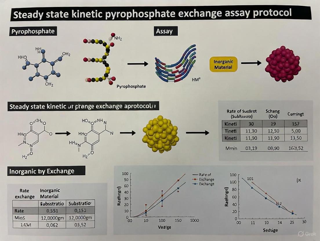
Steady-State Kinetic Pyrophosphate Exchange Assay: Principles, Modern Protocols, and Applications in Biomedical Research
This article provides a comprehensive guide to the steady-state kinetic pyrophosphate (PPi) exchange assay, a fundamental tool for studying enzyme kinetics, particularly aminoacyl-tRNA synthetases (AARSs) and adenylation domains of nonribosomal...
Steady-State Kinetic Pyrophosphate Exchange Assay: Principles, Modern Protocols, and Applications in Biomedical Research
Abstract
This article provides a comprehensive guide to the steady-state kinetic pyrophosphate (PPi) exchange assay, a fundamental tool for studying enzyme kinetics, particularly aminoacyl-tRNA synthetases (AARSs) and adenylation domains of nonribosomal peptide synthetases (NRPS). We cover the foundational principles of the assay, detail both traditional radioactive and contemporary non-radioactive and mass spectrometry-based protocols, and offer practical troubleshooting and optimization strategies. Aimed at researchers, scientists, and drug development professionals, this resource also explores validation techniques and compares the assay's capabilities with other kinetic methods, highlighting its crucial role in enzyme characterization, inhibitor screening, and antibiotic discovery.
Understanding the Pyrophosphate Exchange Assay: Core Principles and Historical Context
The reversible adenylation reaction, central to the function of enzyme classes such as aminoacyl-tRNA synthetases (AARSs) and adenylate-forming enzymes (ANL superfamily), is a fundamental biochemical process. It can be represented by the equation: ATP + Amino Acid ⇌ Aminoacyl-AMP + PPi. The ATP/Pyrophosphate (PPi) Exchange Assay is a classic, powerful kinetic method used to study this activation step, measuring the enzyme's ability to catalyze the reverse reaction by incorporating radiolabeled pyrophosphate into ATP [1] [2]. For decades, this assay has been a cornerstone for characterizing enzyme kinetics, probing amino acid selectivity, and screening for inhibitors in drug development [1]. However, the discontinuation of [³²P]PPi in 2022 created a significant bottleneck for researchers [1]. This application note details a modified protocol that circumvents this issue by using readily available γ-[³²P]ATP, ensuring the continued viability of this essential methodological tool for studying steady-state kinetics [1].
The PPi-ATP exchange assay provides key kinetic parameters that illuminate enzyme efficiency and mechanism. The tables below summarize core principles and representative data for AARS enzymes.
Table 1: Core Kinetic Parameters Measured in PPi-ATP Exchange Studies
| Parameter | Description | Significance in PPi-Exchange Assay |
|---|---|---|
| ( k_{cat} ) | Turnover number: maximum number of substrate molecules converted to product per enzyme active site per unit time. | Reflects the maximum rate of the adenylation (activation) step [3]. |
| ( K_m ) | Michaelis constant: substrate concentration at which the reaction rate is half of ( V_{max} ). | Measures the apparent affinity for the amino acid, ATP, and PPi substrates [3]. |
| ( k{cat}/Km ) | Specificity constant: measures the enzyme's catalytic efficiency. | A high value indicates strong preference for a given substrate, crucial for understanding amino acid selectivity [1] [3]. |
Table 2: Empirical Kinetic Data for E. coli AARSs (Data synthesized from empirical models based on in vitro PPi-exchange and aminoacylation assays) [3]
| Enzyme Class | Enzyme Example | Key Kinetic Characteristic | Burst Kinetics |
|---|---|---|---|
| Class I | Isoleucyl-tRNA synthetase (IleRS), Leucyl-tRNA synthetase (LeuRS) | Burst of aa-tRNA production followed by steady-state rate; product release is often rate-limiting [3]. | Yes [3] |
| Class II | Aspartyl-tRNA synthetase (AspRS) | Steady-state kinetics without a burst phase [3]. | No [3] |
| Class I (tRNA-dependent) | Arginyl-tRNA synthetase (ArgRS), Glutamyl-tRNA synthetase (GluRS) | Amino acid activation requires the presence of cognate tRNA [1] [3]. | Varies |
Detailed Experimental Protocol: The Modified [³²P]ATP/PPi Exchange Assay
This protocol describes the modified assay using γ-[³²P]ATP to measure the adenylation activity of AARSs, based on the method developed to address the discontinuation of [³²P]PPi [1].
Research Reagent Solutions
Table 3: Essential Materials and Reagents
| Item | Function/Description | Example (Supplier/Catalog) |
|---|---|---|
| γ-[³²P]ATP | Radiolabeled substrate; donates ³²P for the reverse reaction to form [³²P]PPi. | γ-[³²P]ATP (Revvity, cat no. BLU002Z) [1] |
| Adenylation Enzyme | The enzyme of interest (e.g., AARS). | Purified recombinant enzyme. |
| Amino Acid Substrate | Specific cognate amino acid for the enzyme. | L-Leucine (Sigma-Aldrich, cat no. 61819) [1] |
| Sodium Pyrophosphate (PPi) | Unlabeled substrate for the reverse reaction. | Sodium pyrophosphate (Sigma, cat no. P-8010) [1] |
| ATP | Unlabeled substrate; accepts the radiolabel from [³²P]PPi. | Adenosine 5'-triphosphate disodium salt hydrate (Sigma, cat no. A2383) [1] |
| Reaction Buffer | Provides optimal pH and ionic conditions. | 20-50 mM HEPES-KOH pH 7.5, MgCl₂, KCl, DTT, BSA [1] |
| Quench Solution | Stops the reaction and denatures the enzyme. | 200 mM sodium acetate, 0.1% SDS, pH 5.0 [1] |
| TLC Plates | Separates [³²P]ATP from [³²P]PPi. | Polyethyleneimine (PEI) cellulose plates (Macherey-Nagel, cat no. 801063) [1] |
| Developing Buffer | Mobile phase for TLC separation. | 0.1 M KH₂PO₄, 4.5 M urea, pH 7.0 with H₃PO₄ [1] |
| Phosphor Storage Screen | Detects and stores the spatial distribution of radioactivity on the TLC plate. | BAS-IP MS (Cytiva, cat no. 28-9564-74) [1] |
| Biomolecular Imager | Visualizes and quantifies the radioactive spots on the phosphor screen. | Amersham Typhoon biomolecular imager (Cytiva) [1] |
Step-by-Step Methodology
Reaction Mixture Setup:
- Prepare the master mix on ice. A standard reaction contains [1]:
- Reaction Buffer (e.g., 20-50 mM HEPES-KOH pH 7.5)
- Magnesium chloride (e.g., 10 mM)
- Potassium chloride (e.g., 50 mM)
- Dithiothreitol (DTT, e.g., 2 mM)
- Bovine Serum Albumin (BSA, e.g., 100 μg/mL)
- Amino acid substrate (at varying concentrations for kinetics)
- Unlabeled ATP
- Unlabeled sodium pyrophosphate
- Critical: Check the pH of the final reaction mixture after all components are added.
- Prepare the master mix on ice. A standard reaction contains [1]:
Initiation and Incubation:
- Pre-incubate the reaction mixture (without enzyme) at the desired temperature (e.g., 37°C) in a microtiter plate or Eppendorf tubes.
- Start the reaction by adding the purified adenylation enzyme and a precise amount of γ-[³²P]ATP.
- Incubate for appropriate time points (e.g., 0, 1, 2, 5, 10 minutes) to capture the initial velocity of the reaction.
Quenching:
- Stop the reaction at each time point by transferring an aliquot into a tube containing the quench solution (200 mM sodium acetate, 0.1% SDS, pH 5.0) [1].
Separation by Thin-Layer Chromatography (TLC):
- Spot each quenched reaction sample onto a PEI-cellulose TLC plate.
- Develop the TLC plate in a glass chamber pre-equilibrated with the developing buffer (0.1 M KH₂PO₄, 4.5 M urea, pH 7.0) until the solvent front nears the top.
- Remove the plate and dry it thoroughly using a hair dryer.
Visualization and Quantification:
- Expose the dried TLC plate to a phosphor storage screen in a cassette for a sufficient time to capture a strong signal.
- Scan the phosphor screen using a biomolecular imager (e.g., Typhoon). The [³²P]PPi (product) and unreacted γ-[³²P]ATP (substrate) will appear as distinct spots with different migration distances (( R_f )).
- Use quantification software (e.g., ImageQuant) to measure the intensity of the [³²P]PPi spot. The amount of [³²P]PPi formed is directly proportional to the rate of the adenylation reaction.
Experimental Workflow and AARS Reaction Pathway
The following diagrams illustrate the core biochemical reaction and the modified experimental workflow.
Diagram 1: AARS adenylation reaction and PPi-ATP exchange equilibrium.
Diagram 2: Modified ATP/PPi exchange assay workflow.
Applications in Kinetic Analysis and Drug Discovery
The modified ATP/PPi exchange assay retains all critical applications of the original method. It is indispensable for steady-state kinetic characterization, allowing researchers to determine ( Km ) and ( k{cat} ) values for amino acid and ATP substrates, which reveals enzyme efficiency and specificity [1] [3]. Furthermore, the assay is ideal for initial velocity studies of the adenylation half-reaction, free from the complicating factors of the subsequent transfer step [1]. This makes it particularly powerful for large-scale inhibitor screens targeting AARSs, which are validated targets for antibiotic development [1] [3]. By measuring the reduction in radiolabeled ATP formation in the presence of test compounds, researchers can rapidly identify potent inhibitors of the adenylation reaction.
Adenylate-forming enzymes represent a functionally conserved yet structurally diverse class of enzymes that catalyze the chemical activation of carboxylate substrates through adenylation. These enzymes facilitate a crucial two-step reaction that begins with the condensation of a carboxylic acid with ATP to form an acyl-adenylate intermediate and inorganic pyrophosphate (PPi). This high-energy intermediate then reacts with a nucleophile (amine, alcohol, or thiol) to generate the final product and AMP [4]. This biochemical strategy mirrors the chemical approach of using activating groups like acid chlorides in organic synthesis, making these enzymes nature's equivalent to sophisticated synthetic chemists [4].
The biological significance of adenylate-forming enzymes spans fundamental metabolic pathways including ribosomal and non-ribosomal peptide synthesis, fatty acid oxidation, enzyme regulation, and secondary metabolite production. Their essential roles make them potential drug targets, exemplified by D-alanyl carrier protein ligase DltA in bacterial systems [4]. Modern classification systems have organized these enzymes based on their common chemistry into three major classes: Class I (including non-ribosomal peptide synthetases, acyl/aryl-CoA synthetases, and luciferases), Class II (aminoacyl-tRNA synthetases), and Class III (NRPS-independent siderophore synthetases) [4].
Enzyme Classification and Kinetic Parameters
Adenylate-forming enzymes demonstrate remarkable diversity in their structural features while conserving core chemical mechanisms. The table below summarizes the key characteristics and kinetic parameters across different enzyme classes.
Table 1: Classification and Properties of Adenylate-Forming Enzymes
| Enzyme Class | Representative Members | Domain Architecture | Metal Ion Coordination | Key Catalytic Residues |
|---|---|---|---|---|
| Class I | NRPS A-domains, Acetyl-CoA synthetases, Luciferases | Large N-terminal + small C-terminal domain connected by flexible hinge [4] | Variable geometry (α-β, β-γ, or α-γ phosphate coordination) [4] | Arg, Lys, or His near α-phosphate; conserved Thr [4] |
| Class II | Aminoacyl-tRNA synthetases (aaRSs) | Multiple domains: zinc-binding, catalytic, anticodon-binding, amino acid-specific [4] | Multiple Mg²⁺ ions coordinating α/β and β/γ phosphates [4] | Strictly conserved Arg (e.g., R262 in LysU) [4] |
| Class III | NRPS-independent siderophore synthetases | Three domains (N-terminal, central, C-terminal) forming deep cavity [4] | Single Mg²⁺ coordinating α-γ phosphate [4] | Conserved His and Arg near α-phosphate [4] |
Table 2: Kinetic Assay Comparison for Adenylate-Forming Enzymes
| Assay Method | Principle | Detection Method | Applications | Advantages/Limitations |
|---|---|---|---|---|
| ATP/[³²P]PPi Exchange | Equilibrium exchange of radiolabel between PPi and ATP using [³²P]PPi [1] | TLC separation and phosphor storage imaging [1] | AARSs, ANL superfamily enzymes [1] | Historically standard but limited by [³²P]PPi discontinuation [1] |
| Modified [³²P]ATP/PPi Assay | Equilibrium exchange using γ-[³²P]ATP as labeled compound [1] | TLC separation and phosphor storage imaging [1] | AARSs, inhibitor screening [1] | Uses readily available γ-[³²P]ATP; good agreement with standard assay [1] |
| Aminoacylation Assay | Cumulative two-step aminoacylation | Radiolabeled amino acids (¹⁴C, ³H, ³²S) or ³²P-labeled tRNA [1] | Functional analysis of complete AARS reaction [1] | Measures overall activity but doesn't isolate activation step [1] |
Experimental Protocols
Modified ATP/PPi Exchange Assay for Amino Acid Activation
Principle: This protocol measures the amino acid activation step of aminoacyl-tRNA synthetases (AARSs) and other adenylate-forming enzymes through an equilibrium-based exchange reaction. The assay follows the conversion of γ-[³²P]ATP to [³²P]PPi as a proxy for the adenylate formation step, providing a crucial tool for kinetic characterization and inhibitor screening [1].
Reagents and Solutions:
- Reaction Buffer: 20-50 mM HEPES-KOH (pH 7.5), magnesium chloride hexahydrate, potassium chloride, dithiothreitol (DTT), bovine serum albumin (BSA)
- Substrates: Sodium pyrophosphate, adenosine 5'-triphosphate disodium salt hydrate, appropriate amino acid (e.g., L-leucine, L-isoleucine)
- Radiolabel: γ-[³²P]ATP
- Quench Solution: Sodium acetate, acetic acid, sodium dodecyl sulphate (SDS)
- TLC Mobile Phase: Urea, potassium dihydrogen phosphate, phosphoric acid
Procedure:
- Reaction Setup: Prepare reaction mixture containing buffer, 2.5 mM ATP, 5 mM MgCl₂, 10 mM KF, 0.1 mg/mL BSA, 0.5 mM sodium pyrophosphate, appropriate amino acid concentration, and enzyme.
- Initiation: Start the reaction by adding γ-[³²P]ATP.
- Incubation: Maintain reactions at 25°C in a dry block heater.
- Quenching: At specific time intervals, remove aliquots and mix with quench solution to stop the reaction.
- Separation: Spot quenched samples on polyethyleneimine TLC plates.
- Chromatography: Develop TLC plates in mobile phase containing 2.5 M urea, 3.75 mM KH₂PO₄, and 3.75 mM H₃PO₄.
- Detection: Dry plates and expose to phosphor storage screens.
- Visualization and Analysis: Image using a Typhoon biomolecular imager and quantify using ImageQuant software [1].
Critical Considerations:
- The assay is suitable for most AARSs that activate amino acids independently of tRNA, except for arginyl-, glutamyl-, glutaminyl-, and class I lysyl-tRNA synthetases which require tRNA for activation [1].
- Enzyme concentration should be optimized to ensure linear initial rates.
- Control reactions without enzyme or without amino acid should be included to account for non-specific activity.
Steady-State Kinetic Analysis
Data Interpretation: Kinetic parameters (KM, Vmax, kcat) are derived by measuring initial velocities at varying substrate concentrations and fitting data to the Michaelis-Menten equation. For the ATP/PPi exchange assay, the measured rate represents the activation step specifically, unlike cumulative aminoacylation assays that combine both steps of the reaction [1] [5].
Research Reagent Solutions
Table 3: Essential Research Reagents for Adenylate-Forming Enzyme Studies
| Reagent/Category | Specific Examples | Function/Application |
|---|---|---|
| Radiolabeled Compounds | γ-[³²P]ATP, [³²P]PPi (custom), ¹⁴C-amino acids | Tracing reaction progress; equilibrium exchange measurements; product formation quantification [1] |
| Enzyme Sources | Recombinant AARSs, NRPS adenylation domains, Acyl-CoA synthetases | Functional assays; substrate specificity profiling; structural studies [4] [1] |
| Essential Cofactors | Mg²⁺ or Mn²⁺ salts, ATP, inorganic pyrophosphate | Essential divalent cation for catalysis [4]; primary substrate; reaction product and exchange substrate [1] |
| Chromatography Materials | Polyethyleneimine TLC plates, urea, phosphate buffers | Separation of ATP from PPi in exchange assays [1] |
| Detection Systems | Phosphor storage screens, Typhoon biomolecular imager, scintillation counters | Visualization and quantification of radiolabeled compounds [1] |
Signaling Pathways and Experimental Workflows
Adenylate-forming enzymes represent a fascinating convergence of chemical logic and biological diversity, employing a conserved activation mechanism across structurally distinct scaffolds. The experimental approaches outlined in this application note, particularly the steady-state kinetic pyrophosphate exchange assays, provide powerful tools for elucidating the function, mechanism, and inhibition of these biologically significant enzymes. The continuous refinement of these protocols, including the adaptation to available reagents as demonstrated by the modified ATP/PPi exchange assay, ensures that researchers can effectively study these essential enzymes for both basic science and drug discovery applications.
The ATP/[32P]Pyrophosphate (PPi) exchange assay stands as a cornerstone technique in the history of enzymology, providing critical insights into the mechanisms of enzyme-catalyzed reactions that involve adenosine triphosphate (ATP) hydrolysis and the formation of enzyme-bound acyl-adenylate intermediates. This classic radioactive method has been indispensable for studying the steady-state kinetics of a diverse family of enzymes, primarily aminoacyl-tRNA synthetases (AARSs) and the adenylation domains (A-domains) of non-ribosomal peptide synthetases (NRPSs) [1] [6]. For over half a century, since its development in the 1960s, this assay has enabled researchers to probe the fundamental process of amino acid activation, a crucial initial step in protein synthesis and natural product biosynthesis [3]. The assay's principle is rooted in the reversibility of the adenylation reaction; it measures the enzyme's ability to catalyze the exchange of radiolabeled pyrophosphate ([32P]PPi) into ATP, thereby serving as a direct proxy for the activation step independently of the subsequent transfer step [1]. Despite recent challenges in reagent availability prompting methodological adaptations, the ATP/[32P]PPi exchange assay remains a powerful tool for kinetic characterization, substrate profiling, and inhibitor screening in both basic science and drug development [1] [6].
Historical Background and Development
The history of the ATP/[32P]PPi exchange assay is deeply intertwined with the maturation of biochemistry and radiochemistry in the mid-20th century. The discovery of radioactivity by Henri Becquerel in 1896 and the subsequent pioneering work of Marie and Pierre Curie, who isolated polonium and radium, laid the foundational knowledge that would eventually make such tracer methods possible [7]. The application of radioactive isotopes in biochemical assays became practical after the seminal work of Ernest Rutherford and Frederick Soddy, who formulated the theory of radioactive decay in 1902, explaining how elements transmute through the emission of radiation [7].
The ATP/[32P]PPi exchange assay emerged as a direct application of these principles to study the kinetics of enzymes like aminoacyl-tRNA synthetases. As noted in historical kinetic analyses, "Since roughly the mid 1960’s, there has been tremendous work on elucidating the kinetic mechanism of tRNA charging by aminoacyl tRNA synthetases (AARS). Pioneering experimental work on AARSs started with the examination of their pyrophosphate exchange kinetics" [3]. This assay provided the first means to isolate and study the initial activation step of the aminoacylation reaction, which occurs as follows: Amino Acid + ATP ⇄ Aminoacyl-AMP + PPi The assay capitalizes on the reversibility of this reaction; in the presence of [32P]PPi, a functioning enzyme will incorporate the radioactive label into the newly synthesized ATP molecule [1] [3]. For decades, this method was the gold standard for measuring the substrate specificity and catalytic efficiency of adenylating enzymes, forming the basis for our understanding of their kinetic mechanisms [6] [3]. Its utility was later extended to the adenylation domains of NRPSs, key enzymes in the biosynthesis of important natural products with activities ranging from antibiotic to antitumor effects [6].
Principle and Mechanism of the Assay
Biochemical Basis
The ATP/[32P]PPi exchange assay is fundamentally an equilibrium-based isotopic exchange method that tracks the incorporation of a radioactive label from pyrophosphate into adenosine triphosphate. The assay is uniquely suited for enzymes that catalyze the reversible formation of an acyl-adenylate intermediate, a reaction mechanism shared by aminoacyl-tRNA synthetases (AARSs), acyl-CoA synthetases, and the adenylation domains of non-ribosomal peptide synthetases (NRPSs) [1] [3]. These enzymes all facilitate a two-step reaction where the first step involves the activation of a carboxylate substrate (e.g., an amino acid or a carboxylic acid) with ATP to form an enzyme-bound acyl-adenylate and inorganic pyrophosphate (PPi).
The core chemical reaction monitored by the assay is: Enzyme + Substrate + ATP ⇄ Enzyme–Substrate–AMP + PPi
In the presence of the enzyme, the reaction rapidly reaches equilibrium. When [32P]PPi is added to the reaction mixture, the enzyme catalyzes the reverse reaction, incorporating the labeled pyrophosphate into ATP. The rate of this exchange is directly proportional to the enzyme's catalytic activity in the adenylation step [1] [3]. It is critical to note that this assay specifically measures the activation step independently of the subsequent transfer step, which, for AARSs, would be the transfer of the aminoacyl moiety to the cognate tRNA [1]. This separation of the two chemical steps is a key advantage, as it allows researchers to study adenylation kinetics without requiring the often laborious production of tRNA [1].
Workflow and Logic
The following diagram illustrates the logical workflow and fundamental components of the classic ATP/[32P]PPi exchange assay.
Key Applications in Research
The ATP/[32P]PPi exchange assay has been instrumental across multiple domains of biochemical research. The following table summarizes its primary applications and the significance in each area.
Table 1: Key Applications of the ATP/[32P]PPi Exchange Assay
| Application Area | Specific Use | Significance |
|---|---|---|
| Enzyme Kinetics & Mechanism | Determination of kinetic parameters (kcat, KM) for the adenylation step [3]. | Provided foundation for understanding AARS complex kinetic mechanisms and classification into Class I/II [3]. |
| Substrate Specificity Profiling | Screening alternative/promiscuous substrates for adenylation domains [6]. | Enabled engineering of NRPSs for novel non-ribosomal peptides with potential drug candidate properties [6]. |
| Drug Discovery & Screening | Identification and characterization of enzyme inhibitors, particularly for AARSs [1]. | Validated AARSs as targets for antibiotic development [1] [3]. |
| Mutant Enzyme Characterization | Assessment of the functional impact of mutations on adenylation activity [8] [6]. | Essential for directed evolution and structure-function studies; example: T278A mutant of TycA A-domain showed 330-fold lower kcat/KM [6]. |
Standard Protocol and Methodologies
Classic Radioactive Assay Protocol
The standard ATP/[32P]PPi exchange assay, as refined over decades, involves a series of carefully optimized steps. The following protocol is synthesized from methodologies used for AARSs and NRPS A-domains [9] [1] [6].
Reaction Mixture Setup: A typical 100 µL reaction mixture contains:
- 50-100 mM Buffer (Tris-HCl, pH 7.5-8.5, or HEPES-KOH, pH 7.5) [9] [1].
- 10 mM MgCl₂ (Magnesium is an essential cofactor) [9].
- 5 mM ATP (Adenosine 5'-triphosphate disodium salt) [9].
- 1-5 mM Amino Acid Substrate (The specific amino acid or alternative substrate being profiled) [10] [9].
- 2-4 mM Na₄PPi containing [32P]PPi (e.g., 2–6 × 10⁵ cpm/mL) [9].
- 5 mM Dithiothreitol (DTT) (For reducing environment) [9].
- 200 mM KCl or NaCl (For ionic strength) [9].
Initiation and Incubation: The reaction is initiated by the addition of the enzyme (e.g., 1 µM final concentration for NRPS A-domains [9] or a determined concentration of AARS). The reaction mixture is incubated at room temperature (or a specified temperature like 25°C) for a set time (e.g., 30 minutes) [9].
Reaction Quenching: The reaction is stopped by adding a charcoal suspension (e.g., 200 µL of 16 g/L activated charcoal in 100 mM Na₄PPi and 3.5% HClO₄) [9]. The strong acid denatures the enzyme, and the charcoal selectively binds nucleotides (ATP and the newly formed [32P]ATP).
Separation and Washing: The sample is centrifuged (e.g., 13,000 rpm for 3 minutes) to pellet the charcoal with the bound nucleotides. The supernatant, containing unincorporated [32P]PPi, is carefully removed. The pellet is typically washed twice with a wash buffer (e.g., 200 µL of 100 mM Na₄PPi, 3.5% HClO₄) to remove any residual free [32P]PPi [9].
Detection and Quantification: The final charcoal pellet is re-suspended in a scintillation cocktail (e.g., 300 µL of wash buffer added to 10 mL of Ultima Gold scintillation fluid). Radioactivity is measured using a scintillation counter (e.g., Beckman LS 6000SC). The amount of radioactivity detected is directly proportional to the amount of [32P]ATP formed, which reflects the enzyme's adenylation activity [9].
The Scientist's Toolkit: Essential Reagents and Materials
Successful execution of the ATP/[32P]PPi exchange assay requires specific reagents and equipment. The following table catalogs the core components of this methodological toolkit.
Table 2: Essential Research Reagent Solutions and Materials for the ATP/[32P]PPi Exchange Assay
| Category | Item | Function / Specification |
|---|---|---|
| Radioactive Substrate | [32P]Pyrophosphate ([32P]PPi) | Radiolabeled tracer for exchange reaction. Note: Commercial production was largely discontinued in 2022, creating a major challenge for the classic protocol [1]. |
| Core Reaction Components | ATP (Adenosine 5'-triphosphate) | Substrate for the adenylation reaction [9]. |
| Amino Acid / Carboxylic Acid | The specific substrate being activated by the enzyme [10]. | |
| MgCl₂ | Essential divalent cation cofactor for most adenylating enzymes [9]. | |
| Dithiothreitol (DTT) | Reducing agent to maintain enzyme stability and active site cysteine residues [9]. | |
| Separation & Detection | Activated Charcoal Suspension | Binds nucleotides (ATP/[32P]ATP) for separation from unincorporated [32P]PPi [9]. |
| Scintillation Cocktail & Vials | For emulsifying the sample and detecting beta radiation from 32P [9]. | |
| Equipment | Scintillation Counter | Instrument to quantify radioactivity (e.g., Beckman LS 6000SC) [9]. |
| Temperature-Controlled Incubator | For maintaining consistent reaction temperature [9]. | |
| Microcentrifuge | For pelleting charcoal after quenching [9]. |
Modern Adaptations and Recent Developments
The landscape of the ATP/[32P]PPi exchange assay has shifted significantly due to the discontinuation of commercial [32P]PPi in 2022, which made the classic protocol "much less convenient" and far less accessible [1]. This challenge has spurred the development of innovative adaptations to maintain the assay's utility.
The [32P]ATP/PPi Assay: A Modern Solution
A primary modern workaround is a modified protocol that inverts the labeling strategy. Instead of using radiolabeled PPi, this new "[32P]ATP/PPi assay" uses the readily available γ-[32P]ATP as the radioactive tracer [1]. In this format, the enzyme catalyzes the forward reaction in the presence of unlabeled PPi, generating [32P]PPi from γ-[32P]ATP. The reaction is quenched at specific time points, and the products are separated via thin-layer chromatography (TLC). The TLC plate is then exposed to a phosphor storage screen, and the results are visualized and quantified using a biomolecular imager (e.g., Typhoon imager) and specialized software (e.g., ImageQuant) [1]. This method has been successfully used to characterize the kinetics of AARSs like isoleucyl-tRNA synthetase (IleRS) and leucyl-tRNA synthetase (LeuRS), with results showing "good agreement with the previously published kinetic constants obtained with the standard ATP/[32P]PPi exchange assay" [1].
Non-Radiometric Alternative Methods
In parallel, fluorescence-based methods have been developed as non-radioactive alternatives for studying nucleotide binding, though they measure binding affinity rather than the catalytic exchange itself. These assays utilize fluorescent ATP analogs like TNP-ATP or MANT-ATP [8]. Upon binding to the enzyme's active site, these analogs exhibit a significant increase in fluorescence intensity and a shift in their emission maximum. This signal change can be monitored in real-time using a spectrofluorometer. While the binding kinetics of these analogs differ from native ATP, the assay is advantageous due to its safety, ease of use, and suitability for high-throughput screening of inhibitors that compete with ATP for binding [8].
Experimental Workflow and Data Analysis
Quantitative Data from Literature
The ATP/[32P]PPi exchange assay has generated a wealth of quantitative kinetic data over the years. The table below summarizes representative data for different enzymes, highlighting the assay's application in measuring key catalytic parameters.
Table 3: Exemplary Kinetic Data Determined via ATP/[32P]PPi Exchange Assay
| Enzyme | Substrate | KM (µM) | kcat (s⁻¹) | kcat/KM (s⁻¹ M⁻¹) | Context / Reference |
|---|---|---|---|---|---|
| TycA A-domain (Wild-type) | Phenylalanine | Not Specified | Not Specified | Baseline | Used as reference for high-throughput screening [6]. |
| TycA A-domain (T278A mutant) | Phenylalanine | Not Specified | Not Specified | 330-fold lower | Demonstrated large dynamic range for mutant screening [6]. |
| E. coli AARSs | Various Cognate Amino Acids | Wide range reported in literature | Wide range reported in literature | Modeled empirically | Data used to parametrize kinetic models of in vivo translation [3]. |
High-Throughput Implementation
To meet the demands of drug discovery and enzyme engineering, the ATP/[32P]PPi exchange assay was successfully adapted to a 96-well format. This optimization allowed for the rapid measurement of "approximately 1000 data points per week" and enabled quantitative high-throughput screening of A-domain substrate specificity [6]. This format was crucial for the directed evolution of NRPS A-domains and for the precise kinetic assessment of mutant enzymes, significantly accelerating research in combinatorial biosynthesis [6]. The workflow for this high-throughput approach is summarized below.
The ATP-pyrophosphate (PPi) exchange assay is a foundational steady-state kinetic method used to study the first half-reaction catalyzed by adenylate-forming enzymes, such as aminoacyl-tRNA synthetases (aaRSs). This assay specifically monitors the enzyme's ability to activate an amino acid by measuring the reverse reaction of adenylate formation. In the presence of enzyme, amino acid, ATP, and radio-labeled inorganic pyrophosphate ([³²P]-PPi), the assay quantifies the incorporation of radioactivity into ATP. The measured rate of [³²P]-ATP formation is directly proportional to the forward rate of aminoacyl-adenylate formation, allowing researchers to determine the key kinetic constants, (k{cat}) and (Km), for the substrates involved in the activation reaction. This protocol is essential for initial enzyme characterization, inhibitor screening, and studies of substrate specificity [11] [2] [12].
Theoretical Foundation and Key Kinetic Parameters
The Underlying Chemical Reaction
The PPi exchange assay monitors the reversibility of the initial adenylation reaction. For aminoacyl-tRNA synthetases, this is the first step in the two-step aminoacylation process [12]:
- Adenylation (Activation):
E + AA + ATP ⇄ E•AA~AMP + PPi - Aminoacyl Transfer:
E•AA~AMP + tRNA^AA ⇄ E + AA-tRNA^AA + AMP
The exchange assay focuses exclusively on the first, reversible step. The presence of excess labeled PPi drives the reverse reaction, leading to the re-formation of ATP from the E•AA~AMP complex. The rate of this exchange is contingent upon the formation of the adenylate intermediate, making it a direct reporter on the activation kinetics [12].
Relationship Between Exchange Data and Kinetic Constants
In steady-state kinetics, the measured velocity ((v)) of the PPi exchange reaction is used to determine the Michaelis-Menten parameters, (k{cat}) and (Km), for the enzyme's substrates (amino acid and ATP). The standard Michaelis-Menten equation is applied:
[v = \frac{V{max}[S]}{Km + [S]}]
Where:
- (V_{max}) is the maximum reaction velocity.
- ([S]) is the substrate concentration (AA or ATP).
- (Km) is the Michaelis constant, representing the substrate concentration at half (V{max}).
- (k{cat}) (the turnover number) is calculated as (V{max} / [Et]), where ([Et]) is the total enzyme concentration.
The parameter (k{cat}/Km) describes the catalytic efficiency of the enzyme for a particular substrate [13] [12].
Experimental Protocol for the PPi Exchange Assay
Research Reagent Solutions
The following table details the essential materials and reagents required to perform a standard PPi exchange assay.
Table 1: Key Research Reagent Solutions for the PPi Exchange Assay
| Reagent / Solution | Function / Description | Key Considerations |
|---|---|---|
| Recombinant Enzyme (aaRS) | The enzyme catalyst for the adenylation reaction. | Purified to homogeneity; concentration accurately determined [12]. |
| Amino Acid Substrate | One of the primary substrates for the activation reaction. | Prepare a dilution series covering a range above and below the expected (K_m) [12]. |
| ATP Solution | The second primary substrate and source of the AMP moiety. | Typically used with Mg²⁺ as a cofactor (e.g., Mg-ATP) [12]. |
| [³²P]-Pyrophosphate (PPi) | Radio-labeled tracer to monitor the reverse reaction. | The key detectable reagent; requires handling per radiation safety protocols [2] [12]. |
| Reaction Buffer | Provides optimal pH and ionic conditions for enzyme activity. | Commonly includes Tris-HCl or HEPES, KCl, MgCl₂, DTT [12]. |
| Stop Solution | Halts the enzymatic reaction at precise time intervals. | Typically contains an acidic agent (e.g., TCA) and unlabeled PPi to dilute label [12]. |
| Charcoal Suspension (ACB) | Binds/binds nucleotides (ATP, AMP) but not inorganic PPi. | Used to separate [³²P]-ATP from unused [³²P]-PPi [12]. |
Step-by-Step Workflow
The following diagram illustrates the core experimental workflow of the PPi exchange assay, from reaction initiation to data acquisition.
Detailed Procedural Steps
- Reaction Setup: Prepare a master mix containing reaction buffer, MgCl₂, and ATP. Aliquot this mix into separate tubes.
- Initiation: Start the enzymatic reaction by simultaneously adding the enzyme and the radio-labeled [³²P]-PPi solution to the master mix. Vortex immediately to ensure homogeneity.
- Timed Incubation: Incubate the reaction mixture at a constant temperature (e.g., 37°C).
- Quenching: At predetermined time intervals (e.g., 0, 30, 60, 120 seconds), withdraw aliquots from the reaction mixture and dispense them into tubes containing a large excess of cold stop solution (e.g., 5% Trichloroacetic acid (TCA) with unlabeled PPi). This step instantly denatures the enzyme and halts the reaction.
- Separation and Quantification: a. Transfer the quenched aliquots to a vessel containing activated charcoal (ACB) suspension. The ACB selectively binds nucleotide phosphates (ATP, ADP, AMP) while leaving inorganic PPi in solution. b. Wash the charcoal-bound nucleotides multiple times with distilled water to remove any residual unincorporated [³²P]-PPi. c. Transfer the charcoal with the bound [³²P]-ATP to a scintillation vial, add scintillation fluid, and measure the radioactivity using a scintillation counter. The measured counts per minute (CPM) are proportional to the amount of [³²P]-ATP formed.
Data Analysis and Interpretation
From Raw Data to Kinetic Constants
The analysis involves transforming the raw radioactivity measurements into reaction velocities and fitting the data to determine (Km) and (k{cat}).
- Calculate Initial Velocity (v): For each substrate concentration, plot the amount of [³²P]-ATP formed (in CPM or converted to moles) versus time. The slope of the linear portion of this progress curve represents the initial velocity, (v), typically expressed in nM/s or pmol/min.
- Plot and Curve Fitting: Plot the initial velocity ((v)) against the varying concentration of one substrate (e.g., amino acid) while keeping the other substrates (ATP, Mg²⁺) at saturating, fixed concentrations.
- Nonlinear Regression: Fit the resulting data points to the Michaelis-Menten equation using nonlinear regression software (e.g., GraphPad Prism). This fit directly yields the (V{max}) and (Km) for that substrate.
- Calculate (k{cat}): Divide the obtained (V{max}) by the total molar concentration of active enzyme sites in the reaction (([Et])) to determine the turnover number: (k{cat} = V{max} / [Et]).
The table below provides a generalized overview of the kinetic parameters that can be expected for aminoacyl-tRNA synthetases, based on empirical models and experimental observations.
Table 2: Representative Kinetic Parameters for E. coli Aminoacyl-tRNA Synthetases from Pyrophosphate Exchange and Aminoacylation Data (Adapted from [11])
| Enzyme Class | Representative Enzymes | Typical (K_m) (Amino Acid) (μM) | Typical (k_{cat}) (s⁻¹) | Key Kinetic characteristic |
|---|---|---|---|---|
| Class I AARS | CysRS, ArgRS, ValRS | Low to mid micromolar range | Variable, can be >10 s⁻¹ | Often displays "burst kinetics" in pre-steady-state aminoacylation [11]. |
| Class II AARS | SerRS, ThrRS, HisRS | Low to mid micromolar range | Variable, can be >10 s⁻¹ | Generally displays no burst kinetics; steady-state production of aa-tRNA [11]. |
Advanced Applications and Considerations
Limitations and Complementary Assays
While powerful, the PPi exchange assay has limitations. It does not provide information on the second half-reaction (aminoacyl transfer to tRNA) and may not be suitable for enzymes that do not rapidly release PPi [2]. For a complete kinetic picture, the PPi exchange assay should be complemented with other methods:
- Aminoacylation Assay: Directly measures the formation of aminoacyl-tRNA, reporting on the combined kinetics of both the activation and transfer steps [12].
- Pre-steady-state Kinetics (Rapid Quench/Stopped-Flow): These methods dissect the reaction into its elementary steps, allowing for the direct measurement of individual rate constants for adenylate formation ((k{chem})) and transfer ((k{tran})) [11] [12].
- Continuous Spectrophotometric Assays: Methods like the coupled hydroxamate–MesG assay offer a non-radioactive, continuous alternative for monitoring the adenylation reaction, which is particularly useful for inhibitor screening [2].
Troubleshooting Common Issues
- Low Signal-to-Noise Ratio: Ensure enzyme is active and not denatured. Optimize substrate concentrations and verify the specific activity of the [³²P]-PPi.
- Non-Linear Progress Curves: If the reaction velocity decreases rapidly, check for enzyme instability, product inhibition, or substrate depletion.
- High Background: Increase the number and volume of washes after charcoal binding to remove all unincorporated [³²P]-PPi thoroughly.
- Inconsistent Replicates: Standardize pipetting, mixing, and quenching times precisely across all samples.
Executing the Assay: Step-by-Step Protocols from Classic to Modern Methods
Within the framework of steady-state kinetic pyrophosphate exchange assay research, the ATP/PPi exchange assay has served as a fundamental tool for elucidating the mechanisms of enzymes that catalyze adenylate-forming reactions [1]. This assay traditionally monitors the amino acid activation step catalyzed by aminoacyl-tRNA synthetases (AARSs), essential enzymes that couple cognate amino acids to their corresponding tRNAs for protein synthesis [11] [1]. For over half a century, the standard method for following this activation involved an ATP/[32P]PPi exchange assay, which relies on the equilibrium exchange of radiolabel between pyrophosphate (PPi) and ATP [1]. However, the commercial discontinuation of [32P]PPi in 2022 created a significant methodological gap for researchers studying AARSs and other enzymes from the ANL superfamily, such as nonribosomal peptide synthetases [1]. This application note details a modernized protocol that circumvents this challenge by using readily available γ-[32P]ATP, ensuring the continued viability of this critical kinetic characterization method in drug development and basic research.
Principle of the Assay
Biochemical Basis
AARSs catalyze aminoacylation in two discrete chemical steps [1]:
- Amino Acid Activation: The amino acid (aa) is activated by ATP to form an aminoacyl-adenylate intermediate (aa-AMP) and inorganic pyrophosphate (PPi).
- Aminoacyl Transfer: The aminoacyl moiety is transferred from aa-AMP to the 3'-end of its cognate tRNA, yielding aminoacylated tRNA (aa-tRNA).
The modern [32P]ATP/PPi exchange assay isolates and monitors the first activation step. In the forward direction of the activation reaction, the enzyme catalyzes the formation of aa-AMP and PPi from an amino acid and ATP. At equilibrium, the enzyme also catalyzes the reverse reaction. The assay leverages this reversibility by providing a radiolabel in the form of γ-[32P]ATP. As the reverse reaction occurs, the enzyme transfers the radiolabeled gamma-phosphate from ATP to PPi, generating [32P]PPi. The rate of this isotopic exchange is directly proportional to the rate of the amino acid activation step, serving as a proxy for AARS activity [1].
Experimental Workflow
The following diagram outlines the core procedural workflow for the [32P]ATP/PPi exchange assay:
Materials and Reagents
Research Reagent Solutions
The following table catalogues the essential materials required to establish this protocol.
Table 1: Essential Research Reagents and Materials
| Item | Function/Application | Specific Example(s) |
|---|---|---|
| γ-[32P]ATP | Radiolabeled substrate; source of the 32P for exchange. | Revvity, cat. no. BLU002Z [1]. |
| Aminoacyl-tRNA Synthetase (AARS) | Enzyme of interest; catalyzes the exchange reaction. | Purified IleRS, LeuRS, or other AARS [1]. |
| Polyethyleneimine (PEI) TLC Plates | Stationary phase for separation of [32P]PPi from γ-[32P]ATP. | Macherey-Nagel, cat. no. 801063 [1]. |
| Adenosine 5'-triphosphate (ATP) | Essential substrate for the activation reaction. | Sigma, cat. no. A2383 [1]. |
| Sodium Pyrophosphate (PPi) | Essential substrate for the reverse reaction. | Sigma, cat. no. P-8010 [1]. |
| Amino Acid(s) | Specific substrate for the AARS being studied. | L-leucine (cat. no. 61819), L-isoleucine (cat. no. 58879) [1]. |
| TLC Developing Buffer | Mobile phase for chromatographic separation. | 0.1 M potassium phosphate, 6 M urea, pH 7.0 [1]. |
| Quench Solution | Stops the enzymatic reaction and denatures the protein. | 400 mM sodium acetate, 2% SDS, pH 5.0 [1]. |
| Phosphor Storage Screen | Captures the spatial distribution of radioactivity on the TLC plate. | Cytiva, BAS-IP MS [1]. |
Preparation of Key Solutions
Assay Buffer (100 mL):
- 20-50 mM HEPES-KOH, pH 7.5
- 10-20 mM MgCl₂
- 5-50 mM KCl
- 1-2 mM dithiothreitol (DTT)
- 0.1-0.5 mg/mL bovine serum albumin (BSA) [1].
TLC Developing Buffer (1 L):
- Dissolve 360 g of urea (6 M final concentration) in approximately 600 mL of deionized water.
- Add 100 mL of 1 M potassium dihydrogen phosphate (KH₂PO₄).
- Adjust the pH to 7.0 using phosphoric acid or KOH.
- Bring the final volume to 1 L with deionized water [1].
Step-by-Step Protocol
Activation Reaction
- Reaction Mixture Setup: On ice, prepare a master mix containing the assay buffer, unlabeled ATP, PPi, and the respective amino acid substrate.
- Initiation: Add the purified AARS enzyme and the γ-[32P]ATP to the master mix to initiate the reaction. A typical final reaction volume is 50-100 µL.
- Incubation: Incubate the reaction mixture at a constant temperature (e.g., 25°C or 37°C) for a predetermined time to allow the exchange reaction to proceed. A time course (e.g., 0, 2, 5, 10, 20 minutes) is recommended for initial experiments [1].
- Quenching: At specific time points, withdraw aliquots from the reaction and immediately mix them with a quench solution (e.g., 400 mM sodium acetate, 2% SDS, pH 5.0) to stop the enzymatic activity [1].
Thin-Layer Chromatography
- Spotting: Using a micropipette, spot a small volume (e.g., 1-2 µL) of the quenched reaction mixture onto a pre-marked origin line on a PEI-cellulose TLC plate.
- Development: Place the spotted TLC plate in a glass chamber pre-equilibrated with the developing buffer (0.1 M potassium phosphate, 6 M urea, pH 7.0). Ensure the liquid level is below the spotted samples.
- Separation: Allow the mobile phase to migrate up the plate until it nearly reaches the top (approximately 30-45 minutes).
- Drying: Carefully remove the plate from the chamber and dry it thoroughly using a hair dryer [1].
The TLC separation capitalizes on the different charges of the substrate and product. PPi, being more negatively charged, migrates slower than ATP in the phosphate-based buffer, resulting in clear separation.
Visualization and Data Analysis
- Exposure: Place the dried TLC plate in an exposure cassette with a phosphor storage screen. The screen should be exposed to the plate for several hours or until a sufficient signal is acquired.
- Scanning: Scan the phosphor screen using a biomolecular imager (e.g., Amersham Typhoon).
- Quantification: Use image analysis software (e.g., ImageQuant) to quantify the intensity of the [32P]PPi spots.
- Kinetic Analysis: Plot the quantified [32P]PPi intensity against time to determine the initial velocity of the exchange reaction. These velocities can then be used in Michaelis-Menten plots to derive kinetic constants (Km, kcat) for the amino acid activation [1].
Applications and Significance
This modernized protocol is critical for several research domains:
- Mechanistic Enzyme Studies: It allows for the detailed kinetic characterization of the amino acid activation step independent of the subsequent transfer step, which is crucial for understanding the complete kinetic mechanism of AARSs [1].
- Drug Discovery and Antibiotic Development: AARSs are prominent targets for antibiotics and other anti-infectives [14]. The assay is ideal for high-throughput screening of inhibitors targeting the activation step of AARSs, as it can be performed in the absence of tRNA, which is laborious to produce [1].
- Investigating Substrate Selectivity: The protocol is a critical tool for determining the initial amino acid selectivity and fidelity of AARSs, which is fundamental to the accurate decoding of genetic information [1] [11].
Table 2: Representative Kinetic Data Obtained via the [32P]ATP/PPi Assay
| Enzyme (AARS) | Variable Substrate | Apparent Km (mM) | Apparent kcat (s⁻¹) | Reference / Validation |
|---|---|---|---|---|
| Isoleucyl-tRNA Synthetase (IleRS) | Isoleucine | Data consistent with values from traditional ATP/[32P]PPi exchange assay [1]. | ||
| Leucyl-tRNA Synthetase (LeuRS) | Leucine | Data consistent with values from traditional ATP/[32P]PPi exchange assay [1]. | ||
| AgnB2 (Self-immunity LeuRS) | ATP | ~10-fold higher Km for ATP in ATP/PPi exchange vs. aminoacylation [14]. |
The kinetic constants (Km and kcat) obtained with this modified assay show excellent agreement with those derived from the traditional ATP/[32P]PPi exchange method, confirming its reliability and accuracy for steady-state kinetic analysis [1].
The study of kinase and synthetase activity through pyrophosphate exchange assays has long been a cornerstone of enzymatic kinetics research. Traditional methods have predominantly relied on radioactive isotopes such as γ-[32P]ATP or [32P]PPi to monitor the transfer of phosphate groups. However, the recent discontinuation of [32P]PPi in 2022 has created a significant methodological challenge for researchers studying aminoacyl-tRNA synthetases (AARS) and other enzymes utilizing ATP/PPi exchange mechanisms [1]. This development has accelerated the need for robust, non-radioactive alternatives that maintain analytical precision while improving safety profiles.
Mass spectrometry-based assays utilizing γ-[18O4]-labeled ATP represent an innovative solution to this challenge. This approach leverages stable isotope labeling combined with contemporary quantitative proteomic workflows to enable precise detection of phosphorylation and aminoacylation events without radioactive materials [15]. The technique has demonstrated particular utility for identifying kinase substrates and studying the complex kinetics of aminoacyl-tRNA synthetases, offering advantages in specificity, safety, and the ability to perform multiplexed experiments.
Principle of the Method
Fundamental Mechanism
The γ-[18O4]-ATP assay operates on the principle of mass differential created by isotopic substitution. When a kinase or aminoacyl-tRNA synthetase transfers the γ-phosphate group from ATP to a substrate, the oxygen atoms in the phosphate moiety are incorporated into the product. By using ATP with 18O-labeled γ-phosphate, the transferred phosphate group exhibits a characteristic mass shift of +4 Da compared to naturally occurring phosphate groups containing 16O [15]. This mass difference is readily detectable by modern mass spectrometers, allowing precise identification of newly formed phosphopeptides amid a complex background of pre-existing cellular phosphorylation.
For aminoacyl-tRNA synthetases, which catalyze the attachment of amino acids to their cognate tRNAs, the assay can be adapted to study the amino acid activation step that involves ATP hydrolysis and PPi exchange [1]. While traditional ATP/[32P]PPi exchange assays measured the reverse reaction at equilibrium, the γ-[18O4]-ATP method follows the forward reaction directly, providing complementary kinetic information under pre-steady-state and steady-state conditions.
Comparative Advantages
Table 1: Comparison of Radioactive and Stable Isotope-Based ATP Assays
| Parameter | Traditional Radioactive Assay | γ-[18O4]-ATP MS Assay |
|---|---|---|
| Detection Method | Scintillation counting | Mass spectrometry |
| Isotope Used | 32P (radioactive) | 18O (stable, non-radioactive) |
| Safety Requirements | Strict radiation safety protocols | Standard laboratory safety |
| Spatial Resolution | Limited to bulk measurements | Site-specific identification |
| Multiplexing Capability | Limited | High (with TMT, SILAC, or label-free) |
| Information Obtained | Overall activity | Specific sites + kinetics |
| Compatibility with Complex Mixtures | Low without separation | High (with enrichment) |
Experimental Workflow
The complete experimental procedure for implementing the γ-[18O4]-ATP mass spectrometry assay can be divided into several critical phases, each requiring specific attention to detail to ensure reproducible and meaningful results.
Sample Preparation and Endogenous Kinase Inactivation
Proper sample preparation is essential for reducing background signal and increasing assay sensitivity. For cellular extracts:
- Harvest cells of interest during exponential growth phase and prepare cytosolic extracts using standard protocols.
- Inactivate endogenous kinases by treating with 1 mM 5′-[p-(fluorosulfonyl)benzoyl]adenosine (FSBA) for 30-60 minutes at room temperature [15]. FSBA acts as an ATP analogue that covalently modifies the ATP-binding site of kinases, irreversibly inhibiting their activity.
- Remove excess FSBA and endogenous ATP using centrifugal filtration devices with appropriate molecular weight cut-offs (typically 10-kDa).
- Confirm inactivation of endogenous kinase activity through control experiments using catalytically inactive kinase mutants [15].
For studies focusing on purified enzymes such as aminoacyl-tRNA synthetases, the inactivation step may be omitted, though buffer exchange to remove endogenous nucleotides remains important.
Kinase/AARS Reaction Setup
The reaction conditions must be optimized for each specific enzyme system:
- Prepare reaction buffer: 50 mM HEPES-KOH (pH 7.4), 150 mM NaCl, 5 mM MgCl2 [16]. Magnesium ions are essential as cofactors for ATP-dependent enzymes.
- Add enzyme: Purified active kinase or AARS (concentration dependent on specific activity; typically 0.1-1 µM).
- Include appropriate controls: Catalytically inactive enzyme (e.g., ABL1-Kin− with K290R mutation) [15] and no-enzyme controls are essential.
- Initiate reaction by adding γ-[18O4]-ATP (typically 20 mM stock solution diluted to final concentration of 100-500 µM) [16].
- Incubate at appropriate temperature (typically 30°C) for multiple time points (e.g., 30, 90, 150 minutes) to capture kinetic information [15].
- Quench reactions by adding quenching solution (e.g., sodium acetate/acetic acid/SDS) or by rapid freezing [1].
Sample Processing for Mass Spectrometry
Proper sample preparation is critical for successful phosphopeptide detection:
- Reduce proteins with 10 mM dithiothreitol (DTT) at 56°C for 30 minutes.
- Alkylate cysteine residues with 55 mM iodoacetamide (IAA) at room temperature for 20 minutes in the dark [16].
- Digest proteins with trypsin (10 ng/µL in 100 mM ammonium bicarbonate) overnight at 37°C [16].
- Enrich phosphopeptides using immobilized metal affinity chromatography (IMAC) or titanium dioxide (TiO2) columns [15].
- Optionally label peptides with tandem mass tags (TMT) to enable multiplexed quantitative comparisons across different time points or conditions [15].
Mass Spectrometry Analysis
Liquid Chromatography and Mass Spectrometry Parameters
The instrumental analysis requires careful optimization to distinguish 18O-labeled peptides:
- Chromatographic separation: Use reverse-phase C18 columns with 75 µm inner diameter and 25 cm length. Employ a gradient of 2-30% acetonitrile in 0.1% formic acid over 120 minutes [15].
Mass spectrometer settings:
- Resolution: ≥70,000 for MS1 scans
- Isolation width: ±0.4 Thomson to minimize co-isolation of light and heavy phosphopeptide isotopologues [15]
- Fragmentation: Higher-energy collisional dissociation (HCD) with normalized collision energy of 28-32%
- Dynamic exclusion: 30 seconds to increase proteome coverage
Data-dependent acquisition: Include top 15-20 most intense ions for MS/MS fragmentation.
Data Analysis and Interpretation
Processing the raw mass spectrometry data requires specialized bioinformatic approaches:
- Database search: Use software such as MaxQuant, Proteome Discoverer, or FragPipe to search MS/MS spectra against the appropriate protein database.
- Variable modifications: Include phosphorylation on serine, threonine, and tyrosine (+79.966 Da for 16O; +83.977 Da for 18O4) [15].
- Quantification: Extract ion chromatograms for light and heavy phosphopeptides and calculate ratios between experimental conditions and time points.
- Quality filters: Apply strict false discovery rates (≤1%) and require the presence of phosphotyrosine-specific immonium ions at m/z 216.0420 (light) and 220.0505 (heavy) for tyrosine phosphorylation sites [15].
Research Reagent Solutions
Table 2: Essential Reagents for γ-[18O4]-ATP Mass Spectrometry Assays
| Reagent | Function | Example Specifications |
|---|---|---|
| γ-[18O4]-ATP | Stable isotope-labeled substrate for kinase/AARS reactions | 20 mM solution in 50 mM HEPES, pH 7.4 [16] |
| FSBA | Irreversible inhibitor of endogenous ATP-binding proteins | 100 mM stock in DMSO, use at 1 mM final concentration [15] |
| HEPES-KOH | Buffering agent for kinase/AARS reactions | 500 mM stock, pH 7.4 [16] |
| MgCl2 | Essential cofactor for ATP-dependent enzymes | 50 mM stock solution [16] |
| Trypsin | Protease for protein digestion | Sequencing grade, 10 ng/µL in 100 mM NH4HCO3 [16] |
| TMT Reagents | Isobaric tags for multiplexed quantification | 6-plex or 11-plex kits for relative quantification across samples [15] |
| IMAC/TiO2 Resin | Phosphopeptide enrichment | Iron or titanium-based resins for selective phosphopeptide capture [15] |
Expected Results and Data Interpretation
Mass Spectrometric Detection of Labeled Peptides
Successful application of the γ-[18O4]-ATP assay yields distinctive spectral features:
- Mass shift detection: Newly phosphorylated peptides appear as doublets separated by 4 Da in MS1 spectra, representing the light (endogenous) and heavy (newly synthesized) forms [15].
- Immonium ion confirmation: Phosphotyrosine-containing peptides show characteristic immonium ions at m/z 216.0420 (light) and 220.0505 (heavy), providing confident site localization [15].
- Temporal patterns: Heavy phosphopeptide signals increase over time in active enzyme samples while remaining constant in inactive controls [15].
Kinetic Analysis
The time-resolved nature of the assay enables detailed kinetic characterization:
- Progress curves: Plot heavy/light ratios versus time to generate reaction progress curves for individual phosphorylation sites.
- Rate determination: Fit progress curves to appropriate kinetic models (e.g., single exponential) to determine site-specific phosphorylation rates [15].
- Substrate ranking: Compare phosphorylation rates across different sites to establish hierarchical phosphorylation patterns.
Applications in Enzymology Research
The γ-[18O4]-ATP mass spectrometry assay has broad applicability in studying diverse ATP-dependent enzymes:
- Kinase substrate identification: The method has successfully identified known and novel substrates for tyrosine kinases such as ABL1, with approximately 30% of identified substrates containing the preferred YxxP consensus motif [15].
- Aminoacyl-tRNA synthetase kinetics: The approach can be adapted to study the amino acid activation step of AARS enzymes, particularly valuable given the recent challenges with radioactive [32P]PPi availability [1].
- Multisite phosphorylation dynamics: The method excels at characterizing complex phosphorylation patterns in intrinsically disordered proteins, which often contain multiple phosphorylation sites that function cooperatively in signaling [16].
Troubleshooting Guide
Table 3: Common Issues and Solutions in γ-[18O4]-ATP Assays
| Problem | Potential Cause | Solution |
|---|---|---|
| Low heavy phosphopeptide signal | Insufficient endogenous kinase inhibition | Increase FSBA concentration or pre-incubation time |
| High background phosphorylation | Incomplete inactivation of endogenous kinases | Add additional ATP-depletion step before FSBA treatment |
| Co-isolation of light/heavy forms | Wide isolation windows in MS | Reduce isolation width to ±0.4 Thomson [15] |
| Poor phosphopeptide recovery | Suboptimal IMAC/TiO2 enrichment | Include peptide cleaning steps and optimize binding/washing conditions |
| Incomplete protein digestion | Protein folding or insufficient protease | Add denaturation step (guanidine HCl) and extend digestion time |
Workflow Visualization
Figure 1: Experimental workflow for γ-[18O4]-ATP mass spectrometry assays, highlighting key steps from sample preparation through data analysis.
The γ-[18O4]-ATP mass spectrometry assay represents a powerful alternative to traditional radioactive methods for studying pyrophosphate exchange and kinase activity. By leveraging stable isotope labeling and modern proteomic workflows, this approach enables precise, site-specific kinetic measurements while eliminating radiation hazards. As research continues to address the complexities of cellular signaling and enzyme kinetics, methodologies such as this will play an increasingly important role in advancing our understanding of ATP-dependent enzymes in health and disease.
The continued refinement of this technology, including potential developments with larger mass shifts (18O3 or 18O4-ATP) and improved instrumentation, promises to further enhance sensitivity and reliability, establishing mass spectrometry as a cornerstone technique for enzymatic kinetics research in the post-radioactive era.
The steady-state kinetic pyrophosphate (PPi) exchange assay is a fundamental tool in enzymology for studying the first step of the aminoacylation reaction catalyzed by aminoacyl-tRNA synthetases (AARSs) and adenylation enzymes from the ANL superfamily (Acyl-CoA synthetases, Nonribosomal peptide synthetases, and Luciferases) [1] [17]. This assay monitors the reverse reaction of amino acid activation, where an enzyme-bound aminoacyl-adenylate (aa-AMP) intermediate is formed, and inorganic pyrophosphate (PPi) is released. In the presence of excess labeled PPi or ATP, the enzyme catalyzes an exchange of the label into the ATP pool, the rate of which serves as a measure of its adenylation activity [1] [17] [18]. The optimization of substrate concentrations—Enzyme, ATP, amino acid, and PPi—is therefore critical for obtaining accurate kinetic parameters ((Km) and (k{cat})) and for reliable high-throughput screening of AARS-targeting inhibitors [1] [19]. This protocol details the components and methods for a modified ATP/PPi exchange assay that uses readily available γ-[32P]ATP, providing a solution to the recent discontinuation of [32P]PPi [1].
Optimization of Reaction Components
The following tables summarize the essential reaction components and their optimized concentration ranges for establishing a robust PPi exchange assay. These ranges are derived from established protocols and have been validated for several AARSs [1] [17].
Table 1: Core Reaction Components for the ATP/PPi Exchange Assay
| Component | Function | Optimized Concentration Range | Notes |
|---|---|---|---|
| AARS Enzyme | Catalyzes the adenylation reaction | 5-100 nM | Concentration should be optimized for specific activity; ensure linear reaction kinetics [1]. |
| ATP | Substrate; provides the AMP moiety for aa-AMP | 0.5-5 mM | A starting concentration of 1-2 mM is typical; (K_m) determination requires a range [1] [17]. |
| Amino Acid | Substrate; activated during the reaction | Varies by enzyme | Use a range encompassing the specific (K_m); typically 1 µM to 1 mM [1] [3]. |
| PPi (Pyrophosphate) | Substrate for the reverse exchange reaction | 0.1-5 mM | A common starting concentration is 1-2 mM; high concentrations can be inhibitory for some enzymes [1] [17]. |
| MgCl₂ | Essential cofactor for ATP binding and catalysis | 5-20 mM | Typically in slight molar excess over total nucleotide phosphate concentration [1] [17]. |
Table 2: Buffering and Supporting Reagents
| Component | Function | Optimized Concentration | Notes |
|---|---|---|---|
| HEPES-KOH or Tris-HCl | Buffering system, pH maintenance | 20-50 mM, pH 7.5 | The buffer capacity must be sufficient for the chosen amino acid concentration [1]. |
| KCl | Modulates ionic strength | 50-150 mM | Can affect enzyme stability and binding kinetics [1]. |
| Dithiothreitol (DTT) | Reducing agent; maintains enzyme cysteine residues | 1-5 mM | Prevents oxidation and maintains enzyme activity during the assay [1]. |
| Bovine Serum Albumin (BSA) | Stabilizes enzyme; prevents surface adhesion | 0.1-1 mg/mL | Particularly important for dilute enzyme solutions [1]. |
| γ-[32P]ATP | Radiolabeled tracer for exchange monitoring | 0.1-1 µCi per reaction | Enables sensitive detection of the formed [32P]PPi [1]. |
Detailed Experimental Protocol
Reagent Preparation
- Reaction Buffer (10X Stock): Prepare a solution containing 200-500 mM HEPES-KOH (pH 7.5), 500 mM KCl, 100 mM MgCl₂, and 10-50 mM DTT. Adjust the pH to 7.5 at room temperature using KOH. Store aliquots at -20°C [1].
- Substrate Stocks:
- ATP Solution: 100 mM adenosine 5'-triphosphate disodium salt hydrate in ultrapure water. Aliquot and store at -20°C.
- Amino Acid Solution: Prepare a 100 mM stock of the required L-amino acid in ultrapure water or a suitable buffer. Filter sterilize and store at 4°C or -20°C.
- PPi Solution: 100 mM sodium pyrophosphate in ultrapure water. Adjust pH to 7.5. Aliquot and store at -20°C.
- BSA Solution: 10 mg/mL bovine serum albumin in ultrapure water. Store at -20°C.
- Radiolabeled Tracer: Dilute the commercial γ-[32P]ATP to a working concentration as per experimental requirements. Always use appropriate shielding and follow radiation safety protocols [1].
- Quench Solution: 200 mM sodium acetate and 2% (w/v) sodium dodecyl sulphate (SDS) in water, adjusted to pH 2.0 with acetic acid. This solution stops the enzymatic reaction and denatures the protein [1].
Assay Procedure
- Reaction Setup: On ice, prepare a master mix containing 1X Reaction Buffer, BSA (0.1 mg/mL), ATP, amino acid, and the AARS enzyme. The final volume should account for the subsequent addition of PPi and γ-[32P]ATP.
- Initiation: Pre-incubate the master mix at the desired reaction temperature (e.g., 25°C or 37°C) for 2 minutes. Initiate the reaction by simultaneously adding unlabeled PPi and γ-[32P]ATP. Mix thoroughly but gently.
- Incubation: Allow the reaction to proceed for a predetermined time (e.g., 5, 10, 20 minutes) at the constant temperature. It is critical that the reaction rate is linear with respect to both time and enzyme concentration [1].
- Quenching: At designated time points, withdraw aliquots of the reaction mixture and immediately mix with a 2-3 fold volume excess of the pre-chilled Quench Solution to terminate the reaction [1].
- Separation and Detection:
- Spot the quenched reaction samples onto a polyethyleneimine (PEI) cellulose thin-layer chromatography (TLC) plate [1].
- Separate [32P]PPi from γ-[32P]ATP using a developing buffer composed of 0.1 M urea and 0.5 M potassium dihydrogen phosphate, adjusted to pH 3.5 with phosphoric acid [1].
- Air-dry the TLC plate and expose it to a phosphor storage screen.
- Visualize and quantify the radioactive spots using a phosphorimager (e.g., Typhoon biomolecular imager) with associated software (e.g., ImageQuant) [1].
- Data Analysis: The rate of the reaction is proportional to the amount of [32P]PPi formed. Calculate the velocity and determine kinetic constants by fitting the data to the Michaelis-Menten equation using non-linear regression.
Workflow Visualization
The following diagram illustrates the logical workflow and core reaction mechanism of the ATP/PPi exchange assay.
The Scientist's Toolkit: Research Reagent Solutions
Table 3: Essential Research Reagents and Materials
| Item | Function/Description | Example (Supplier/Cat. No.) |
|---|---|---|
| Aminoacyl-tRNA Synthetase | Enzyme of interest; can be purified recombinant protein. | N/A (Recombinant expression) |
| γ-[32P]ATP | Radiolabeled substrate for sensitive detection of exchange. | BLU002Z (Revvity) [1] |
| PEI-Cellulose TLC Plates | Stationary phase for separation of nucleotide phosphates. | 801063 (Macherey-Nagel) [1] |
| Phosphor Storage Screen | For recording 2D spatial distribution of radioactivity. | BAS-IP MS 2040 (Cytiva) [1] |
| Phosphorimager | Instrument for visualizing and quantifying radiolabel on TLC plates. | Amersham Typhoon (Cytiva) [1] |
| ATP Sulfurylase | Key enzyme in alternative, non-radioactive PPi detection assays. | (New England Biolabs) [20] |
| Firefly Luciferase | Enzyme for bioluminescent detection of ATP in coupled assays. | (Biothema, Sigma-Aldrich) [20] [21] |
Advanced Applications and Alternative Methodologies
While the radioactive TLC-based method is highly sensitive, alternative approaches have been developed to address the challenges of radiolabel use.
- Coupled Enzymatic Assay with Bioluminescent Detection: This method converts the PPi generated in any polymerization reaction (including adenylation) into ATP using ATP sulfurylase. The newly synthesized ATP is then quantified with high sensitivity using firefly luciferase, which produces light proportional to the ATP concentration [20] [21]. This assay is robust and applicable to complex biological matrices like plasma [20].
- Mass Spectrometry-Based PPi Exchange Assay: A non-radioactive method that uses γ-18O4-labeled ATP to monitor the exchange. The adenylation enzyme catalyzes the back-exchange of unlabeled PPi, leading to the formation of γ-16O4-ATP, which is detected as an 8 Da mass shift by MALDI-TOF or LC/ESI-MS. This method is highly sensitive and avoids radioactivity entirely [17].
Within the broader investigation of steady-state kinetics in enzymatic studies, the pyrophosphate exchange assay is a fundamental method for characterizing the first step of aminoacylation catalyzed by aminoacyl-tRNA synthetases (AARSs). These enzymes activate amino acids using ATP, forming an aminoacyl-adenylate intermediate and releasing inorganic pyrophosphate (PPi) [1] [22]. The assay historically monitored the reverse reaction of this equilibrium—the enzyme-catalyzed incorporation of radiolabeled ([^{32}P])PPi into ATP [1] [3]. However, with the discontinuation of commercially available ([^{32}P])PPi, a modified protocol using the readily available γ-([^{32}P])ATP has been developed, herein referred to as the ([^{32}P])ATP/PPi exchange assay [1]. This application note details a step-by-step workflow for this modernized assay, from reaction setup to product separation and analysis via thin-layer chromatography (TLC), providing researchers with a robust tool for kinetic characterization and inhibitor screening.
Research Reagent Solutions
The following table lists the essential materials required to perform the ATP/PPi exchange assay.
Table 1: Key Research Reagents and Materials
| Item | Function/Description | Example (Source) |
|---|---|---|
| Reaction Buffer [1] | Provides optimal pH and ionic conditions for enzymatic activity. Typically includes HEPES-KOH, MgCl₂, KCl, DTT, and BSA. | HEPES (Fisher Scientific, cat no. BP310-100) |
| γ-([^{32}P])ATP [1] | Radiolabeled substrate; donates the (^{32}P) label for exchange with PPi. | γ-([^{32}P])ATP (Revvity, cat no. BLU002Z) |
| Sodium Pyrophosphate (PPi) [1] | Unlabeled substrate; one of the reactants in the reverse (exchange) reaction. | Sodium pyrophosphate (Sigma, cat no. P‐8010) |
| Adenosine 5'‐Triphosphate (ATP) [1] | Unlabeled substrate; the target product of the exchange reaction. | ATP disodium salt hydrate (Sigma, cat no. A2383) |
| Amino Acid [1] | Cognate substrate for the specific AARS being studied. | e.g., L-leucine (Sigma-Aldrich, cat no. 61819) |
| Quench Solution [1] | Stops the reaction and prepares the sample for TLC. Typically contains sodium acetate, acetic acid, and SDS. | Lab-prepared formulation. |
| TLC Plates [1] [23] | Stationary phase for separating ([^{32}P])ATP from ([^{32}P])PPi. | Polyethyleneimine (PEI) cellulose plates (Macherey-Nagel, cat no. 801063) |
| Developing Buffer [1] | Mobile phase for TLC; separates nucleotides based on polarity. | e.g., Potassium dihydrogen phosphate in high-molarity urea, adjusted with phosphoric acid. |
Methodology
Activation Reaction Setup
Prepare Reaction Mixture: In a 1.5 mL Eppendorf tube or a microtiter plate well, assemble the standard reaction mixture on ice. A typical mixture includes [1]:
- 20–50 mM HEPES-KOH, pH 7.5
- Magnesium chloride hexahydrate
- Potassium chloride
- Dithiothreitol (DTT)
- Bovine serum albumin (BSA)
- Unlabeled ATP and sodium pyrophosphate (PPi)
- The cognate L-amino acid
- The AARS enzyme
Check pH: Before adding the enzyme, verify the pH of the reaction mixture using pH strips to ensure optimal activity [1].
Initiate Reaction: Start the enzymatic reaction by adding the AARS enzyme. Vortex the mixture gently to ensure homogeneity.
Incubate: Transfer the reaction tube or plate to a dry block heater pre-set to the desired temperature (e.g., 37°C) to initiate and maintain the reaction [1].
Quench the Reaction: At precise time intervals, remove aliquots from the reaction mixture and immediately mix them with the Quench Solution (e.g., a solution containing sodium acetate, acetic acid, and sodium dodecyl sulphate) [1]. This step halts enzymatic activity instantly.
Product Separation via Thin-Layer Chromatography (TLC)
Prepare TLC Plate: Using a pencil (ink can dissolve in the mobile phase), draw a faint origin line approximately 1 cm from the bottom of the polyethyleneimine (PEI)-cellulose plate [1] [24]. Spot the quenched reaction samples onto this line in small, concentrated spots [24].
Saturate the Chamber: Pour the developing buffer (mobile phase) into the TLC chamber and place a filter paper against the wall to aid saturation. Seal the chamber and allow it to equilibrate for 20-30 minutes to become saturated with solvent vapor [24].
Develop the Chromatogram: Place the spotted TLC plate vertically into the chamber, ensuring the origin line and the spots are above the level of the solvent. Close the chamber. The solvent will travel up the plate via capillary action, separating the compounds [24].
Dry the Plate: Once the solvent front has migrated a sufficient distance (e.g., 3/4 of the plate height), carefully remove the plate from the chamber. Mark the solvent front with a pencil and dry the plate completely using a hair dryer [1].
Visualization and Data Analysis
Expose Phosphor Screen: In a darkroom, place the dried TLC plate against a phosphor storage screen within an exposure cassette [1]. The radioactive products (([^{32}P])ATP and ([^{32}P])PPi) will expose the screen.
Image and Quantify: After an appropriate exposure time, scan the phosphor screen using a biomolecular imager (e.g., Amersham Typhoon) [1]. The software will visualize the spots and allow for quantification of their intensity.
Calculate Rf Values: The Retention factor ((Rf)) is calculated for each spot using the formula [23] [24]: ( Rf = \dfrac{\text{distance traveled by compound}}{\text{distance traveled by solvent front}} ) The product of the exchange reaction, ([^{32}P])ATP, will have a different (R_f) value than the unreacted substrate, ([^{32}P])PPi, allowing for clear identification and quantification.
Workflow and Data Analysis Diagrams
The following diagram illustrates the complete experimental workflow from reaction setup to data analysis.
Diagram 1: ATP/PPi Exchange Assay Workflow
The data analysis process involves distinguishing the reaction product from the substrate based on their separation on the TLC plate, as shown in the following logical pathway.
Diagram 2: TLC Data Analysis Logic
Quantitative Data and Expected Results
The modified ([^{32}P])ATP/PPi exchange assay yields kinetic constants that are in good agreement with those obtained from the traditional ATP/([^{32}P])PPi method [1]. The key quantitative outputs from the TLC analysis are the (R_f) values, which are used to identify the compounds, and the spot intensities, which are used for kinetic calculations.
Table 2: Expected TLC Separation and Kinetic Parameters
| Parameter | Description | Application in Kinetics |
|---|---|---|
| (R_f) of ([^{32}P])ATP [1] | The retention factor for the adenosine triphosphate spot. Used to identify the product of the exchange reaction. | The intensity of this spot is proportional to the amount of exchange reaction that has occurred. |
| (R_f) of ([^{32}P])PPi [1] | The retention factor for the inorganic pyrophosphate spot. Used to identify the remaining substrate. | The decrease in intensity correlates with the consumption of substrate over time. |
| Exchange Rate [1] [22] | The equilibrium rate of ([^{32}P])ATP formation, measured under steady-state conditions. | A direct measure of the amino acid activation step velocity. |
| Michaelis Constant ((K_m)) [3] | The substrate concentration at which the reaction rate is half of (V_{max}). | Determined for amino acid, ATP, or PPi to assess substrate affinity. |
| Turnover Number ((k_{cat})) [3] | The maximum number of substrate molecules converted to product per enzyme active site per unit time. | A measure of the catalytic efficiency of the activation step. |
Troubleshooting and Technical Notes
- TLC Spot Shape: If spots are streaked or poorly defined, the sample may have been overloaded, or the TLC chamber may not have been properly saturated with solvent vapor [24]. Ensure saturation and apply small, concentrated spots.
- No Separation: If no separation between ATP and PPi is observed, the composition of the mobile phase may need optimization. Systems using potassium dihydrogen phosphate in high-molarity urea have been successfully employed for this assay [1].
- Low Signal: This could be due to insufficient enzyme activity, suboptimal reaction conditions (pH, Mg²⁺ concentration), or insufficient exposure time of the phosphor screen. Titrate enzyme concentration and check the pH of the reaction mixture [1].
- Safety: Always use appropriate personal protective equipment and plexiglass shielding when handling radioactive materials [1].
Within the framework of steady-state kinetic analysis, the pyrophosphate exchange assay serves as a fundamental tool for investigating the catalytic mechanisms of a wide range of enzymes, particularly those that hydrolyze nucleotide triphosphates or catalyze the formation of amino acid adenylates [3] [25]. This protocol details the application of this assay for the quantitative determination of kinetic parameters, enabling researchers to decipher catalytic efficiency and substrate specificity. The assay is especially pivotal for characterizing the initial amino acid activation step catalyzed by aminoacyl-tRNA synthetases (AARSs) and adenylation domains in nonribionosomal peptide synthetases (NRPSs) [3] [25]. By providing a sensitive means to monitor the reverse reaction of pyrophosphate (PPi) incorporation into ATP, this method yields invaluable data on the reaction's initial velocity under various substrate concentrations. The ensuing data, when subjected to appropriate steady-state kinetic analysis, allows for the extraction of key parameters such as Kₘ (Michaels constant) and kcat (catalytic turnover number). This document provides a comprehensive guide to performing these experiments and analyzes the resulting data, framed within the context of advanced research on enzyme kinetics.
Theoretical Foundations and Key Applications
The pyrophosphate exchange assay is grounded in the principle of measuring the incorporation of radioactively labeled inorganic pyrophosphate (³²P-PPi) into adenosine triphosphate (ATP) during the reversible reaction catalyzed by many enzymes [3] [26]. For enzymes like aminoacyl-tRNA synthetases (AARSs), the reaction mechanism is a two-step process. The first step, which this assay probes, is the activation of the amino acid to form an enzyme-bound aminoacyl-adenylate (AMP-aa), with the concomitant release of PPi.
The core reaction is: Amino Acid + ATP ⇌ (Enzyme-Aminoacyl-AMP) + PPi
In the presence of added ³²P-PPi, the reverse reaction leads to the formation of ³²P-ATP, which can be quantified following separation from unincorporated ³²P-PPi [3]. The rate of this exchange is directly proportional to the concentration of the enzyme-bound adenylate intermediate, thereby providing a direct measure of the rate of the initial activation step. This assay is uniquely powerful for several key applications in enzymology and drug discovery. It is extensively used to determine substrate specificity profiles of adenylation domains, as seen in studies of NRPSs like AusA, where it helped delineate preferences for valine in the A1 domain and a relaxed specificity for aromatic amino acids in the A2 domain [25]. Furthermore, by measuring initial velocities across a range of substrate concentrations, researchers can derive the steady-state kinetic parameters Kₘ and kcat, which are indispensable for comparing catalytic efficiency across different enzyme variants or substrates. It also serves as a primary tool in high-throughput screening for inhibitors that target the adenylation step, a validated mechanism for several therapeutic areas.
The diagram below illustrates the logical workflow connecting the core enzymatic reaction to the measurable output of the assay and the subsequent kinetic analysis.
Experimental Protocol: A Step-by-Step Guide
Reagent Setup and Reaction Conditions
The following table lists the essential components required to establish a robust pyrophosphate exchange assay.
Table 1: Key Research Reagent Solutions for Pyrophosphate Exchange Assay
| Reagent/Solution | Function and Specification |
|---|---|
³²P-Labeled Pyrophosphate (³²P-PPi) |
Radioactive tracer enabling sensitive detection of the reverse reaction and ATP formation. Typically used at a specific activity of 100-1000 cpm/pmol [26] [25]. |
| Adenosine Triphosphate (ATP) | Essential co-substrate for the adenylation reaction. Concentration is varied to determine kinetic parameters [25]. |
| Amino Acid Substrate(s) | The specific substrate activated by the enzyme (e.g., L-Valine for AusA A1 domain). Concentration is varied to determine Kₘ [25]. |
| Purified Enzyme (AARS/NRPS) | The enzyme of interest, such as a purified aminoacyl-tRNA synthetase or a nonribosomal peptide synthetase module (e.g., AusA) [25]. |
| Termination Solution | Acidic solution (e.g., 5% Trichloroacetic acid) or EDTA-containing buffer to stop the enzymatic reaction at precise time points [26]. |
| Charcoal Suspension ( Activated Charcoal ) | Used to adsorb the newly synthesized ³²P-ATP from the reaction mixture, allowing separation from unincorporated ³²P-PPi [26]. |
Step-by-Step Procedure
- Reaction Mixture Setup: For each assay condition, prepare a master mix containing the appropriate buffer (e.g., 50 mM Tris-HCl, pH 7.5, with MgCl₂), the unlabeled amino acid substrate, and ATP. The concentrations of the amino acid and ATP should be varied according to the experimental design for kinetic analysis.
- Initiation of Reaction: Start the reaction by adding the purified enzyme to the master mix, followed immediately by the addition of
³²P-PPi. Mix the components rapidly and incubate at the desired temperature (e.g., 37°C). - Termination: At precisely timed intervals (e.g., 0, 2, 5, 10, and 20 minutes), withdraw aliquots from the reaction mixture and transfer them into a solution that stops the enzymatic activity, such as a solution containing trichloroacetic acid or a large molar excess of unlabeled PPi and EDTA [26].
- Separation of
³²P-ATP: Apply the terminated reaction aliquot to a slurry of activated acid-washed charcoal. The³²P-ATPbinds to the charcoal, while the unincorporated³²P-PPiremains in the supernatant. - Washing and Quantification: Wash the charcoal pellet multiple times with distilled water to remove any residual free
³²P-PPi. The radioactivity associated with the charcoal, which corresponds to the amount of³²P-ATPformed, is then quantified using a liquid scintillation counter [26].
Critical Control Experiments
- No-Enzyme Control: A reaction mixture without the addition of the enzyme must be included to account for any non-specific binding of radioactivity to the charcoal.
- No-Substrate Control: A reaction lacking the amino acid substrate is essential to verify that the observed exchange is dependent on the specific catalytic activity of the enzyme.
- Time Course: Initial experiments must establish the linear range of the reaction with respect to both time and enzyme concentration to ensure that initial velocity measurements are obtained.
Data Analysis and Calculation of Kinetic Parameters
Data Processing and Normalization
The raw data from the scintillation counter (in counts per minute, cpm) must be converted into meaningful reaction rates. First, subtract the background counts from the no-enzyme control. The resulting net cpm is then converted to the molar amount of ³²P-ATP formed using the specific activity of the ³²P-PPi in the reaction. The initial velocity (v₀) is expressed as the amount of ATP formed per unit time (e.g., µmol/min).
DerivingKₘandkcat
With initial velocities (v₀) measured at varying concentrations of one substrate (e.g., amino acid) while keeping other substrates (ATP, PPi) at saturating levels, the data can be fitted to the Michaelis-Menten equation to obtain the kinetic parameters Kₘ and Vmax.
Michaelis-Menten Equation: v₀ = (Vmax * [S]) / (Kₘ + [S])
Non-linear regression analysis is the preferred method for fitting the data directly to this equation. From this fit, Vmax is derived. The catalytic turnover number, kcat, is then calculated as kcat = Vmax / [E], where [E] is the total molar concentration of active enzyme.
Table 2: Exemplary Kinetic Parameters from Pyrophosphate Exchange and Product Formation Assays
| Enzyme / Domain | Substrate | Assay Type | Kₘ (µM) |
kcat (min⁻¹) |
kcat/Kₘ (µM⁻¹min⁻¹) |
Citation Context |
|---|---|---|---|---|---|---|
| AusA (A1 Domain) | L-Valine | PPi Exchange | Not Specified | Not Specified | - | Used to probe substrate specificity [25]. |
| AusA (A2 Domain) | L-Tyrosine | PPi Exchange | Not Specified | Not Specified | - | Used to probe substrate specificity [25]. |
| AusA (Full Enzyme) | L-Valine, L-Tyrosine | Product Formation | Up to 60-fold lower than in PPi Exchange | 0.4 - 1.3 | Reported | Provides comprehensive steady-state kinetics for the overall reaction [25]. |
| E. coli AARSs | Various Amino Acids | Pyrophosphate Exchange & Aminoacylation | Literature-derived values | Literature-derived values | Reported | Empirical model parametrized based on in vitro Kₘ and kcat [3]. |
Important Considerations in Data Interpretation
It is critical to note that the kinetic parameters obtained from the pyrophosphate exchange assay (Kₘ, PPi-Exchange) reflect only the initial adenylation step. As highlighted in the characterization of AusA, these values can differ significantly—sometimes by up to 60-fold for Kₘ—from the parameters derived from a product formation assay, which measures the overall catalytic cycle [25]. Therefore, the choice of assay must align with the specific catalytic step being investigated. Furthermore, for enzymes exhibiting non-Michaelis-Menten kinetics, such as those with substrate inhibition or allosteric regulation, more complex steady-state models must be employed [27]. The workflow for data analysis, from raw data processing to final parameter determination, is summarized below.
Troubleshooting and Optimization: Enhancing Assay Sensitivity and Reliability
Steady-state kinetic pyrophosphate (PPi) exchange assays are fundamental tools for studying the catalytic mechanisms of enzymes such as aminoacyl-tRNA synthetases (AARSs) and adenylation domains in nonribosomal peptide synthetases (NRPS). These assays measure the enzyme-catalyzed reversible exchange of radioactive or isotopically labeled PPi into ATP, providing insights into the initial amino acid activation step of protein synthesis. However, researchers frequently encounter significant experimental challenges, primarily high background noise and low specific signal, which can compromise data accuracy and reliability. This application note details the common pitfalls associated with these assays and provides validated solutions and optimized protocols to enhance data quality, drawing from recent methodological advancements in the field.
Common Pitfalls and Strategic Solutions
High background and low signal in PPi exchange assays often stem from issues related to reagent stability, detection specificity, and enzymatic contamination. The following table summarizes the primary challenges and their corresponding solutions.
Table 1: Common Pitfalls and Corresponding Solutions in PPi Exchange Assays
| Pitfall | Impact on Assay | Recommended Solution |
|---|---|---|
| Use of Unstable Radiolabeled [³²P]PPi | High background due to hydrolysis; discontinued commercial availability [1]. | Adopt the modified [³²P]ATP/PPi exchange assay using stable γ-[³²P]ATP [1]. |
| Non-specific PPi Detection | Indirect measurement leads to high background and complex data analysis [17] [28]. | Implement direct PPi detection via fluorescence probes (e.g., 4-isoACBA–Zn(II) complex) or mass spectrometry [17] [28]. |
| Radiolabel Scintillation Counting | High background signal complicating analysis [17]. | Replace with Thin-Layer Chromatography (TLC) separation or mass spectrometric analysis [1] [17]. |
| Enzymatic or Hydrolytic PPi Degradation | Low signal due to loss of the reaction product, PPi [28]. | Use a highly specific fluorescence sensor to confirm PPi is the direct catalytic product [28]. |
| Isotopic Scrambling in ¹⁸O-Labeled ATP | Background signal from non-bridging oxygen exchange, complicating MS analysis [17]. | Calculate exchange based on the ratio of unlabeled ATP to all ATP species, normalizing to the theoretical equilibrium [17]. |
Optimized Experimental Protocols
Protocol 1: Modified ATP/PPi Exchange Assay Using γ-[³²P]ATP
This protocol addresses the primary pitfall of [³²P]PPi unavailability and instability by using the readily available γ-[³²P]ATP [1].
Materials:
- Reaction Buffer: 20–50 mM HEPES-KOH (pH 7.5), magnesium chloride, potassium chloride, dithiothreitol, bovine serum albumin [1].
- Substrates: Sodium pyrophosphate, ATP, relevant amino acid (e.g., L-leucine, L-isoleucine).
- Radiolabel: γ-[³²P]ATP (e.g., cat no. BLU002Z; Revvity) [1].
- Quench Solution: Sodium acetate, acetic acid, sodium dodecyl sulphate (SDS) [1].
- Separation & Detection: Polyethyleneimine (PEI) TLC plates, phosphor storage screen, biomolecular imager (e.g., Typhoon) [1].
Workflow:
- Reaction Setup: In a microtiter plate, mix the reaction buffer, ATP, amino acid, sodium pyrophosphate, and the AARS enzyme.
- Initiation & Incubation: Start the reaction by adding γ-[³²P]ATP. Incubate at an appropriate temperature (e.g., 37°C) for a determined time course [1].
- Reaction Quenching: At specific time points, stop the reaction by adding a quench solution containing SDS and sodium acetate [1].
- Product Separation: Spot the quenched reaction onto a PEI-cellulose TLC plate. Separate [³²P]PPi from γ-[³²P]ATP using a mobile phase of urea, KH₂PO₄, and phosphoric acid [1].
- Visualization & Quantification: Expose the TLC plate to a phosphor storage screen. Visualize and quantify the spots using a biomolecular imager and software like ImageQuant [1].
Modified ATP/PPi Exchange Assay Workflow
Protocol 2: Non-Radioactive PPi Exchange Assay Using γ–¹⁸O₄–ATP and Mass Spectrometry
This method eliminates radioactivity and its associated background by employing stable isotope-labeled ATP and mass spectrometric detection [17].
Materials:
- Labeled Substrate: γ–¹⁸O₄–ATP (commercially available or chemically synthesized) [17].
- Enzyme: Adenylation enzyme (e.g., 200 nM TycA, TrpRS) [17].
- Other Components: Amino acid substrate (1 mM), MgCl₂ (5 mM), unlabeled PPi (5 mM) [17].
- Quenching & Analysis: Acetone, 9-aminoacridine (MALDI matrix), graphitic matrix column (Hypercarb) for ESI-LC/MS [17].
Workflow:
- Reaction Setup: Incubate the adenylation enzyme with 1 mM γ–¹⁸O₄–ATP, 1 mM amino acid, 5 mM MgCl₂, and 5 mM PPi for 5–30 minutes [17].
- Reaction Quenching: Quench the reaction by mixing with an equal volume of acetone or 9-aminoacridine in acetone [17].
- Sample Clean-up (for ESI-LC/MS): Use a graphitic carbon column (e.g., Hypercarb) to separate ATP from salts and buffers. Elute with an isocratic gradient of 17.5% acetonitrile in 20 mM ammonium acetate [17].
- Mass Spectrometric Analysis: Monitor the reaction by MALDI-TOFMS or ESI-LC/MS. Detect the 8 Da mass shift from γ–¹⁸O₄–ATP to γ–¹⁶O₄–ATP [17].
- Data Quantification: Calculate enzyme activity as the integrated peak ratio of γ–¹⁶O₄–ATP to the total ATP species (γ–¹⁶O₄–ATP + γ–¹⁸O₄–ATP). Account for any slow non-enzymatic hydrolysis of the label by normalization [17].
Non-Radioactive MS-Based PPi Exchange Workflow
The Scientist's Toolkit: Key Reagent Solutions
The following table outlines essential reagents for implementing robust PPi exchange assays, along with their critical functions and considerations.
Table 2: Key Research Reagent Solutions for PPi Exchange Assays
| Reagent | Function in Assay | Key Considerations |
|---|---|---|
| γ-[³²P]ATP | Radiolabeled substrate for the modified ATP/PPi exchange assay; donates the ³²P label for PPi formation [1]. | Readily available and stable. Requires standard radiation safety protocols and TLC separation [1]. |
| γ–¹⁸O₄–ATP | Stable isotope-labeled substrate for non-radioactive MS-based assays; mass shift indicates PPi exchange [17]. | Commercially available. Monitor for slow non-bridging oxygen exchange over long incubations [17]. |
| 4-isoACBA–Zn(II) Complex | Fluorescence sensor for direct, real-time detection of PPi; fluorescence recovers upon PPi binding [28]. | Highly specific for PPi over Pi and NTPs. Works in neutral pH buffers appropriate for most enzymes [28]. |
| Polyethyleneimine (PEI) TLC Plates | Stationary phase for separating [³²P]PPi from γ-[³²P]ATP in the radioactive assay [1]. | Essential for resolving anionic substrates and products. Requires specific mobile phase conditions [1]. |
| Sodium Pyrophosphate (PPi) | Key substrate whose exchange into ATP is the measured parameter of enzyme activity [1] [17]. | Use high-purity stocks. Prepare fresh solutions to avoid hydrolysis to orthophosphate (Pi), which can cause background [28]. |
High background and low signal in steady-state kinetic pyrophosphate exchange assays are manageable challenges. By understanding their root causes—such as the instability of traditional radiolabels and the limitations of indirect detection—researchers can adopt robust solutions. The protocols detailed herein, leveraging a modified radioactive assay with γ-[³²P]ATP or advanced non-radioactive methods with mass spectrometry and fluorescence sensing, provide clear paths to obtaining high-quality, reliable kinetic data. Integrating these optimized reagents and methodologies into your research workflow will significantly enhance the characterization of adenylating enzymes in both fundamental and drug discovery contexts.
Within the framework of steady-state kinetic analysis, the pyrophosphate (PPi) exchange assay stands as a fundamental methodology for elucidating the catalytic mechanisms of enzymes that utilize adenosine triphosphate (ATP) to activate substrates. This assay is particularly crucial for characterizing the first half-reaction catalyzed by aminoacyl-tRNA synthetases (AARSs), the enzymes responsible for charging tRNAs with their cognate amino acids for protein synthesis [11] [1]. The reaction involves the enzyme (E)-catalyzed activation of an amino acid (aa) with ATP, forming an enzyme-bound aminoacyl-adenylate intermediate (E•AMP-aa) and releasing inorganic pyrophosphate (PPi) [11]. The assay operates at equilibrium and measures the rate of isotopic exchange between labeled PPi and ATP, which serves as a direct proxy for the amino acid activation rate [1]. The accurate determination of kinetic parameters such as ( k{cat} ) and ( Km ) for this activation step is heavily dependent on the precise optimization of the reaction milieu, primarily the Mg²⁺ concentration, pH, and temperature [1]. This application note provides detailed protocols for optimizing these key parameters to ensure robust and reproducible data from pyrophosphate exchange assays.
Research Reagent Solutions: Essential Materials for the PPi Exchange Assay
The following table catalogs the core reagents required to establish and perform a standard pyrophosphate exchange assay, based on methodologies from recent literature [1].
Table 1: Key Research Reagents for Pyrophosphate Exchange Assays
| Reagent | Function/Description | Example |
|---|---|---|
| γ-[³²P]ATP | Radiolabeled substrate; donates the labeled phosphate group for the back-exchange reaction with PPi to form [³²P]ATP [1]. | γ-[³²P]ATP (cat. no. BLU002Z, Revvity) |
| Sodium Pyrophosphate (PPi) | Unlabeled reaction substrate; its exchange with ATP is the measured event of the assay [1]. | Sodium pyrophosphate (cat. no. P‐8010, Sigma) |
| Adenosine Triphosphate (ATP) | High-energy substrate; provides the adenosine monophosphate (AMP) moiety for the adenylate intermediate [1]. | Adenosine 5′‐triphosphate disodium salt hydrate (cat. no. A2383, Sigma) |
| Reaction Buffer | Maintains consistent pH and ionic strength; typically a HEPES-KOH system is used [1]. | HEPES-KOH, pH 7.5 |
| Magnesium Chloride (MgCl₂) | Essential cofactor; forms the biologically active complex with ATP (MgATP) and is often required for enzyme stability and activity [1]. | Magnesium chloride hexahydrate |
| Dithiothreitol (DTT) | Reducing agent; maintains a reducing environment to prevent oxidation of cysteine residues in the enzyme [1]. | Dithiothreitol |
| Bovine Serum Albumin (BSA) | Protein stabilizer; added to the reaction mixture to prevent adhesion of the enzyme to labware and to stabilize dilute enzyme solutions [1]. | Bovine Serum Albumin (cat. no. B9200S, New England Biolabs) |
| Quench Solution | Halts the enzymatic reaction at precise time intervals; contains SDS to denature the enzyme and acetate to acidify the mixture [1]. | Sodium acetate, acetic acid, and sodium dodecyl sulphate (SDS) |
| Polyethyleneimine (PEI) Cellulose TLC Plates | Stationary phase for separating [³²P]ATP from [³²P]PPi after the reaction is quenched [1]. | TLC PEI Cellulose plates (cat. no. 801063, Macherey‐Nagel) |
Quantitative Optimization Parameters for Enzymatic Assays
While systematic studies providing direct side-by-side comparisons of all parameters for PPi exchange are limited in the retrieved literature, the following table synthesizes established conditions and general principles for optimizing enzymatic activity from recent studies on related systems.
Table 2: Optimization Parameters for Enzyme Activity Assays
| Parameter | Typical / Optimized Range | Experimental Impact and Considerations |
|---|---|---|
| Mg²⁺ Concentration | Commonly 5 mM [1] | Critical Cofactor: Serves as an essential cofactor for many ATP-dependent enzymes like AARSs. Mg²⁺ chelates ATP to form the true substrate, MgATP. Insufficient Mg²⁺ can limit reactivity, while excess can be inhibitory or promote non-specific binding [1]. |
| pH | 7.5 (e.g., in HEPES-KOH) [1] | Ionization State: Directly influences the ionization state of amino acid residues in the enzyme's active site, substrate binding, and the overall reaction chemistry. The optimal pH is enzyme-specific and must be determined empirically. |
| Temperature | 25°C - 37°C (Standard assays) [1]; Up to 47°C - 80°C (Thermophilic enzymes) [17] [29] | Reaction Rate & Stability: Higher temperatures generally increase reaction rates but can also accelerate enzyme denaturation. The choice depends on the enzyme's source (mesophilic vs. thermophilic) and stability. |
| Enzyme Concentration | 200 nM [17] | Linear Kinetics: Must be within a range where the initial velocity is linear with respect to enzyme concentration to ensure steady-state conditions. |
| Incubation Time | 5 - 30 minutes [17] | Initial Rate Conditions: The reaction must be monitored during its linear phase before significant substrate depletion or product accumulation occurs. |
Detailed Experimental Protocol for ATP/PPi Exchange Assay
This protocol is adapted from the recent methodology that uses readily available γ-[³²P]ATP, developed after the discontinuation of [³²P]PPi [1].
Reaction Setup and Optimization
- Master Mix Preparation: Prepare a master mix on ice containing the following components to ensure consistency across replicates:
- Reaction Buffer: 20-50 mM HEPES-KOH, pH 7.5. The final concentration should be determined based on the amino acid's buffering capacity.
- MgCl₂: 5 mM final concentration is a standard starting point [1].
- KCl: Can be included (e.g., 50-100 mM) to adjust ionic strength if needed.
- DTT: 1-2 mM to maintain a reducing environment.
- BSA: 0.1-0.5 mg/mL to stabilize the enzyme.
- ATP: 1 mM final concentration.
- Amino Acid Substrate: Concentration should be varied depending on the experiment (e.g., for ( K_m ) determination, use a range around the expected value).
- γ-[³²P]ATP: A trace amount (e.g., 0.1-0.5 μCi per reaction) to monitor the exchange.
- PPi: 5 mM final concentration [1].
- Initiation: Distribute the master mix into individual reaction tubes. Initiate the enzymatic reaction by adding the AARS enzyme (e.g., 200 nM final concentration) and mix thoroughly by pipetting. A control reaction without the enzyme must be included to account for non-specific background.
- Incubation and Time Course: Incubate the reactions at the desired temperature (e.g., 25°C or 37°C). To measure initial velocities, withdraw aliquots at multiple time points (e.g., 0, 2, 5, 10, 15, and 30 minutes).
Quenching and Analysis
- Quenching: At each time point, mix a portion of the reaction (e.g., 2 μL) with an equal volume of quench solution (200 mM sodium acetate, pH 4.0, 1% SDS) in a microcentrifuge tube. The SDS denatures the enzyme, instantly halting the reaction [1].
- Chromatography: Spot the quenched reaction onto a Polyethyleneimine (PEI) cellulose Thin-Layer Chromatography (TLC) plate. Separate [³²P]ATP from [³²P]PPi using a developing buffer, for example, a mixture of 2 M urea and 0.75 M KH₂PO₄, pH 3.5 [1].
- Visualization and Quantification: After development and drying, expose the TLC plate to a phosphor storage screen. Visualize and quantify the spots corresponding to ATP and PPi using a biomolecular imager (e.g., Typhoon Imager) and dedicated software (e.g., ImageQuant) [1]. The rate of exchange is calculated based on the ratio of [³²P]ATP to the total radioactivity ([³²P]ATP + [³²P]PPi).
The workflow for this protocol is summarized in the following diagram:
Figure 1: PPi Exchange Assay Workflow. This diagram outlines the key steps, from reaction setup to data quantification.
Advanced Optimization and Troubleshooting
Strategic Parameter Adjustment
- Mg²⁺ Titration: Perform a titration of MgCl₂ from 0.5 mM to 20 mM while holding ATP concentration constant (e.g., at 1-5 mM) to identify the optimal concentration for maximal activity. This accounts for the stoichiometric requirement of Mg²⁺ for ATP and potential additional roles in catalysis [1].
- pH Profiling: Characterize enzyme activity across a pH range from 6.5 to 9.0 using suitable buffers (e.g., MES for pH 6.0-7.0, HEPES for pH 7.0-8.0, Tris for pH 8.0-9.0) to determine the pH optimum. This can provide insights into the catalytic mechanism.
- Temperature Kinetics: Assess activity at various temperatures to construct an Arrhenius plot, which can be used to calculate the activation energy of the reaction. For thermophilic enzymes, this range can extend to 80°C or higher [29].
Troubleshooting Common Issues
- High Background Signal: Ensure the quench solution is freshly prepared and effectively stops the reaction. Check the purity of the non-radioactive ATP and PPi stocks.
- Low Signal-to-Noise Ratio: Verify the specific activity of the γ-[³²P]ATP and the enzyme's specific activity. Increase enzyme concentration or incubation time within the linear range.
- Non-linear Kinetics: Confirm that the enzyme concentration and incubation times are within the initial velocity phase. Enzyme or substrate instability might require shorter time points or different buffer conditions.
The reliable characterization of enzymes via the pyrophosphate exchange assay is fundamentally dependent on a meticulously optimized experimental environment. By systematically adjusting and controlling the concentrations of critical cofactors like Mg²⁺, the pH, and the reaction temperature, researchers can acquire robust kinetic data that accurately reflects an enzyme's catalytic efficiency and mechanism. The protocols and strategies outlined herein provide a concrete foundation for researchers to optimize these assays, thereby supporting advanced research in enzymology and drug discovery, particularly in the screening of AARS-targeting inhibitors.
Within the context of steady-state kinetic pyrophosphate exchange assays, the stability of adenosine triphosphate (ATP) and inorganic pyrophosphate (PPi) is paramount for obtaining accurate and reproducible kinetic data. Aminoacyl-tRNA synthetases (AARSs) catalyze a two-step aminoacylation reaction fundamental to protein synthesis. The first step, amino acid activation, involves the formation of an aminoacyl-adenylate intermediate with the concomitant release of PPi [1]. The ATP/[32P]PPi exchange assay has been a cornerstone technique for decades, measuring the reverse reaction of this activation step at equilibrium to study AARS kinetics and screen for inhibitors [1] [3]. However, the integrity of this assay is critically dependent on the stability of its core substrates, ATP and PPi. Non-enzymatic hydrolysis of these molecules, driven by factors such as temperature, pH, and the presence of divalent cations, introduces significant background noise and depletes substrate pools, leading to substantial inaccuracies in the determination of kinetic parameters like ( Km ) and ( k{cat} ) [30] [31]. This application note details the primary sources of non-enzymatic hydrolysis of ATP and PPi and provides validated protocols to mitigate these effects, ensuring data reliability in pyrophosphate exchange assays.
Background and Theoretical Framework
The Pyrophosphate Exchange Assay and Its Challenges
The standard ATP/[32P]PPi exchange assay relies on the AARS-catalyzed reversion of the activation step. At equilibrium, the enzyme facilitates the exchange of radiolabeled [32P]PPi into ATP, which can be separated and quantified [1]. A significant practical challenge emerged when the primary radiolabeled substrate, [32P]PPi, was discontinued in 2022, prompting the development of modified assays using readily available γ-[32P]ATP [1]. In both the classic and modified formats, the chemical instability of ATP and PPi presents a fundamental obstacle.
Non-enzymatic hydrolysis is any chemical cleavage of a bond that occurs without the specific catalysis of an enzyme. In metabolic networks, such reactions are widespread and are categorized as class III non-enzymatic reactions, which occur in parallel to enzyme-catalyzed reactions [31]. In the context of an AARS assay, these non-enzymatic reactions consume substrates and generate products (e.g., ADP, AMP, Pi) that can interfere with the enzymatic reaction being measured, thereby skewing the kinetic analysis.
Common Pathways of Hydrolysis
ATP and PPi are polyphosphates and are inherently susceptible to hydrolysis. The primary routes of instability include:
- Metal-Ion Catalyzed Hydrolysis: Divalent cations, particularly Mg2+, which is essential for AARS activity, can also catalyze the non-enzymatic hydrolysis of ATP [32] [31]. The binding of Mg2+ to the phosphate groups of ATP can polarize the phosphoanhydride bonds, making them more vulnerable to nucleophilic attack by water.
- pH-Dependent Hydrolysis: Both ATP and PPi are stable within a narrow pH range. Hydrolysis is accelerated under both highly acidic and alkaline conditions.
- Enzymatic Contamination: The presence of trace amounts of contaminating enzymes, such as ATPases, alkaline phosphatases (TNAP), and nucleotide pyrophosphatase/phosphodiesterases (ENPP1), in protein preparations or buffers can lead to rapid substrate degradation [30] [33]. For instance, tissue-nonspecific alkaline phosphatase (TNAP) can sequentially hydrolyze ATP to ADP, AMP, and ultimately, inorganic phosphate (Pi), without producing PPi [30].
The following diagram illustrates the competing enzymatic and non-enzymatic pathways for ATP and PPi in a typical assay system.
Factors Affecting Substrate Stability and Stabilization Strategies
A systematic approach is required to identify and mitigate the factors leading to ATP and PPi degradation. The table below summarizes the primary risk factors and corresponding stabilization strategies.
Table 1: Factors Affecting ATP and PPi Stability and Recommended Stabilization Strategies
| Factor | Effect on ATP/PPi | Stabilization Strategy |
|---|---|---|
| Divalent Cations (e.g., Mg²⁺) | Catalyzes non-enzymatic hydrolysis of phosphoanhydride bonds [32] [31]. | - Use optimal, not excess, concentrations (e.g., 10 mM MgCl₂).- Add Mg²⁺ to the reaction mix last, just before initiation.- Consider chelators like EDTA (0.1-1 mM) in control experiments to assess metal-catalyzed hydrolysis. |
| pH & Buffer Composition | Accelerated hydrolysis outside pH 7.0-8.5; some buffers may contain contaminants. | - Use a high-quality HEPES-KOH buffer (e.g., 20-50 mM, pH 7.5) [1].- Avoid phosphate buffers that can complicate PPi measurement.- Confirm final reaction pH after adding all components. |
| Temperature | Higher temperatures increase hydrolysis rate. | - Perform reactions at or below standard assay temperature (e.g., 30°C or 37°C).- Keep stock solutions on ice and store at -20°C or -80°C.- Avoid repeated freeze-thaw cycles. |
| Enzymatic Contamination | Contaminating ATPases, phosphatases (TNAP), and nucleotide pyrophosphatases (ENPP1) in enzyme preps or buffers degrade substrates [30] [33]. | - Use high-purity, molecular biology-grade reagents and water.- Include "no-enzyme" and "heat-denatured enzyme" controls.- Pre-treat AARS preparations with apyrase (to degrade nucleotides) followed by removal/inactivation.- Use specific inhibitors (e.g., levamisole for TNAP) [30]. |
| Storage Conditions | Long-term storage leads to gradual decomposition. | - Prepare PPi and ATP stock solutions fresh weekly in small aliquots.- Store aliquots at ≤ -20°C in a non-frost-free freezer.- Use single-use aliquots to prevent freeze-thaw cycles. |
The Scientist's Toolkit: Research Reagent Solutions
The following table lists key reagents essential for implementing the stabilization strategies and executing a robust pyrophosphate exchange assay.
Table 2: Essential Research Reagents for Substrate Stabilization and Assay Execution
| Reagent | Function/Description | Stabilization/Specific Use Case |
|---|---|---|
| HEPES-KOH Buffer | A high-quality, non-interfering buffering agent (pKa ~7.5). | Maintains physiological pH with minimal metal ion complexation, reducing metal-catalyzed hydrolysis [1]. |
| Magnesium Chloride (MgCl₂) | Essential divalent cation for AARS enzymatic activity. | Required for catalysis, but a known hydrolysis promoter. Use at precise concentrations (e.g., 10 mM) and add last [1] [32]. |
| Dithiothreitol (DTT) | A reducing agent that maintains sulfhydryl groups in AARS enzymes. | Prevents enzyme inactivation due to oxidation, ensuring that observed activity is not confounded by enzyme instability. |
| Bovine Serum Albumin (BSA) | A stabilizing protein added to reaction mixtures. | Stabilizes dilute enzyme preparations, prevents surface adhesion, and does not typically introduce significant phosphatase activity. |
| Levamisole | An uncompetitive inhibitor of tissue-nonspecific Alkaline Phosphatase (TNAP) [30] [33]. | Used as a diagnostic tool (e.g., at 5 mM) to identify and inhibit TNAP-mediated ATP hydrolysis in crude enzyme preparations [30]. |
| Sodium Pyrophosphate (Na₄P₂O₇) | The substrate (PPi) for the exchange assay. | Prepare fresh solutions in nuclease-free water and adjust pH to 7.5 to prevent base-catalyzed degradation. |
| γ-[³²P]ATP | Radiolabeled tracer for the modified ATP/PPi exchange assay [1]. | The readily available substitute for [³²P]PPi. Handle according to radiation safety protocols; store as recommended. |
| Apyrase | An enzyme that degrades ATP and ADP to AMP. | Can be used to pre-treat non-AARS enzyme preparations to remove nucleotide contaminants, followed by its removal/inactivation before the main assay. |
Recommended Experimental Protocols
Protocol 1: Preparation and Stabilization of Stock Solutions
Objective: To prepare ATP and PPi stock solutions with minimized pre-assay hydrolysis. Materials:
- High-purity ATP (disodium salt) and Sodium Pyrophosphate (Na₄P₂O₇)
- Nuclease-free, ultra-pure water
- HEPES-KOH buffer (1 M, pH 7.5)
- 0.5 M EDTA solution (pH 8.0)
- Ice bath, microcentrifuge tubes, pipettes
Procedure:
- PPi Stock Solution (100 mM):
- Weigh the appropriate mass of Na₄P₂O₇.
- Dissolve in ice-cold nuclease-free water to the desired concentration.
- Adjust the pH to 7.5 using dilute KOH.
- Aliquot into single-use volumes (e.g., 20 µL) and store at -20°C or below.
- ATP Stock Solution (100 mM):
- Weigh the appropriate mass of ATP.
- Dissolve in ice-cold nuclease-free water. Gentle vortexing may be required.
- Adjust the pH to 7.0-7.5 to prevent acid-catalyzed degradation.
- Aliquot and store as for PPi.
Critical Notes:
- Aliquoting: This is the most critical step to prevent repeated freeze-thaw cycles, which dramatically accelerate decomposition.
- pH Verification: Always check the pH of a diluted mock reaction mixture to ensure the final assay conditions are correct.
- EDTA Consideration: For assessing metal-catalyzed hydrolysis, a control with 1 mM EDTA can be included. Note that EDTA will chelate Mg²⁺ and inhibit AARS activity, so it is for diagnostic purposes only.
Protocol 2: Modified ATP/PPi Exchange Assay with Hydrolysis Controls
Objective: To measure AARS amino acid activation kinetics while monitoring and correcting for non-enzymatic hydrolysis. Materials:
- Reaction Buffer: 50 mM HEPES-KOH (pH 7.5), 20 mM KCl, 10 mM MgCl₂, 2 mM DTT, 0.1 mg/mL BSA [1]
- Substrates: Amino acid, ATP, non-radiolabeled PPi, γ-[³²P]ATP
- Quench Solution: 2 M sodium acetate (pH 5.0), 2% SDS, 20 mM non-radiolabeled PPi
- Thin-Layer Chromatography (TLC) materials: Polyethyleneimine (PEI) cellulose plates
- Developing Buffer: 0.1 M urea, 0.5 M KH₂PO₄ (pH to 3.5 with H₃PO₄)
- Phosphorimager for visualization and quantification
Procedure:
- Reaction Setup:
- Prepare a master mix on ice containing Reaction Buffer, amino acid, ATP, and non-radiolabeled PPi.
- Distribute the master mix into reaction tubes.
- Add MgCl₂ last, just before initiating the reaction with the AARS enzyme.
- Include control reactions:
- No-Enzyme Control: To measure non-enzymatic hydrolysis and background.
- No-Amino Acid Control: To measure any amino acid-independent activity.
- +Levamisole (5 mM): If using crude AARS preps, to inhibit contaminating TNAP [30].
Initiation and Quenching:
- Start the reaction by adding AARS enzyme.
- Incubate at the desired temperature (e.g., 30°C).
- At designated time points (e.g., 0, 5, 10, 20, 30 min), remove an aliquot and mix with a large excess of Quench Solution.
Separation and Quantification:
- Spot the quenched reaction mixture onto a PEI-cellulose TLC plate.
- Develop the TLC plate in the developing buffer to separate ATP (origin) from PPi (migrated spot).
- Dry the plate and expose it to a phosphor storage screen.
- Visualize and quantify the spots using a biomolecular imager (e.g., Typhoon) and software (e.g., ImageQuant) [1].
- The rate of [³²P]PPi formation is proportional to the AARS activation rate.
The complete workflow for this protocol is summarized below.
Troubleshooting Guide
Table 3: Troubleshooting Common Issues in Pyrophosphate Exchange Assays
| Problem | Potential Cause | Solution |
|---|---|---|
| High Background in No-Enzyme Control | Non-enzymatic hydrolysis of ATP is too high. | - Verify Mg²⁺ concentration is not excessive.- Ensure stock solutions are fresh and pH is correct.- Include an EDTA control to diagnose metal-catalyzed hydrolysis. |
| Low or No Signal | Substrates have degraded; enzyme is inactive; contaminating phosphatases are consuming PPi. | - Test substrate freshness and enzyme activity independently.- Include a positive control with a known active enzyme prep.- Use levamisole or apyrase pretreatment to remove contaminants. |
| Signal is Linear then Plateaus | Depletion of a substrate (ATP or amino acid) due to hydrolysis or consumption. | - Increase substrate concentration.- Shorten reaction time points to stay in the initial linear rate phase.- Verify enzyme concentration is not too high. |
| High Variability Between Replicates | Inconsistent pipetting of viscous or unstable components; enzyme instability. | - Ensure all solutions are thoroughly mixed before use.- Prepare a single master mix for all reactions.- Keep enzyme and substrate stocks on ice at all times. |
Maintaining the stability of ATP and PPi is not merely a preliminary consideration but a continuous requirement throughout the design and execution of steady-state kinetic pyrophosphate exchange assays. By understanding the mechanisms of non-enzymatic hydrolysis—including metal-ion catalysis, pH instability, and enzymatic contamination—researchers can implement the practical strategies and detailed protocols outlined in this document. The consistent application of these methods, including the use of proper controls and the modified assay protocol with γ-[32P]ATP, will significantly enhance the accuracy and reliability of kinetic data for AARS enzymes. This rigorous approach is fundamental for advancing research in enzymology and for the robust screening of AARS-targeting inhibitors in drug discovery.
The transition from low-throughput, manual biochemical assays to automated, high-throughput screening (HTS) platforms represents a critical juncture in modern drug discovery. This is particularly true for targeting intricate biological processes such as the aminoacylation of tRNAs by aminoacyl-tRNA synthetases (AARS), which are vital for protein synthesis and have emerged as attractive targets for therapeutic intervention, especially in infectious diseases and oncology. The traditional steady-state kinetic pyrophosphate exchange assay, while invaluable for mechanistic enzymology, is notoriously low-throughput, relying on manual separation of radiolabeled substrates and products by ion-exchange chromatography [34] [11]. This methodology is fundamentally incompatible with the rapid profiling of thousands of compounds required for HTS campaigns [34]. Herein, we detail the strategic adaptation of the classic pyrophosphate exchange assay into a robust, homogenous, HTS-compatible format using Time-Resolved Fluorescence Resonance Energy Transfer (TR-FRET). This application note provides a comprehensive protocol for researchers and drug development professionals aiming to deploy this assay for the identification of novel AARS inhibitors.
Assay Reformattion: From Radioactive Assays to TR-FRET
The core biochemical principle remains the detection of ATP formed from AMP and pyrophosphate (PPi) in the reverse (exchange) reaction catalyzed by AARS enzymes. However, the detection method shifts from measuring radiolabeled ATP to quantifying ADP generated in a coupled reaction system.
Principle of the HTS-Compatible TR-FRET Assay
The adapted assay is a coupled enzyme reaction. The primary reaction is the AARS-catalyzed amino acid activation, which consumes ATP and produces AMP and PPi. The secondary, indicator reaction couples this to the conversion of ADP to ATP. The ADP generated is detected using a commercial TR-FRET-based ADP Detection Kit (e.g., Adapta Universal Kinase Assay Kit). This system utilizes an antibody labeled with a Europium (Eu) cryptate donor fluorophore that specifically binds ADP, and an ADP analog labeled with a light-emitting acceptor dye (e.g., Alexa Fluor 647). When ADP is present, it binds to the antibody, bringing the donor and acceptor into close proximity and enabling a TR-FRET signal. As the primary AARS reaction proceeds, ADP concentration increases, leading to a corresponding increase in the TR-FRET signal [34]. This homogenous, "mix-and-read" format eliminates the need for separation steps and is ideal for automation.
Key Advantages of the TR-FRET Format
- Homogenous Format: No washing or separation steps are required, significantly increasing speed and facilitating automation.
- High Sensitivity: Time-resolved measurement minimizes background fluorescence from compounds and biological samples, yielding a high signal-to-noise ratio.
- Robustness: The assay format is highly reproducible, with quality metrics such as Z'-factors routinely exceeding 0.7, indicating an excellent assay for HTS [34].
- Miniaturization: The assay can be efficiently miniaturized to 384-well or 1536-well formats, drastically reducing reagent costs and enabling the screening of large compound libraries [35].
Experimental Protocol: A Step-by-Step Guide
Reagent Preparation
- Buffer: 50 mM HEPES (pH 7.5), 10 mM MgCl₂, 1 mM DTT, 0.01% BSA.
- Enzyme: Recombinantly expressed and purified AARS enzyme (e.g., catalytic domain). A working concentration must be determined empirically but typically ranges from 1-20 nM.
- Substrates: Prepare stocks of ATP, amino acid, and PPi in assay buffer. The Kₘ for each substrate should be determined beforehand to inform screening concentrations.
- Test Compounds: Dissolve small-molecule compounds in 100% DMSO. For a primary screen, a single concentration (e.g., 10 µM) is typically used. For dose-response studies, prepare a serial dilution series.
- TR-FRET Detection Reagents: Reconstitute the anti-ADP Eu-antibody and Alexa Fluor 647-labeled ADP tracer according to the manufacturer's instructions.
HTS Workflow and Protocol
The following procedure is optimized for a 384-well plate format with a final reaction volume of 20 µL.
Step 1: Reaction Setup
- Dispense Compounds: Using an automated liquid handler, transfer 100 nL of compound in DMSO or DMSO alone (for controls) to the wells of a white, low-volume, 384-well assay plate.
- Add Enzyme/Substrate Mix: Add 10 µL of a master mix containing the AARS enzyme, ATP, and amino acid in assay buffer to all wells. The final concentration of ATP and amino acid should be at or below their Kₘ values to maximize sensitivity to inhibition.
- Initiate Reaction: Add 10 µL of PPi solution in assay buffer to all wells to initiate the enzymatic reaction.
- Incubate: Seal the plate and incubate at room temperature for a predetermined time (e.g., 60 minutes) to allow the AARS-catalyzed reaction to proceed within the linear range.
Step 2: ADP Detection
- Stop and Detect: Add 5 µL of the TR-FRET detection mixture (containing the Eu-antibody and Alexa Fluor 647-ADP tracer) to all wells. The EDTA in the detection buffer stops the kinase reaction by chelating Mg²⁺.
- Incubate and Read: Incubate the plate for 60 minutes at room temperature in the dark. Measure the TR-FRET signal using a compatible plate reader (e.g., PerkinElmer EnVision). Excitation is at 320-340 nm, and emission is measured at 615 nm (donor) and 665 nm (acceptor).
Step 3: Controls and Data Analysis
- Include Controls: Each plate must contain control wells for:
- Positive Control (100% Inhibition): Reaction stopped at time zero or containing a known potent inhibitor.
- Negative Control (0% Inhibition): Reaction containing DMSO only (full enzyme activity).
- Background Control: All reaction components except the enzyme.
- Calculate Inhibition: Calculate the percent inhibition for each test compound using the formula:
- % Inhibition = [1 - (SignalCompound - SignalPositive) / (SignalNegative - SignalPositive)] × 100
- Quality Control: Calculate the Z'-factor for each assay plate to ensure robustness:
- Z' = 1 - [3×(SDpositive + SDnegative) / |Meanpositive - Meannegative|]. A Z' > 0.5 indicates an excellent assay [34].
Workflow Visualization
The following diagram illustrates the key stages of the high-throughput screening protocol:
Research Reagent Solutions Toolkit
The table below summarizes the essential materials and reagents required for successful implementation of this HTS assay.
Table 1: Essential Research Reagents and Materials for the TR-FRET-based Pyrophosphate Exchange HTS Assay
| Item | Function / Role in the Assay | Example / Notes |
|---|---|---|
| Recombinant AARS Enzyme | The enzymatic target of the screening campaign. Catalyzes the pyrophosphate exchange reaction. | Use purified catalytic domain for improved stability and solubility [34]. |
| TR-FRET ADP Detection Kit | Homogeneous, antibody-based detection of ADP produced in the coupled assay. | e.g., Adapta Universal Kinase Assay Kit (Thermo Fisher Scientific) [34]. |
| ATP, PPi, Amino Acid | Substrates for the AARS-catalyzed pyrophosphate exchange reaction. | Concentration should be optimized around the Kₘ for each substrate. |
| Small-Molecule Library | Source of potential inhibitors for screening. | Focused libraries (e.g., kinase-focused) can increase hit rates for nucleotide-binding sites [34]. |
| Low-Volume 384-Well Plates | Reaction vessel compatible with automation and miniaturization. | White, solid-bottom plates for optimal fluorescence signal detection. |
| Automated Liquid Handler | For precise, high-speed dispensing of compounds and reagents. | Essential for ensuring reproducibility and throughput. |
| Multimode Plate Reader | Instrument capable of measuring time-resolved fluorescence. | Must have TR-FRET optical filters (excitation ~340 nm, emission ~615 nm & 665 nm). |
Data Analysis and Hit Validation
Primary Screening and Hit Identification
Following the primary HTS campaign, data must be rigorously analyzed to identify "hit" compounds. A hit selection criterion is established, typically based on a threshold of percent inhibition (e.g., >50% inhibition) or a statistical measure like Z-score (e.g., >4) [35]. Confirmed hits from the primary screen are then subjected to dose-response analysis to determine their potency (IC₅₀ values).
Table 2: Key Quantitative Parameters from a Representative HTS Campaign for an AARS Inhibitor
| Parameter | Value / Outcome | Protocol Step / Significance |
|---|---|---|
| Screening Concentration | 10 µM | Primary screen to identify initial "hits" [35]. |
| Assay Format | 384-well | Standard HTS format, balancing throughput and reagent use. |
| Assay Robustness (Z'-factor) | 0.82 ± 0.06 | Indicates an excellent and robust assay suitable for HTS [34]. |
| Hit Rate | ~0.74% | Percentage of compounds showing significant activity in the primary screen [35]. |
| Follow-up: Kₐ determination | e.g., ITC, Kₐ = 1.37 ± 0.03 µM | Validates direct binding and affinity of the hit compound to the target enzyme [34]. |
| Mechanism of Inhibition | Competitive with ATP | Informs on the compound's mode of action and guides further optimization [34]. |
Secondary Assays for Hit Confirmation
Hit compounds from the TR-FRET screen require validation using orthogonal, non-activity-based assays.
- Isothermal Titration Calorimetry (ITC): Used to directly confirm binding between the hit compound and the AARS enzyme and to determine the binding affinity (Kd), as demonstrated in the screening of PPIP5K inhibitors [34].
- Orthogonal Kinetic Assay: The inhibitory activity of confirmed hits should be verified using a traditional method, such as HPLC analysis of the formation of the aminoacyl-adenylate or the aminoacylated tRNA product, to rule out artifacts from the coupled TR-FRET system [34] [11].
The adaptation of the steady-state pyrophosphate exchange assay into a TR-FRET-based HTS platform represents a significant advancement in the field of enzymology and drug discovery. The protocol outlined herein provides a clear, actionable roadmap for scientists to transition from a low-throughput, manual method to an automated, robust, and information-rich screening system. This approach enables the rapid identification and characterization of novel inhibitors against AARS enzymes and other challenging targets that rely on pyrophosphate exchange or ATP-consuming reactions, ultimately accelerating the pace of therapeutic development.
The relentless pursuit of lower detection limits represents a fundamental driver of progress in analytical science, enabling researchers to quantify biological interactions and pharmaceutical compounds with unprecedented precision. Achieving sub-picomole (sub-pmol) sensitivity, corresponding to amounts less than 10-12 moles, is crucial for elucidating subtle molecular mechanisms, diagnosing diseases at earlier stages, and monitoring therapeutic drug levels with high fidelity. This application note details two cutting-edge methodologies that successfully push detection boundaries into the sub-picomole regime: 19F Photo-Chemically Induced Dynamic Nuclear Polarization (Photo-CIDNP) for detecting antiviral drugs and Quantum Dot Molecular Beacons (QD-MBs) integrated with CRISPR-Cas systems for nucleic acid sensing. These protocols are framed within the context of steady-state kinetic pyrophosphate exchange assay research, providing a pathway to observe enzymatic activities and their products at concentrations previously considered undetectable [11] [36] [37].
The significance of these advances is profound for researchers, scientists, and drug development professionals. The ability to detect sub-pmol amounts of a substance non-invasively, as demonstrated for the antiviral drug favipiravir, opens new avenues for in vivo drug monitoring and pharmacokinetic studies. Similarly, the capacity to detect nucleic acid targets at sub-picomolar concentrations without amplification revolutionizes molecular diagnostics, making highly sensitive testing feasible in field-ready applications. These methods leverage sophisticated physical phenomena (hyperpolarization) and nanomaterial properties (FRET) to overcome the inherent sensitivity limitations of conventional spectroscopic and diagnostic techniques [36] [37].
Experimental Protocols for Sub-Picomole Detection
Protocol 1: 19F MRI Detection of Favipiravir via Photo-CIDNP
Principle: This technique amplifies the inherently weak magnetic resonance signal of fluorine-19 (19F) nuclei in a target molecule through a light-induced hyperpolarization process. The method is particularly suited for 19F-containing compounds, such as the antiviral drug favipiravir, as the human body contains no natural 19F background, allowing for background-free detection [36].
Key Materials:
- Target molecule: Favipiravir (or other suitable 19F-containing compound)
- Chromophore: Riboflavin-5′-monophosphate (FMN)
- Solvent: Aqueous, biocompatible buffer
- MRI System: A 0.6 T MR system or similar, capable of 1H and 19F acquisition
- Light Source: High-power LED system for sample irradiation at the appropriate wavelength for FMN excitation
Detailed Procedure:
- Sample Preparation: Prepare an aqueous solution containing favipiravir and the FMN chromophore.
- System Setup: Place the sample within the MRI magnet. Precisely position an optical fiber connected to the LED light source to ensure uniform illumination of the sample volume.
- Parameter Adjustment: Use the simultaneous 1H and 19F signal acquisition capability to optimize sequence parameters (e.g., repetition time, echo time) and illumination power. The high-resolution 1H images are first acquired to verify fiber and sample positioning [36].
- Data Acquisition:
- Without Illumination (Control): Acquire a 19F MR image or spectrum. Expect no visible signal or a very low signal-to-noise ratio (SNR) requiring thousands of acquisitions and long measurement times (e.g., 341 minutes) [36].
- With Illumination: Irradiate the sample with the LED to initiate the photo-CIDNP process. Acquire the 19F MR image or spectrum immediately after or during illumination. The hyperpolarization effect generates a dramatically amplified signal, allowing for clear detection in a single or a few acquisitions (e.g., 5.3 minutes) [36].
- Image Analysis: Employ a histogram-based segmentation procedure to analyze spatially varying degrees of hyperpolarization and calculate the SNR in different regions of the image.
The experimental workflow for this protocol, from sample preparation to data analysis, is visualized in the following diagram:
Protocol 2: Sub-pM Nucleic Acid Detection using QD-MB and CRISPR-Cas
Principle: This assay integrates the programmability and specificity of CRISPR-Cas systems with the superior optical properties of Quantum Dots (QDs) to create a highly sensitive Förster Resonance Energy Transfer (FRET)-based biosensor. Activation of a Cas nuclease (e.g., LwaCas13a) by a target nucleic acid triggers the cleavage of a dye-labeled hairpin substrate, disrupting FRET and producing a ratiometric signal change [37].
Key Materials:
- FRET Pair: CdSe/CdS/ZnS QDs (e.g., 4.1 nm diameter, 525 nm emission) as donor; Cy3 organic fluorophore as acceptor.
- Conjugation Molecule: Peptide-PNA conjugate containing a His-tag for anchoring to the QD surface.
- CRISPR Components: LwaCas13a enzyme and corresponding guide RNA (gRNA).
- Substrate: Dye-labeled RNA or DNA hairpin (RHP/DHP).
- Passivation Agents: To minimize unintended Cas conjugation to QDs.
- Detection Platform: High-throughput microplate reader or a simplified lamp-and-smartphone setup.
Detailed Procedure:
- QD-MB Assembly:
- Conjugate the dye-labeled nucleic acid hairpin (RHP/DHP) to the surface of the QD via a chimeric peptide/peptide nucleic acid (PNA) that includes a His-tag for binding to the ZnS surface of the QD.
- In this pre-cleavage state, the QD fluorescence is quenched by the surrounding dyes via FRET.
- Assay Incubation:
- Mix the assembled QD-MB complex with the Cas13a-gRNA ribonucleoprotein complex.
- Introduce the target RNA to the reaction.
- Incubate the mixture to allow the target to activate the Cas enzyme, which then performs trans-cleavage of the hairpin substrate.
- Signal Readout:
- Upon cleavage, the dye acceptors are released from the QD complex, leading to the recovery of QD donor fluorescence and a reduction in sensitized dye emission.
- Measure the ratiometric emission data of the QD donor and Cy3 acceptor using a microplate reader. Alternatively, use a lamp-and-smartphone setup, where the broad excitation of the QDs allows for simple illumination and the ratio of red (dye) to green (QD) channels in the RGB image can be quantified.
- Data Analysis: Plot the photoluminescence (PL) ratio (or the channel ratio from smartphone images) against target concentration to generate a calibration curve and determine the limit of detection (LOD).
- QD-MB Assembly:
The following diagram illustrates the key steps and components of the QD-MB CRISPR-Cas assay:
Quantitative Performance Data
The performance of the described methodologies is quantified through key metrics such as detection limits, signal enhancement, and sensitivity gains, as summarized in the table below.
Table 1: Summary of Sub-Picomole Detection Performance
| Methodology | Target Analyte | Reported Limit of Detection (LOD) | Key Performance Metric | Experimental Conditions |
|---|---|---|---|---|
| 19F Photo-CIDNP MRI [36] | Favipiravir (Antiviral Drug) | ~500 pmol per voxel (estimated minimal detectable amount) | Signal Enhancement (SE) factor ~103; 64-fold reduction in data acquisition time | 0.6 T magnetic field; LED illumination; single-shot acquisition |
| QD-MB CRISPR-Cas (Plate Reader) [37] | RNA (via LwaCas13a) | < 1 pM (concentration) | Two-order-of-magnitude improvement in sensitivity from prior QD-MB systems | 60-minute assay; non-amplified; high-throughput plate reader |
| QD-MB CRISPR-Cas (Smartphone) [37] | RNA (via LwaCas13a) | < 10 pM (concentration) | Enables portable, field-ready applications | Lamp-and-smartphone camera readout |
The Scientist's Toolkit: Research Reagent Solutions
Successful implementation of these sensitive protocols relies on a carefully selected set of reagents and materials. The following table details the core components and their functions.
Table 2: Essential Research Reagents and Materials for Sub-Picomole Detection Assays
| Item | Function / Role in the Assay | Key Characteristics / Examples |
|---|---|---|
| FMN (Flavin Mononucleotide) [36] | Chromophore that absorbs light to generate the radical pair initiating the photo-CIDNP hyperpolarization process. | Biocompatible; photo-excitable; forms radical pairs with target molecules. |
| His-tagged Peptide-PNA [37] | Conjugation molecule that self-assembles onto the QD surface, anchoring the dye-labeled nucleic acid hairpin via a His-tag. | Contains six contiguous histidines (His-tag) for high-affinity (KD ~1 nM) coordination to ZnS QD surface; cost-effective and robust. |
| CdSe/CdS/ZnS Quantum Dots (QDs) [37] | Fluorescent donor in the FRET pair; provides the initial signal that is modulated by target detection. | High brightness, broad excitation, narrow emission, photostability; 4.1 nm diameter, 525 nm emission peak. |
| Cy3 Fluorophore [37] | Fluorescent acceptor in the FRET pair; quenches the QD fluorescence when in proximity and signals cleavage upon release. | Bright, commercially available conjugated to DNA/RNA; forms a FRET pair with QDs (Förster radius R0 ~5.5 nm). |
| LwaCas13a Enzyme [37] | CRISPR-associated nuclease that provides specificity and signal amplification via trans-cleavage activity upon target recognition. | RNA-targeting; exhibits "collateral" trans-cleavage of nearby RNA molecules upon activation. |
| Zwitterionic Surface Ligand (CL4) [37] | Coats the QD surface to ensure colloidal stability in aqueous buffers and minimize non-specific interactions. | Short ligand; helps maintain small donor-acceptor distances and improves stability. |
Validation and Comparative Analysis: Ensuring Accuracy and Choosing the Right Tool
The steady-state kinetic pyrophosphate (PPi) exchange assay is a fundamental tool for studying the first step of the aminoacylation reaction catalyzed by aminoacyl-tRNA synthetases (AARSs). This assay measures an enzyme's ability to activate an amino acid by tracking the incorporation of radiolabeled pyrophosphate into ATP. While invaluable for initial kinetic characterization and inhibitor screening, data from this method must be validated against other kinetic techniques to construct a complete and mechanistically accurate picture of AARS function. This note details the protocols for the PPi exchange assay and describes how its results are correlated with aminoacylation and pre-steady-state kinetics, forming a multi-faceted approach to elucidate enzymatic mechanisms.
The Pyrophosphate Exchange Assay: Principle and Protocol
The PPi exchange assay monitors the first step of the AARS-catalyzed reaction: the formation of an enzyme-bound aminoacyl-adenylate (AA-AMP) intermediate, which releases inorganic pyrophosphate (PPi). The assay is based on the reversibility of this activation step.
Principle of the Assay: In the presence of enzyme, amino acid, and ATP, the released PPi is exchanged with radiolabeled PPi ([³²P]PPi) in the reaction medium. This leads to the formation of [γ-³²P]ATP, which can be quantified to determine the rate of the adenylation reaction [38]. A modified version of this assay, developed in response to the discontinuation of [³²P]PPi, uses readily available γ-[³²P]ATP as the labeled compound to follow the same equilibrium exchange, and is herein referred to as the [³²P]ATP/PPi assay [38].
Detailed Protocol: ATP/PPi Exchange Assay
Reaction Setup: The typical reaction mixture contains the following components [38] [12]:
- Buffer: An appropriate buffer (e.g., HEPES or Tris-HCl, pH 7.5), often supplemented with MgCl₂ (or KCl), and EDTA.
- AARS Enzyme: Purified aminoacyl-tRNA synthetase.
- Amino Acid Substrate: The cognate amino acid for the AARS.
- ATP: Typically at a concentration of 2-10 mM.
- Radiolabel: Historically, [³²P]PPi; in the modern protocol, γ-[³²P]ATP [38].
Procedure:
- Prepare the master mix containing buffer, ATP, amino acid, and the radiolabel (γ-[³²P]ATP for the modern assay).
- Pre-incubate the master mix and the enzyme separately at the desired reaction temperature (e.g., 37°C) for 1-2 minutes.
- Initiate the reaction by adding the enzyme to the master mix and mix thoroughly.
- Allow the reaction to proceed for a predetermined time (timepoints from 30 seconds to 30 minutes may be taken).
- Terminate the reaction by adding a quenching agent, such as a solution containing activated charcoal (Norit) in HCl or trichloroacetic acid (TCA). This acidifies the mixture and causes the nucleotides (ATP and AMP) to adsorb to the charcoal, while PPi remains in solution.
- Separation and Quantification: The charcoal-bound [γ-³²P]ATP is collected by vacuum filtration onto glass-fiber filters. Unincorporated radiolabel is washed away. The radioactivity on the filters is then measured using a scintillation counter [12] [26].
Data Analysis: The rate of ATP formation is calculated from the measured counts per minute (CPM). Kinetic parameters (Km for amino acid/ATP and kcat) can be derived by varying the concentration of one substrate while keeping others constant.
Correlation with the Aminoacylation Assay
The aminoacylation assay measures the overall, two-step reaction culminating in the formation of aminoacyl-tRNA, providing a direct functional readout that must correlate with the activation step.
Principle of the Aminoacylation Assay:
This assay follows the complete reaction as shown below, detecting the formation of aminoacyl-tRNA (AA-tRNAAA) [12].
E + AA + ATP ⇄ E•AA~AMP + PPi
E•AA~AMP + tRNAAA ⇄ E + AA-tRNAAA + AMP
Detailed Protocol: Aminoacylation Assay
Reaction Setup: The reaction mixture is similar to the PPi exchange assay but includes the cognate tRNA [12].
- tRNA Preparation: tRNA can be purified from overexpressing cells (containing natural modifications) or synthesized by in vitro transcription. Homogeneity is critical for accurate kinetics [12].
- Radiolabel: Typically uses a [³⁵S] or [¹⁴C]-labeled amino acid.
Procedure:
- Prepare a master mix containing buffer, ATP, MgCl₂, [¹⁴C]amino acid, and tRNA.
- Initiate the reaction by adding the AARS enzyme.
- Aliquot the reaction mixture at timed intervals and spot onto filter pads pre-soaked in trichloroacetic acid (TCA) or acidified Norit solution.
- Wash the filters extensively with cold TCA to remove unincorporated radiolabeled amino acid.
- Quantify the acid-insoluble AA-tRNA product by scintillation counting [12].
Correlation and Interpretation: A strong positive correlation between high activity in the PPi exchange assay and high aminoacylation efficiency is expected. Discrepancies can be highly informative. For instance, normal adenylation kinetics coupled with impaired aminoacylation may point to a defect in the second, transfer step or in tRNA recognition. This complementary data is essential for pinpointing the specific stage of the aminoacylation pathway that is affected by a mutation or an inhibitor.
Validation with Pre-Steady-State Kinetics
While steady-state assays provide catalytic efficiency (kcat/Km), they mask the individual rate constants of the underlying elementary steps. Pre-steady-state kinetics is required to dissect the reaction mechanism and identify rate-limiting steps [39].
Principle of Pre-Steady-State Kinetics: These experiments investigate the first few turnovers of an enzymatic reaction, on timescales from milliseconds to seconds, to directly observe the formation and decay of reaction intermediates [12] [40]. The most common approaches are:
- Rapid Chemical Quench: The reaction is stopped at very short time points after initiation, and the amounts of substrate and product are quantified to directly measure the chemical step (kchem) [12] [41].
- Stopped-Flow Fluorescence: Intrinsic protein fluorescence changes (often from tryptophan residues) that correlate with substrate binding or chemical steps are monitored in real-time [12].
Protocol Outline: Rapid Chemical Quench Experiment
- Objective: To measure the intrinsic rate of the aminoacyl transfer step (kchem) to tRNA [41].
Procedure:
- A syringe containing AARS pre-mixed with amino acid and ATP is rapidly mixed with a second syringe containing tRNA in a specialized instrument.
- After a precise, varying delay (milliseconds to seconds), the reaction is quenched by mixing with a strong acid (e.g., TCA) or base.
- The quenched samples are analyzed to determine the amount of AA-tRNA formed at each time point, typically using HPLC or electrophoresis to separate the product [12] [39].
Correlation and Interpretation: A key finding from pre-steady-state analysis is the distinction between the two classes of AARSs. Class I synthetases (e.g., GlnRS, CysRS, ValRS) often exhibit burst kinetics, where a rapid initial phase of product formation (the burst) is followed by a slower, linear steady-state phase. The burst amplitude represents the concentration of active enzyme, and its rate (kburst) approximates the rate of the chemical step (kchem). The slower linear phase is governed by the rate-limiting release of the AA-tRNA product. In these cases, the steady-state kcat is significantly lower than kchem [41]. In contrast, Class II synthetases (e.g., HisRS, AlaRS, ProRS) typically do not show burst kinetics. For these enzymes, a step prior to aminoacyl transfer, often the amino acid activation step measured by the PPi exchange assay, is rate-limiting [41]. This fundamental kinetic difference validates the functional relevance of the structural classification of AARSs.
Integrated Data Analysis and Comparison
The table below summarizes the kinetic parameters obtained from the three methods and their interrelationships.
Table 1: Correlation of Kinetic Parameters from Different Assays for AARSs
| Method | Primary Measured Parameter | Derived Kinetic Constants | Information Gained | Correlation with Other Methods |
|---|---|---|---|---|
| PPi Exchange | Rate of [γ-³²P]ATP formation | kcat (adenylation), KMAA, KMATP | Efficiency of amino acid activation (Step 1) | High rate suggests good aminoacylation potential. Validated by pre-steady-state if adenylation is rate-limiting for Class II AARSs. |
| Aminoacylation | Rate of AA-tRNA formation | kcat (overall), KMAA, KMtRNA | Efficiency of the overall two-step reaction | Should be supported by efficient adenylation. A lower kcat vs. PPi exchange kcat suggests a slow transfer step. |
| Pre-Steady-State (Burst Phase) | Rate of AA-tRNA formation in the first turnover | kchem (chemical step rate constant) | Intrinsic speed of the aminoacyl transfer (Step 2) | For Class I AARSs, kchem >> steady-state kcat; explains why PPi exchange can be faster than overall aminoacylation. |
| Pre-Steady-State (Single Turnover) | Rate of AA-AMP or AA-tRNA formation at E > S | Rate constant for activation (kact) or transfer (kchem) | Direct measure of a specific elementary step | Can directly measure the adenylation rate, providing a ground truth for the PPi exchange assay. |
The following diagram illustrates the logical workflow for integrating these three kinetic methods to dissect the AARS mechanism.
The Scientist's Toolkit: Key Reagents and Materials
Successful execution of these kinetic assays requires high-quality, specific reagents. The following table lists essential materials.
Table 2: Essential Research Reagents for AARS Kinetic Studies
| Reagent/Material | Function and Importance in Assays |
|---|---|
| Purified AARS Enzyme | The enzyme of interest. Should be highly purified, with concentration determined by an active-site titration burst assay where possible for maximum accuracy [41]. |
| Cognate tRNA | Substrate for the aminoacylation and transfer step. Can be purified from native sources (contains modifications) or produced by in vitro transcription (homogeneous) [12]. |
| Radiolabeled Compounds | γ-[³²P]ATP: For the modern [³²P]ATP/PPi exchange assay [38]. [³²P]PPi: For the traditional ATP/[³²P]PPi exchange assay. [¹⁴C] or [³⁵S]-Amino Acid: For the aminoacylation assay. |
| Nucleotide Substrates | ATP (and optionally GTP for elongation factor studies). Required for the adenylation reaction [38]. |
| Rapid Kinetics Instrumentation | Rapid Chemical Quench Flow Instrument: For pre-steady-state experiments to measure elementary steps on millisecond timescales [12]. Stopped-Flow Spectrofluorometer: For real-time monitoring of fluorescence changes during reaction [12]. |
| Elongation Factor (EF-Tu/eEF1A) | Used in functional validation studies to investigate the physiological release of AA-tRNA from the synthetase, particularly relevant for Class I AARSs [41]. |
The steady-state pyrophosphate exchange assay remains a cornerstone technique for the initial functional characterization of AARSs, especially in high-throughput inhibitor screening [38] [42]. However, its full power is realized only when integrated with other methodologies. By correlating data from the aminoacylation assay, researchers can confirm that efficient amino acid activation translates to successful protein synthesis. Furthermore, validation through pre-steady-state kinetics is indispensable for uncovering the true mechanistic nature of the catalytic cycle, such as identifying rate-limiting steps and revealing fundamental class-dependent differences between these essential enzymes [41]. This multi-method framework provides a robust, validated foundation for advanced research in translation, enzymology, and drug discovery.
Steady-state kinetic analysis of enzymatic ATP-dependent adenylation reactions, such as those catalyzed by aminoacyl-tRNA synthetases (aaRSs) and nonribosomal peptide synthetases (NRPS), traditionally relies on the pyrophosphate (PPi) exchange assay. This assay monitors the reverse reaction where enzyme-bound aminoacyl-AMP reacts with labeled pyrophosphate to regenerate ATP. While this assay has been instrumental for characterizing synthetase activity and specificity for over half a century, technological advancements have introduced powerful alternatives that overcome limitations of traditional methods. This application note provides a comprehensive comparative analysis of radioactive, mass spectrometry (MS)-based, and spectrophotometric PPi exchange assays, enabling researchers to select the most appropriate methodology for their specific experimental needs. The choice between these methods represents a critical decision point in synthetase research, balancing factors of sensitivity, safety, cost, and throughput [17] [43].
The adenylation reaction catalyzed by synthetases involves the initial activation of a carboxylate substrate (e.g., amino acid) with ATP to form an enzyme-bound aminoacyl-adenylate (AA-AMP) and inorganic pyrophosphate (PPi). The PPi exchange assay capitalizes on the reversibility of this adenylation step, where in the presence of excess labeled PPi, the enzyme catalyzes the back-exchange of the label into ATP [43]. The general reaction is represented as: E + AA + ATP ⇄ (Mg²⁺) E • AA∼AMP + PPi
The experimental readout differs significantly across the three methodological approaches. The traditional radioactive assay indirectly measures the incorporation of ³²P-labeled PPi into ATP, which is subsequently captured on activated charcoal for scintillation counting [17]. In contrast, MS-based methods directly monitor the mass shift resulting from the exchange of unlabeled PPi into γ-¹⁸O₄-ATP, detecting the formation of γ-¹⁶O₄-ATP [17]. Spectrophotometric approaches typically employ coupled enzyme systems that convert the generated PPi into a detectable colorimetric signal through secondary reactions [44].
The workflow below illustrates the general decision-making process for selecting an appropriate assay method based on key experimental requirements:
Comparative Analysis of Methodologies
Quantitative Performance Comparison
The following table summarizes the key performance characteristics and requirements for each assay methodology:
| Parameter | Radioactive Assay | MS-Based Assay | Spectrophotometric Assay |
|---|---|---|---|
| Detection Limit | 50 pmol exchange (0.01%) [17] | 0.01% exchange (600 fmol with ESI-LC/MS/MS) [17] | Varies with method; ~μM range for phosphate detection [44] |
| Throughput Potential | Moderate (requires separation steps) [43] | High (MALDI-TOF: 30 sec; ESI-LC/MS: <5 min) [17] | High (96-well plate format) [44] |
| Key Equipment | Scintillation counter [43] | MALDI-TOFMS or ESI-LC/MS [17] | Plate reader (405 nm or 620 nm) [44] |
| Assay Volume | ~100 μL [17] | 6 μL [17] | ~100 μL [44] |
| Detection Principle | Indirect (charcoal capture of [³²P]-ATP) [17] | Direct (mass shift monitoring) [17] | Direct (phosphate detection) or indirect (coupled enzymes) [44] |
| Safety Considerations | Requires handling of ³²P (0.2 μCi/experiment) [17] | Minimal (non-radioactive) [17] | Minimal (non-radioactive) [44] |
| Primary Applications | Steady-state kinetics, substrate specificity [43] | Enzyme selectivity, kinetics, high-throughput screening [17] | Enzyme activity screening, inhibitor studies [44] |
Advantages and Limitations
Radioactive Assay: The established gold standard offers exceptional sensitivity, detecting as little as 0.01% exchange (50 pmol) [17]. This method has been extensively validated over decades and is particularly valuable for studying enzymes with very low activity or when working with minimal protein quantities. However, significant drawbacks include the requirement for specialized licensing and facilities for radioactive work, substantial costs for radioactive waste disposal, potential health hazards to personnel, and relatively lengthy procedures due to multiple liquid handling and separation steps [17].
MS-Based Assay: This innovative approach provides sensitivity comparable to radioactive methods while eliminating radiation hazards. The direct detection of the mass shift enables unambiguous confirmation of the reaction products. The method's versatility allows analysis via either MALDI-TOFMS for rapid screening (30-second analysis time) or ESI-LC/MS/MS for ultra-sensitive quantification (0.01% exchange detection) [17]. Limitations include the requirement for sophisticated instrumentation that may not be readily available in all laboratories, potential ion suppression effects in MALDI-TOFMS, and the need for ¹⁸O-labeled ATP, which is commercially available but more expensive than regular ATP [17].
Spectrophotometric Assay: These methods offer the significant advantages of simplicity, safety, and compatibility with high-throughput screening in 96-well plate formats. Most laboratories possess the necessary equipment (plate readers), and the non-hazardous nature makes them suitable for routine use. The EnzChek Pyrophosphate Kit exemplifies a coupled enzyme approach where PPi is converted to phosphate, which is then detected using a purine nucleoside phosphorylase (PNP)-coupled reaction that generates a measurable chromophore [44]. Limitations typically include lower sensitivity compared to the other methods and potential interference from compounds that affect the coupled enzyme systems [44].
Detailed Experimental Protocols
Traditional Radioactive PPi Exchange Assay
This protocol is adapted from established methods for aminoacyl-tRNA synthetase characterization [43].
Reagents and Solutions:
- Reaction Buffer: 50 mM HEPES (pH 7.5), 10 mM MgCl₂, 30 mM KCl, 1 mM DTT
- Substrate Solution: 2 mM ATP, 1 mM amino acid (substrate of interest)
- Radiolabeled Tracer: [³²P]-PPi (0.2 μCi per reaction)
- Enzyme: Purified synthetase (concentration dependent on activity)
- Quenching Solution: 5% (w/v) activated charcoal in 0.1 M HCl, 20 mM PPi
- Wash Solution: 0.1 M HCl, 20 mM PPi
- Scintillation Cocktail
Procedure:
- Prepare the reaction mixture on ice containing:
- 40 μL Reaction Buffer
- 5 μL Substrate Solution
- 5 μL [³²P]-PPi
- Initiate the reaction by adding 50 μL of enzyme solution (diluted in reaction buffer).
- Incubate at appropriate temperature (e.g., 25°C or 37°C) for predetermined time intervals (typically 5-30 minutes).
- Terminate the reaction by adding 100 μL of quenching solution.
- Centrifuge at 12,000 × g for 5 minutes to pellet charcoal-adsorbed nucleotides.
- Wash the pellet twice with 500 μL wash solution, centrifuging between washes.
- Resuspend the final pellet in 500 μL scintillation cocktail.
- Quantify radioactivity by scintillation counting.
- Calculate exchange rates by comparing to appropriate controls and standards [43].
Mass Spectrometry-Based PPi Exchange Assay
This protocol utilizes γ-¹⁸O₄-ATP to monitor exchange without radioactivity [17].
Reagents and Solutions:
- Reaction Buffer: 50 mM HEPES (pH 7.5), 10 mM MgCl₂, 30 mM KCl
- Substrate Mix: 1 mM γ-¹⁸O₄-ATP, 1 mM amino acid, 5 mM PPi
- Enzyme: 200 nM adenylation domain in reaction buffer
- Quenching Solution: Acetone (for MALDI-TOFMS) or 9-aminoacridine in acetone (for ESI-LC/MS)
Procedure:
- Prepare 6 μL reactions containing:
- 3 μL Reaction Buffer
- 1.5 μL Substrate Mix
- 1.5 μL Enzyme solution
- Incubate at appropriate temperature for 5-30 minutes.
- Terminate reactions by adding equal volume of quenching solution.
- For MALDI-TOFMS Analysis:
- Spot 1 μL of quenched reaction onto MALDI plate with 9-aminoacridine matrix
- Acquire mass spectra in negative ion mode
- Monitor mass shift from γ-¹⁸O₄-ATP (M-H⁻ ~ 509 m/z) to γ-¹⁶O₄-ATP (M-H⁻ ~ 501 m/z)
- For ESI-LC/MS Analysis:
- Inject quenched reaction onto Hypercarb column (Thermo)
- Elute isocratically with 17.5% ACN/82.5% 20 mM ammonium acetate
- Detect in negative ion mode
- Quantify exchange by calculating integrated peak ratio of γ-¹⁶O₄-ATP to total ATP species [17].
Spectrophotometric PPi Detection Assay
This protocol utilizes a commercial kit for PPi quantification without separation steps [44].
Reagents and Solutions:
- EnzChek Pyrophosphate Kit components (Molecular Probes):
- 20× Reaction Buffer
- MESG Substrate
- Purine Nucleoside Phosphorylase (PNP)
- Inorganic Pyrophosphatase
- Standard Curve: KH₂PO₄ diluted in assay buffer (0-100 μM)
- Enzyme Reaction: Completed adenylation reaction mixture
Procedure:
- Prepare the working solution according to manufacturer's instructions:
- Combine 20× Reaction Buffer, MESG, PNP, and inorganic pyrophosphatase
- Add 80 μL working solution to 20 μL adenylation reaction mixture in 96-well plate
- Incubate at room temperature for 15-20 minutes
- Measure initial absorbance at 330 nm (MESG substrate)
- Monitor conversion of MESG to product at 360 nm
- Generate standard curve with phosphate standards (0-100 μM)
- Calculate PPi concentration in unknown samples by comparison to standard curve [44]
The workflow for the MS-Based assay is illustrated below, highlighting its streamlined nature:
The Scientist's Toolkit: Essential Research Reagents
| Reagent/Category | Specific Examples | Function/Application |
|---|---|---|
| Nucleotide Substrates | ATP, γ-¹⁸O₄-ATP [17] | Primary substrate for adenylation reaction; labeled form enables MS detection |
| Radiolabeled Tracers | [³²P]-PPi [17] | Enables sensitive detection of exchange reaction via scintillation counting |
| Chromogenic Substrates | p-Nitrophenyl phosphate (pNPP) [45], MESG [44] | Generate detectable color change upon enzymatic processing |
| Detection Reagents | Malachite green reagent [45], EnzChek Pyrophosphate Kit [44] | Enable colorimetric or spectrophotometric detection of phosphate/PPi |
| Essential Cofactors | MgCl₂ [17] [43] | Required divalent cation for most synthetase activities |
| Separation Media | Activated charcoal [17], Hypercarb columns [17] | Separate reactants from products for detection |
| MS Matrices | 9-aminoacridine [17] | Enable ionization for MALDI-TOFMS analysis |
Application Notes and Method Selection Guide
Case Study: TycA Adenylation Domain Characterization
The MS-based PPi exchange assay was successfully applied to characterize the substrate selectivity of TycA, a phenylalanine-activating adenylation domain from tyrocidine biosynthesis. When tested against a full panel of proteinogenic amino acids and D-phenylalanine, the method correctly identified both L-phenylalanine and D-phenylalanine as substrates, demonstrating 70-100% exchange. This result validated the method against previously established radioactive assays and demonstrated its utility for rapid substrate profiling [17].
Strategic Method Selection
The optimal choice of assay methodology depends on several factors specific to the research context:
Select the Radioactive Assay when:
- Maximum sensitivity is required for low-activity enzymes
- Established laboratory protocols and radioactive facilities are available
- Results must be directly comparable to historical data
- Budget constraints preclude MS instrumentation access [17] [43]
Choose the MS-Based Assay when:
- Avoiding radioactive materials is a priority
- Ultra-sensitive detection with unambiguous product identification is needed
- Rapid analysis of multiple samples is desirable
- Instrumentation and technical expertise are available [17]
Opt for the Spectrophotometric Assay when:
- High-throughput screening of enzyme variants or inhibitors is planned
- Laboratory safety restrictions limit radioactive material use
- MS instrumentation is not accessible
- Moderate sensitivity is sufficient for the experimental goals [44]
The evolution of PPi exchange assays from exclusively radioactive formats to include MS-based and spectrophotometric alternatives provides researchers with a versatile toolkit for studying adenylation enzymes. Each method offers distinct advantages: radioactive assays provide unparalleled sensitivity, MS-based methods combine high sensitivity with safety and direct product detection, while spectrophotometric approaches enable simplified, high-throughput screening. The choice among these methodologies should be guided by specific experimental requirements including sensitivity needs, available instrumentation, safety considerations, and throughput requirements. As MS technology becomes more accessible and spectrophotometric methods increase in sensitivity, these non-radioactive alternatives are likely to see expanded adoption in both academic and industrial settings for characterizing the growing family of adenylation enzymes with roles in primary and secondary metabolism.
Adenylation enzymes catalyze the fundamental activation of carboxylic acids, a critical step in diverse biological processes ranging from primary metabolism to the biosynthesis of complex natural products [2] [46]. These enzymes, including aminoacyl-tRNA synthetases (aaRSs) and domains of non-ribosomal peptide synthetases (NRPSs), utilize adenosine triphosphate (ATP) to form a high-energy acyl-adenylate intermediate, releasing inorganic pyrophosphate (PPi) in the first half-reaction [12] [46]. Profiling the substrate selectivity of these enzymes is paramount for understanding metabolic pathways, deciphering biosynthetic logic, and developing inhibitors for therapeutic intervention.
The ATP-PPi exchange assay is a cornerstone method for characterizing the substrate specificity and catalytic efficiency of adenylation enzymes. This steady-state kinetic assay capitalizes on the partial reversibility of the adenylation reaction; in the presence of enzyme, a carboxylic acid substrate, and ATP, the addition of labeled pyrophosphate enables the back-exchange of the label into ATP, which serves as a quantifiable proxy for adenylation activity [17] [12]. For decades, this relied on the use of radioactive [32P]-PPi, a sensitive but hazardous methodology [17]. This Application Note details both the established radioactive protocol and a modern, non-radioactive mass spectrometry-based alternative, providing researchers with robust tools for elucidating enzyme specificity.
The Principle of the PPi Exchange Assay
Biochemical Foundation
Adenylate-forming enzymes catalyze a two-step reaction. The initial adenylation half-reaction involves the condensation of a carboxylic acid (e.g., an amino acid or fatty acid) with ATP to form an enzyme-bound acyl-adenylate (E•AA~AMP) and PPi [2] [12]. This step is readily reversible. The ATP-PPi exchange assay monitors this reversibility by providing the enzyme with its carboxylic acid substrate, ATP, and isotopically labeled pyrophosphate ([32P]-PPi or [18O]-PPi in the form of γ–18O4-ATP). A selective enzyme will catalyze the incorporation of the labeled pyrophosphate into ATP, forming labeled ATP. The amount of labeled ATP formed over time is directly proportional to the enzyme's activity and specificity for the provided carboxylic acid substrate [17] [12].
The following diagram illustrates the core logic and workflow of the PPi exchange assay for profiling enzyme specificity:
Comparison of Traditional and Modern Methodologies
The core principle of the PPi exchange assay has been implemented in different ways, evolving from a radioactive to a mass spectrometry-based format. The table below summarizes the key characteristics of these two primary methodologies.
Table 1: Key Methodologies for the PPi Exchange Assay
| Feature | Traditional Radioactive ([³²P]) Assay | Modern Non-Radioactive (MS-Based) Assay |
|---|---|---|
| Detection Principle | Scintillation counting of [³²P]-ATP captured on activated charcoal [17] [12]. | Mass spectrometric detection of an 8 Da mass shift from γ–18O4-ATP to γ–16O4-ATP [17]. |
| Key Reagent | [³²P]-Pyrophosphate ([³²P]-PPi) [12]. | γ–18O4-ATP (commercially available or chemically synthesized) [17]. |
| Sensitivity | High (detects ~0.01% exchange or 50 pmol) [17]. | Very High (ESI-LC/MS/MS detects ~0.01% exchange or 600 fmol) [17]. |
| Throughput | Moderate, limited by liquid handling of radioactivity and separation steps [17]. | High, amenable to automation and rapid analysis (e.g., 30 sec via MALDI-TOFMS) [17]. |
| Primary Drawbacks | Use of high-energy β-emitter radioisotope; extensive radioactive liquid handling; indirect measurement [17]. | Potential for slow non-enzymatic 18O lability over long incubations (>2-5 hrs); requires access to a mass spectrometer [17]. |
| Typical Applications | Historical standard; characterization of aaRSs and NRPS A-domains [12]. | Substrate profiling of NRPS A-domains (e.g., TycA, ValA) and aaRSs; ideal for inhibitor screening [17]. |
Experimental Protocols
Protocol A: Traditional Radioactive ATP-[³²P]PPi Exchange Assay
This protocol is adapted from classical methods used for aminoacyl-tRNA synthetases and NRPS adenylation domains [12].
I. Materials and Reagents
- Purified adenylation enzyme.
- Candidate carboxylic acid substrates.
- ATP, MgCl₂.
- [³²P]-Pyrophosphate (e.g., 0.2 μCi per experiment) [17].
- Stopping solution: 1.5 M HClO₄, 400 mM PPi, 4% (w/v) activated charcoal.
- Wash buffer: 10 mM Na₄P₂O₇ in 10 mM HCl.
II. Procedure
- Reaction Setup: For a 100 μL reaction, combine the following on ice:
- Assay Buffer (e.g., 50 mM HEPES-KOH, pH 7.5)
- 5 mM MgCl₂ [17]
- 1-2 mM ATP
- 1 mM candidate substrate
- [³²P]-PPi (e.g., 0.2 μCi)
- Purified enzyme (amount to be determined empirically)
Incubation: Initiate the reaction by adding enzyme. Vortex briefly and incubate at the appropriate temperature (e.g., 25°C or 37°C) for a predetermined time (5-30 minutes).
Termination and Capture: Terminate the reaction by adding a equal volume of cold stopping solution (HClO₄ with charcoal). Incubate on ice for 10-15 minutes to allow ATP adsorption to the charcoal.
Separation and Washing: Pellet the charcoal by centrifugation (e.g., 13,000 × g, 5 min). Carefully aspirate the supernatant (contains unincorporated [³²P]-PPi). Resuspend the pellet in 1 mL of cold wash buffer, vortex, and centrifuge. Repeat this wash step 2-3 times to thoroughly remove unincorporated label.
Quantification: Transfer the final washed charcoal pellet to a scintillation vial with scintillation cocktail. Quantify the adsorbed [³²P]-ATP using a scintillation counter.
Protocol B: Non-Radioactive Mass Spectrometry-Based ATP-PPi Exchange Assay
This protocol, utilizing γ–18O4-ATP, offers a safe and highly sensitive alternative [17].
I. Materials and Reagents
- Purified adenylation enzyme (e.g., 200 nM final).
- Candidate carboxylic acid substrates.
- γ–18O4-ATP (commercially available).
- Unlabeled sodium pyrophosphate (PPi, e.g., 5 mM final).
- MgCl₂ (e.g., 5 mM final).
- Quenching solution: Acetone or 9-aminoacridine in acetone (for MALDI-TOFMS).
II. Procedure
- Reaction Setup: In a low-volume reaction (e.g., 6 μL), combine:
Incubation: Incubate the reaction at the desired temperature for a set time (5 minutes to 30 minutes). Shorter times are preferable to minimize non-enzymatic 18O loss.
Reaction Quenching: Quench the reaction by mixing with an equal volume of cold acetone or 9-aminoacridine matrix solution (for direct MALDI-TOFMS analysis).
Sample Analysis and Data Processing:
- For MALDI-TOFMS: Spot the quenched reaction mixture directly onto a target plate. Acquire mass spectra and monitor the ratio of γ–16O4-ATP (m/z 505.99) to total ATP (γ–18O4-ATP, m/z 513.99) [17].
- For ESI-LC/MS: Inject the quenched reaction onto a graphitized carbon (Hypercarb) column. Elute isocratically with 17.5% acetonitrile/82.5% 20 mM ammonium acetate. Analyze in negative ion mode, monitoring the same mass shift [17].
- Calculate % Exchange: ( [γ–16O4-ATP] / ([γ–16O4-ATP] + [γ–18O4-ATP]) ) × (100/0.833) to account for the theoretical 5:1 16O/18O equilibrium [17].
The workflow for the modern MS-based assay and its key advantage in substrate profiling is shown below:
Research Reagent Solutions
Successful execution of the PPi exchange assay relies on key reagents and materials. The following table details essential components for setting up these experiments.
Table 2: Essential Research Reagents for PPi Exchange Assays
| Reagent / Material | Function / Description | Example Sources / Notes |
|---|---|---|
| Adenylation Enzyme | The enzyme of interest, purified to homogeneity. | Recombinantly expressed and purified from E. coli or other hosts (e.g., VibE, EntE, FadD28, TycA) [2] [17]. |
| γ–18O4-ATP | Heavy isotope-labeled ATP for non-radioactive MS assay. | Commercially available or synthesized chemically [17]. |
| [³²P]-Pyrophosphate | Radioactive tracer for traditional exchange assay. | Requires institutional radioisotope handling protocols and licensing. |
| Inorganic Pyrophosphatase | Coupling enzyme for continuous spectrophotometric assays that detect PPi release. | Sold by Sigma-Aldrich (e.g., Product #I1643) [2]. |
| 7-Methylthioguanosine (MesG) | Chromogenic substrate for continuous coupled assays detecting PPi release. | Obtained from specialized chemical suppliers (e.g., Berry and Associates) [2]. |
| Activated Charcoal | Solid-phase for capturing ATP in radioactive assay. | High-purity, acid-washed charcoal is required for consistent results. |
| 9-Aminoacridine | Matrix for MALDI-TOFMS analysis of nucleotides. | Prepared in acetone for optimal detection of ATP species [17]. |
Applications and Data Interpretation
The primary application of the PPi exchange assay is the systematic profiling of an adenylation enzyme's substrate specificity. This is achieved by running parallel assays with a panel of potential carboxylic acid substrates under identical conditions. For example, the assay was used to validate the selectivity of the TycA adenylation domain from the tyrocidine biosynthetic pathway, confirming its activation of both L- and D-phenylalanine and its discrimination against other proteinogenic amino acids [17]. The output is a substrate selectivity profile, which can be presented as percent exchange or relative activity for each tested substrate, clearly identifying preferred, tolerated, and non-substrates.
This assay is also indispensable for steady-state kinetic characterization. By varying the concentration of one substrate (e.g., the carboxylic acid) while keeping others saturating, one can determine the apparent kinetic parameters ( Km ) and ( k{cat} ), providing a quantitative measure of catalytic efficiency for different substrates [12]. Furthermore, the PPi exchange assay format is highly adaptable to inhibitor screening, where the reduction in exchange rate in the presence of a compound indicates inhibition of the adenylation reaction [2]. This is particularly valuable for targeting essential adenylation enzymes in pathogens, such as those in Mycobacterium tuberculosis [2] [46].
Aminoacyl-tRNA synthetases (AARS) are essential enzymes responsible for charging tRNA molecules with their cognate amino acids, a critical first step in protein synthesis [47] [48]. Their indispensable role in bacterial viability, combined with significant structural differences between bacterial and human AARS enzymes, makes this enzyme family a promising target for antibacterial drug discovery [48]. The global health threat of antimicrobial resistance (AMR), which causes approximately 700,000 deaths annually with projections rising to 10 million by 2050, has intensified the search for novel antibiotics with new mechanisms of action [47] [49]. This application note details the implementation of kinetic assays, particularly the steady-state pyrophosphate exchange assay, for screening and characterizing AARS-targeted inhibitors in antibacterial drug discovery.
AARS as Antibacterial Targets: Biological Rationale
Catalytic Mechanism and Essential Function
AARS enzymes catalyze a two-step aminoacylation reaction. The first step (adenylation) involves the condensation of an amino acid with ATP to form an aminoacyl-adenylate (aa-AMP) intermediate and inorganic pyrophosphate (PPi). In the second step, the activated amino acid is transferred to the 3'-end of the appropriate tRNA molecule [47] [48]. The overall reaction is summarized as:
Amino acid + ATP + tRNA → Aminoacyl-tRNA + AMP + PPi [48]
This catalytic function is fundamental to the accurate decoding of genetic information during protein synthesis. Inhibition of AARS enzymes leads to the accumulation of uncharged tRNA, which subsequently triggers the stringent response, halting bacterial growth and attenuating virulence in vivo [48].
Structural Classification and Selectivity Opportunities
The 20 AARS enzymes are divided into two structurally distinct classes (Table 1), a classification based on the architecture of their catalytic domains [47] [48]. This structural diversity, combined with significant evolutionary divergence between prokaryotic and eukaryotic AARS, provides multiple opportunities for developing selective antibacterial agents that minimize host toxicity [48].
Table 1: Classification of Aminoacyl-tRNA Synthetases
| Class I | Active Site Structure | Conserved Motifs | ATP Conformation | Class II | Active Site Structure | Conserved Motifs | ATP Conformation |
|---|---|---|---|---|---|---|---|
| Subclass Ia | Rossmann fold | HIGH, KMSKS | Extended | Subclass IIa | Antiparallel β-sheet | Motifs 1, 2, 3 | Bent |
| IleRS, LeuRS, ValRS, etc. | ProRS, HisRS, GlyRS, etc. | ||||||
| Subclass Ib | Rossmann fold | HIGH, KMSKS | Extended | Subclass IIb | Antiparallel β-sheet | Motifs 1, 2, 3 | Bent |
| GlnRS, GluRS, LysRS-I | AsnRS, AspRS, LysRS-II | ||||||
| Subclass Ic | Rossmann fold | HIGH, KMSKS | Extended | Subclass IIc | Antiparallel β-sheet | Motifs 1, 2, 3 | Bent |
| TyrRS, TrpRS | PheRS |
Steady-State Kinetic Pyrophosphate Exchange Assay: Principle and Workflow
The ATP-pyrophosphate exchange assay is a cornerstone method for studying the first adenylation step of the AARS-catalyzed reaction. It measures the reverse reaction, where enzyme-bound aa-AMP reacts with labeled pyrophosphate to form radioactive ATP [17] [50]. This assay is highly sensitive and specifically detects the formation of the adenylate intermediate.
Assay Principle and Reaction Workflow
The following diagram illustrates the workflow and principle of the pyrophosphate exchange assay for AARS activity and inhibition screening:
The assay capitalizes on the reversibility of the initial adenylation step. In the presence of amino acid, ATP, and radioactively or isotopically labeled pyrophosphate (*PPi), functional AARS will catalyze the incorporation of the labeled pyrophosphate into ATP, forming *ATP. The amount of *ATP formed is directly proportional to the enzyme's adenylation activity [17]. When an inhibitor is present, it disrupts the formation of the aa-AMP intermediate, thereby reducing the measured exchange.
Detailed Protocol: Radioactive PPi Exchange Assay
Principle: Measures the AARS-catalyzed incorporation of radioactive [32P]-PPi into ATP [17].
Materials:
- Purified AARS enzyme
- Corresponding cognate amino acid
- ATP
- [32P]-Pyrophosphate (specific activity ~0.2 μCi/experiment)
- Charcoal slurry (for solid-phase capture)
- Scintillation cocktail and counter
Procedure:
- Reaction Setup: In a final volume of 100 μL, combine:
- 50 mM HEPES or Tris buffer (pH 7.5-8.0)
- 5 mM MgCl₂
- 1 mM ATP
- 1 mM amino acid
- 5 mM [32P]-PPi (0.2 μCi)
- Purified AARS enzyme (200 nM recommended starting point)
Incubation: Incubate the reaction mixture at 37°C for 5-60 minutes. The incubation time should be optimized to ensure linear initial velocity conditions.
Termination and Capture: Stop the reaction by adding 1 mL of 5% (w/v) activated charcoal slurry in 0.1 M HCl and 2 mM sodium pyrophosphate.
Washing: Vacuum-filter the mixture through a glass microfiber filter. Wash the filter extensively with distilled water to remove unincorporated [32P]-PPi.
Quantification: Transfer the filter to a scintillation vial, add scintillation cocktail, and measure the radioactivity using a scintillation counter.
Data Analysis: Calculate the rate of ATP formation from the measured counts per minute (CPM). Enzyme activity is expressed as the amount of ATP formed per unit time.
Advanced Methodologies and Alternative Assays
Non-Radioactive Mass Spectrometry-Based PPi Exchange
Principle: This method substitutes [32P]-PPi with unlabeled PPi and uses γ-18O4-ATP as the substrate. The back-exchange of unlabeled PPi with γ-18O4-ATP results in a measurable 8 Da mass shift, which is detected by mass spectrometry [17].
Procedure:
- Reaction Setup: Incubate 200 nM AARS with 1 mM γ-18O4-ATP, 1 mM amino acid, 5 mM MgCl₂, and 5 mM unlabeled PPi for 5-30 minutes.
- Quenching: Terminate the reaction by mixing with an equal volume of acetone or 9-aminoacridine in acetone (for MALDI-TOFMS).
- Analysis:
- MALDI-TOFMS: Mix quenched reaction directly with matrix for analysis (30 seconds per sample).
- ESI-LC/MS: Desalt quenched reaction using a graphitic carbon column (Hypercarb) with isocratic elution (17.5% ACN/82.5% 20mM ammonium acetate). Detect in negative ion mode (~5 minutes per sample).
- Quantification: Calculate the fraction of exchange as the integrated peak ratio of γ-16O4-ATP to the total ATP species [17].
Table 2: Comparison of PPi Exchange Assay Formats
| Parameter | Radioactive Assay | MS-Based Assay (ESI-LC/MS) | MS-Based Assay (MALDI-TOFMS) |
|---|---|---|---|
| Detection Principle | [32P] scintillation counting | Mass shift (18O4 to 16O4) | Mass shift (18O4 to 16O4) |
| Limit of Detection | 0.01% exchange (50 pmol) | 0.1% exchange (6 pmol) | 1% exchange (60 pmol) |
| Throughput | Medium (manual handling) | High (5 min/sample) | Very High (30 sec/sample) |
| Safety Considerations | Requires radioactive handling | Non-radioactive | Non-radioactive |
| Key Instrumentation | Scintillation counter | LC-MS system | MALDI-TOF mass spectrometer |
Continuous Spectrophotometric Assay with Hydroxamate Formation
Principle: This assay couples the adenylation reaction to the highly reactive surrogate acceptor hydroxylamine, which reacts with the acyl-adenylate intermediate to form a hydroxamate. The released PPi is then measured using a coupled enzyme system [2].
Procedure:
- Reaction Setup: Combine AARS enzyme with amino acid, ATP, MgCl₂, and hydroxylamine.
- Coupled System: Include inorganic pyrophosphatase (to convert PPi to Pi) and purine nucleoside phosphorylase (to catalyze the reaction of Pi with 7-methylthioguanosine (MesG)).
- Detection: Monitor the conversion of MesG to guanine and 7-methylthioguanine, which causes a spectrophotometric shift (λmax = 343 nm to λmax = 360 nm, Δε = 11,200 M−1cm−1).
- Application: Particularly useful for characterizing AARS enzymes that acylate protein substrates and/or fail to undergo rapid ATP-PPi exchange [2].
Case Study: Tyrosyl-tRNA Synthetase Kinetic Analysis
A detailed kinetic study of Bacillus stearothermophilus tyrosyl-tRNA synthetase provides a relevant case study for AARS inhibitor screening. Investigation of D-tyrosine activation revealed sigmoidal kinetics with respect to ATP concentration under single-turnover conditions, a deviation from standard Michaelis-Menten kinetics [50]. This finding highlights the importance of thorough kinetic characterization when screening potential inhibitors, as mechanistic nuances can significantly impact inhibitor design and efficacy predictions. Such detailed analysis can help identify allosteric inhibitors or those that exploit unusual kinetic mechanisms.
The Scientist's Toolkit: Essential Research Reagents
Table 3: Key Research Reagent Solutions for AARS-Targeted Screening
| Reagent / Solution | Function / Application | Example Specifications / Notes |
|---|---|---|
| Purified AARS Enzymes | Direct target for inhibition studies | Recombinantly expressed; bacterial vs. human for selectivity studies |
| γ-18O4-ATP | Substrate for non-radioactive MS-based exchange assay | Commercially available or chemically synthesized [17] |
| 7-Methylthioguanosine (MesG) | Chromogenic substrate for continuous spectrophotometric assay | Δε = 11,200 M−1cm−1 upon conversion [2] |
| Adenylation Enzyme Panel | For selectivity profiling across multiple AARS targets | Includes both Class I and Class II AARS |
| Hydroxamine Solution | Surrogate nucleophile for hydroxamate formation assay | Enables detection of acyl-adenylate formation [2] |
| Inorganic Pyrophosphatase | Coupling enzyme for continuous assays | Converts PPi to Pi for downstream detection [2] |
| SmartChip Real-Time PCR System | High-throughput profiling of antibiotic resistance genes | 384-plex capability for monitoring resistance development [51] |
The steady-state kinetic pyrophosphate exchange assay remains a fundamental tool in the screening and characterization of AARS-targeted antibacterial compounds. While the traditional radioactive format offers high sensitivity, newer methodologies including mass spectrometry-based detection and continuous spectrophotometric assays provide powerful alternatives that circumvent the need for radioactive materials and enable higher throughput. The implementation of these assays, as part of a comprehensive screening platform, holds significant promise for identifying novel AARS inhibitors to combat the growing threat of antimicrobial resistance. Successful discovery efforts will benefit from integrating these kinetic assays with structural biology and resistance profiling to develop inhibitors with optimal efficacy and reduced potential for resistance emergence.
Limitations and Scope: When to Use PPi Exchange Over Other Kinetic Assays
The pyrophosphate (PPi) exchange assay is a classical, powerful tool for studying the initial adenylation half-reaction catalyzed by adenylation enzymes (AEs), such as aminoacyl-tRNA synthetases (AARSs) and non-ribosomal peptide synthetases (NRPSs) [2] [52]. This assay capitalizes on the inherent reversibility of the adenylation step, where an enzyme (E), a carboxylic acid substrate (e.g., an amino acid), and ATP form an enzyme-bound acyl-adenylate intermediate (E-AMP-aa) and release inorganic pyrophosphate (PPi). The core principle of the assay is to measure the enzyme's ability to catalyze the reverse reaction, incorporating labeled pyrophosphate (32PPi or γ–18O4-ATP) back into ATP [52] [3]. By quantifying this exchange, researchers can directly determine the kinetics and substrate specificity of the adenylation reaction. Despite the development of numerous alternative kinetic methods, the PPi exchange assay remains uniquely suited for addressing specific experimental questions, provided its limitations are well understood.
Key Comparisons: PPi Exchange Versus Alternative Assays
Selecting the appropriate kinetic assay is critical for obtaining meaningful data. The table below summarizes the core characteristics of the PPi exchange assay against other common methodologies.
Table 1: Comparison of Key Kinetic Assays for Adenylation Enzymes
| Assay Type | Detection Principle | Key Measured Parameter | Primary Application | Key Advantage | Key Limitation |
|---|---|---|---|---|---|
| PPi Exchange [2] [52] | Detection of labeled ATP from 32PPi or γ–18O4-ATP. | Rate and specificity of the adenylation (half-)reaction. | Characterizing substrate specificity and kinetics of the reversible adenylation step. | Directly probes the adenylation reaction; high sensitivity. | Requires a rapid and reversible adenylation step; does not report on the second acylation step. |
| Coupled Hydroxamate-MesG (Continuous) [2] | PPi is converted to Pi, then measured via a chromogenic shift with MesG. | Real-time rate of the overall forward reaction (adenylation + acylation). | Characterizing the full reaction cycle when a small molecule surrogate (e.g., hydroxylamine) is used. | Continuous, real-time measurement; non-radioactive. | Requires a reactive surrogate acceptor; not suitable for protein/polymer-accepting AEs. |
| Aminoacylation (tRNA Charging) [3] | Detection of aminoacyl-tRNA formation, often with a labeled amino acid. | Rate and kinetics of the complete two-step reaction. | Studying the physiological function of AARSs and tRNA charging dynamics. | Reports on the biologically relevant final product. | More complex system; kinetics can be influenced by both adenylation and transfer steps. |
| Commercial PPi Kits (Colorimetric) [53] | PPi is converted to Pi, which is detected with a chromogenic probe (MESG). | Direct quantification of free PPi concentration in solution. | Quantifying PPi production in enzymatic reactions (e.g., polymerases). | Highly sensitive (∼1 µM), convenient, and non-radioactive [53]. | Measures net PPi production, not the exchange reaction; unsuitable for characterizing AE substrate specificity. |
When to Employ the PPi Exchange Assay
The decision to use a PPi exchange assay should be guided by the enzyme system under investigation and the specific research question. The following scenarios represent its most appropriate applications.
Determining Substrate Specificity and Promiscuity
The PPi exchange assay is unparalleled for the rapid profiling of an adenylation domain's substrate specificity. It is the method of choice for screening a large panel of potential carboxylic acid substrates (e.g., amino acids, fatty acids) to identify which ones are activated by the enzyme [2] [6] [52]. This is particularly valuable in combinatorial biosynthesis and natural product research, where engineering A-domains to accept non-native substrates is a common goal [6]. The assay's format allows for high-throughput quantitative screening, enabling researchers to precisely assess the kinetic parameters (kcat, KM) of activation for different substrates [6].
Studying Enzymes with Stoichiometric or Slow Acylation
For many adenylation enzymes, particularly those that acylate a protein or carrier protein (CP) substrate, the overall reaction is stoichiometric with respect to the acceptor molecule. This makes steady-state kinetic analysis of the full reaction cycle impossible [2]. In such cases, the PPi exchange assay provides a vital workaround, as it only requires the substrates for the first adenylation half-reaction (carboxylic acid and ATP) and is therefore fully catalytic. Furthermore, some adenylating enzymes, such as certain bacterial siderophore synthetases, do not undergo rapid ATP-PPi exchange and fail to release PPi readily [2]. For these specific enzymes, the PPi exchange assay is not suitable, and alternative methods like the hydroxamate assay must be employed.
Mechanistic and Inhibitor Characterization
The PPi exchange assay is a powerful tool for probing the chemical mechanism of the adenylation step. It can be used to study the order of substrate binding, identify essential catalytic residues via site-directed mutagenesis, and characterize the mode of action of inhibitors that target the adenylation active site [2] [52]. By measuring the effect of potential inhibitors on the exchange rate, researchers can distinguish between compounds that directly interfere with adenylate formation and those that target subsequent steps in the catalytic cycle.
Key Limitations and When to Choose an Alternative
A clear understanding of the limitations of the PPi exchange assay is essential to avoid its misapplication.
- Does Not Probe the Acylation Step: The most significant limitation is that the assay provides no information about the second acylation half-reaction, where the acyl moiety is transferred to the ultimate acceptor molecule (e.g., a tRNA, a carrier protein, or a small molecule) [2]. An enzyme may efficiently adenylate a substrate but be incapable of transferring it. Alternative: If the research goal is to understand the complete catalytic cycle or the selectivity of the transfer step, a full aminoacylation assay or the coupled hydroxamate assay (if applicable) must be used.
- Requires a Reversible Adenylation Step: The assay is fundamentally dependent on the enzyme's ability to catalyze the reverse reaction, incorporating PPi back into ATP. If the enzyme forms an exceptionally stable acyl-adenylate intermediate that does not readily participate in the reverse reaction, the exchange will be slow or undetectable, leading to false negatives [2].
- Handling of Radioactive Materials: The traditional 32PPi-based method requires extensive handling of radioactive materials and specialized waste disposal, posing a safety hazard and increasing operational complexity [52]. Alternative: Mass spectrometry-based methods using γ–18O4-ATP offer a highly sensitive, non-radioactive option that can detect exchange levels as low as 0.01% (600 fmol) [52].
- Not a Direct Measure of the Forward Reaction: The assay measures the reverse reaction as a proxy for adenylation. While it is an excellent indicator, kinetic parameters derived from the exchange assay (kcat, KM) are for the adenylation equilibrium and may not perfectly reflect the kinetics of the forward adenylation step within the context of the full catalytic cycle.
Essential Research Reagent Solutions
The following table lists key reagents and kits essential for performing PPi exchange and related assays.
Table 2: Key Research Reagents for PPi Exchange and Related Assays
| Reagent / Kit | Function / Description | Key Features |
|---|---|---|
| γ–18O4-ATP [52] | A heavy-isotope-labeled ATP substrate for non-radioactive MS-based PPi exchange assays. | Enables detection of PPi exchange via mass shift (8 Da); eliminates need for radioactivity. |
| EnzChek Pyrophosphate Assay Kit [53] | A commercial kit for the direct, colorimetric quantification of free PPi in solution. | Highly sensitive (1 µM), uses enzyme-coupled reaction (PPi → 2Pi) with chromogenic MESG probe [53]. |
| 7-Methylthioguanosine (MesG) [2] | A chromogenic substrate for purine nucleoside phosphorylase (PNP), used in continuous coupled assays. | Produces a spectrophotometric shift upon phosphate release; enables real-time kinetics. |
| ATP Sulfurylase [20] | An enzyme used in some detection assays to convert PPi into ATP, which can then be quantified via luciferase. | Useful for highly sensitive, bioluminescence-based detection of PPi in complex matrices like plasma [20]. |
Experimental Workflow and Protocol Selection
The decision-making process for employing a PPi exchange assay and the core workflow for the modern mass spectrometry-based method are summarized in the diagrams below.
Decision Guide for PPi Exchange Assay Use
MS-Based PPi Exchange Workflow
Detailed Protocol: Non-Radioactive MS-Based PPi Exchange
This protocol is adapted from the method validated for adenylation domain characterization [52].
- Reaction Setup: In a low-volume reaction (e.g., 6 µL), combine 200 nM of the purified adenylation enzyme, 1 mM γ–18O4-ATP, 1 mM candidate carboxylic acid substrate, 5 mM MgCl2, and 5 mM unlabeled PPi in a suitable assay buffer.
- Incubation and Quenching: Incubate the reaction mixture at the appropriate temperature (e.g., 30°C for 5-30 minutes). Quench the reaction by mixing with an equal volume of ice-cold acetone or a suitable MALDI matrix like 9-aminoacridine in acetone.
- Sample Clean-up and Analysis:
- For ESI-LC/MS: Inject the quenched reaction onto a graphitic carbon column (e.g., Hypercarb) to separate ATP from salts and buffer. Elute isocratically with 17.5% acetonitrile / 82.5% 20 mM ammonium acetate buffer. Detect eluted ATP in negative ion mode.
- For MALDI-TOF MS: Spot the quenched reaction mixture directly onto a target plate for analysis.
- Data Quantification: Monitor the consumption of γ–18O4-ATP and the formation of γ–16O4-ATP, identified by an 8 Da mass decrease. Calculate the fraction of exchange as the integrated peak ratio of γ–16O4-ATP to the sum of all ATP species (γ–16O4-ATP + γ–18O4-ATP), normalized for the theoretical equilibrium ratio [52].
Conclusion
The steady-state kinetic pyrophosphate exchange assay remains an indispensable, highly sensitive tool for dissecting the mechanisms of adenylate-forming enzymes. While the classic radioactive method provides a robust and sensitive benchmark, the development of modern alternatives like the [32P]ATP/PPi assay and mass spectrometry-based methods with γ–18O4-ATP offers flexibility and safety without compromising data quality. Mastery of this assay, including its foundational principles, optimized protocols, and troubleshooting nuances, is critical for researchers in enzymology and drug development. Future directions will likely focus on further miniaturization and automation for high-throughput applications, particularly in the screening of novel therapeutics targeting essential enzymes like aminoacyl-tRNA synthetases in pathogenic bacteria.
References
- 1. Kinetic characterization of amino acid activation by ... [https://pmc.ncbi.nlm.nih.gov/articles/PMC11961388/]
- 2. A Continuous Kinetic Assay for Adenylation Enzyme ... [https://pmc.ncbi.nlm.nih.gov/articles/PMC2900519/]
- 3. An empirical model of aminoacylation kinetics for E. coli class I ... [https://journals.plos.org/ploscompbiol/article?id=10.1371/journal.pcbi.1013353]
- 4. Adenylate-forming enzymes - PMC - PubMed Central [https://pmc.ncbi.nlm.nih.gov/articles/PMC3313645/]
- 5. Enzyme kinetics [https://en.wikipedia.org/wiki/Enzyme_kinetics]
- 6. An optimized ATP/PP(i)-exchange assay in 96-well format ... [https://pubmed.ncbi.nlm.nih.gov/17294409/]
- 7. A Brief History of Radiochemistry from Discovery to Modern ... [https://openmedscience.com/a-brief-history-of-radiochemistry-from-discovery-to-modern-applications/]
- 8. Probing Protein Kinase-ATP Interactions Using a ... [https://pmc.ncbi.nlm.nih.gov/articles/PMC5789786/]
- 9. ATP-[32P]PPi exchange assay of full-length ClbH, ClbH-A1, ... [https://bio-protocol.org/exchange/minidetail?id=2076109&type=30]
- 10. ATP-[32P]PPi exchange assay. [https://bio-protocol.org/exchange/minidetail?id=10045580&type=30]
- 11. An empirical model of aminoacylation kinetics for E. coli ... [https://pmc.ncbi.nlm.nih.gov/articles/PMC12367143/]
- 12. Methods for Kinetic and Thermodynamic Analysis of ... [https://pmc.ncbi.nlm.nih.gov/articles/PMC2288706/]
- 13. Analyzing Kinetic Binding Data - Assay Guidance Manual - NCBI [https://www.ncbi.nlm.nih.gov/books/NBK569501/]
- 14. Structural characterization of antibiotic self-immunity tRNA ... [https://www.nature.com/articles/ncomms12928]
- 15. Identifying Kinase Substrates via a Heavy ATP ... [https://www.nature.com/articles/srep28107]
- 16. Detection of Multisite Phosphorylation of Intrinsically ... [https://link.springer.com/protocol/10.1007/978-1-0716-0524-0_42]
- 17. Adenylation enzyme characterization using γ-18O4-ATP ... [https://pmc.ncbi.nlm.nih.gov/articles/PMC2710614/]
- 18. Kinetic demonstration of the intermediate role of aminoacyl ... [https://www.sciencedirect.com/science/article/pii/S0021925819414786]
- 19. Considerations for the design and reporting of enzyme ... [https://www.sciencedirect.com/science/article/pii/S2213020913000025]
- 20. A new enzymatic assay to quantify inorganic ... [https://pmc.ncbi.nlm.nih.gov/articles/PMC9839608/]
- 21. Quantification of pyrophosphate as a universal approach to ... [https://www.sciencedirect.com/science/article/abs/pii/S0003269715000998]
- 22. In vitro assays for the determination of aminoacyl-tRNA ... [https://pmc.ncbi.nlm.nih.gov/articles/PMC2270698/]
- 23. Thin Layer Chromatography [https://chem.libretexts.org/Ancillary_Materials/Demos_Techniques_and_Experiments/General_Lab_Techniques/Thin_Layer_Chromatography]
- 24. Thin Layer Chromatography: The Basics [https://bitesizebio.com/28541/thin-layer-chromatography-basics/]
- 25. Characterization of AusA: A Dimodular Nonribosomal Peptide ... [https://pmc.ncbi.nlm.nih.gov/articles/PMC3577359/]
- 26. A simplified assay of enzymes catalyzing ATP- ... [https://www.sciencedirect.com/science/article/abs/pii/0003269777906169]
- 27. Steady-state analysis of enzymes with non-Michaelis ... [https://pmc.ncbi.nlm.nih.gov/articles/PMC5787813/]
- 28. A new method to investigate the catalytic mechanism of ... [https://www.nature.com/articles/s41598-017-08368-1]
- 29. Enhancement of Activity of Thermophilic Inorganic ... [https://pmc.ncbi.nlm.nih.gov/articles/PMC12562645/]
- 30. of Extracellular Hydrolysis by Vascular Smooth Muscle Cells... ATP [https://pmc.ncbi.nlm.nih.gov/articles/PMC8000465/]
- 31. The widespread role of non-enzymatic reactions in cellular ... [https://pmc.ncbi.nlm.nih.gov/articles/PMC4728180/]
- 32. ATP Can Efficiently Stabilize Protein through a Unique ... [https://pmc.ncbi.nlm.nih.gov/articles/PMC8549052/]
- 33. Alkaline Phosphatases | Purinergic Signalling [https://link.springer.com/article/10.1007/s11302-005-5435-6]
- 34. A High-Throughput Screening-Compatible Strategy for the ... [https://pmc.ncbi.nlm.nih.gov/articles/PMC5063353/]
- 35. High-throughput screening and characterization of novel ... [https://www.nature.com/articles/s41598-025-19090-8]
- 36. Detection of sub-nmol amounts of the antiviral drug ... [https://www.nature.com/articles/s41598-024-51454-4]
- 37. Quantum dot molecular beacons achieve sub-10 pM ... [https://pmc.ncbi.nlm.nih.gov/articles/PMC12313975/]
- 38. Kinetic characterization of amino acid activation by ... [https://pubmed.ncbi.nlm.nih.gov/39344714/]
- 39. Pre-Steady-State Kinetics of Enzymatic Reactions Studied ... [https://www.sciencedirect.com/science/article/abs/pii/S0076687902540052]
- 40. Steady-state, Pre-steady-state, and Single-turnover Kinetic ... [https://pmc.ncbi.nlm.nih.gov/articles/PMC3855924/]
- 41. Distinct Kinetic Mechanisms of the Two Classes ... [https://www.sciencedirect.com/science/article/abs/pii/S002228360600725X]
- 42. Discovery of human TyrRS inhibitors by structure-based virtual ... [https://pubs.rsc.org/en/content/articlehtml/2019/ra/c9ra00458k]
- 43. Methods for kinetic and thermodynamic analysis of ... [https://www.sciencedirect.com/science/article/abs/pii/S1046202307001727]
- 44. Pyrophosphate Assay [https://bio-protocol.org/exchange/minidetail?id=788756&type=30]
- 45. Protein Tyrosine Phosphatase Assays - PMC [https://pmc.ncbi.nlm.nih.gov/articles/PMC3097125/]
- 46. Fatty acyl-AMP ligases in bacterial natural product biosynthesis [https://pubs.rsc.org/en/content/articlehtml/2025/np/d4np00073k]
- 47. Aminoacyl-tRNA Synthetases as Valuable Targets for Antimicrobial Drug Discovery - PMC [https://pmc.ncbi.nlm.nih.gov/articles/PMC7916415/]
- 48. Prospects for Aminoacyl-tRNA Synthetase Inhibitors as New Antimicrobial Agents - PMC [https://pmc.ncbi.nlm.nih.gov/articles/PMC1315952/]
- 49. High throughput platform technology for rapid target ... [https://www.nature.com/articles/s41467-024-49710-2]
- 50. Activation of d-Tyrosine by Bacillus stearothermophilus ... [https://www.sciencedirect.com/science/article/pii/S0021925820597494]
- 51. High-throughput surveillance of ARGs [https://www.takarabio.com/applications/molecular-diagnostics/applications/high-throughput-surveillance-of-args?srsltid=AfmBOoocDy6jIo2rivjEZoMyouTvYnN-rh3oQzesMEe54pVPKnn3qB-P]
- 52. Adenylation enzyme characterization using γ-18O4-ATP pyrophosphate exchange - PMC [https://www.ncbi.nlm.nih.gov/pmc/articles/PMC2710614/]
- 53. EnzChek™ Pyrophosphate Assay Kit 100 Assays | Buy Online | Invitrogen™ [https://www.thermofisher.com/order/catalog/product/E6645]