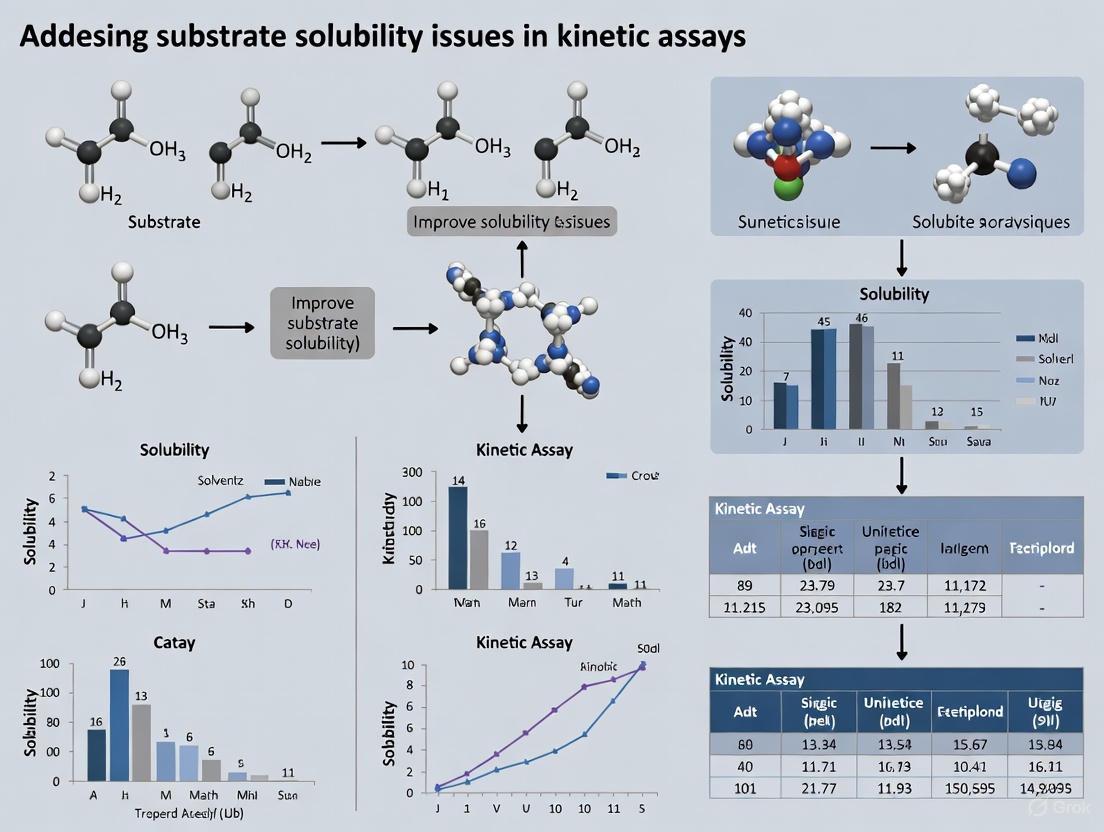
Solving Substrate Solubility in Kinetic Assays: From Foundational Principles to Advanced Strategies for Drug Development
Substrate solubility is a critical, yet often overlooked, parameter that directly impacts the reliability and reproducibility of kinetic assays in enzymology and drug discovery.
Solving Substrate Solubility in Kinetic Assays: From Foundational Principles to Advanced Strategies for Drug Development
Abstract
Substrate solubility is a critical, yet often overlooked, parameter that directly impacts the reliability and reproducibility of kinetic assays in enzymology and drug discovery. This article provides a comprehensive guide for researchers and drug development professionals facing this ubiquitous challenge. It begins by establishing the foundational science of solubility and its direct consequences on assay parameters like KM and kcat. The piece then transitions to practical, methodological solutions for enhancing solubility, including chemical modifications and advanced formulation techniques. A dedicated troubleshooting section offers a systematic framework for diagnosing and optimizing problematic assays, while the final section outlines rigorous protocols for validating solubility and comparatively analyzing different methodological approaches. By integrating traditional techniques with cutting-edge computational tools, this resource aims to equip scientists with a holistic strategy to ensure accurate kinetic data from lead optimization to development.
The Solubility Imperative: Understanding the Fundamental Impact on Kinetic Data
Why Solubility is a Rate-Limiting Step in Drug Discovery and Development
Troubleshooting Guide: Frequently Asked Questions
1. Why is solubility so critical in early drug discovery? Solubility is a fundamental property that directly influences a drug's absorption into the systemic circulation. After oral administration, a drug must first dissolve in gastrointestinal fluids before it can permeate the intestinal wall. Poor solubility often results in low and variable bioavailability, meaning the drug does not reach its target site in sufficient concentration to be therapeutically effective. This makes it a primary rate-limiting step in developing new pharmaceuticals. [1]
2. A significant portion of our drug pipeline consists of BCS Class II compounds. What are the main strategies to enhance their solubility and dissolution? A large number of new chemical entities (70-90%) fall into the BCS Class II category, characterized by high permeability but poor solubility. For these drugs, the rate of dissolution is often the slowest step governing overall absorption. Key strategies to overcome this include:
- Particle Size Reduction (e.g., Nano-suspensions): Increasing the surface area of the drug particles to enhance dissolution rate.
- Complexation: Using cyclodextrins or other agents to form soluble complexes with the drug molecule.
- Advanced Carrier Systems: Employing Metal-Organic Frameworks (MOFs) like MIL-101(Cr) for in-situ loading, which can dramatically increase drug loading and reclassify drugs from "poorly soluble" to "soluble". [1] [2]
3. How can we accurately predict solubility to accelerate synthetic planning? Traditional methods like the Abraham Solvation Model have limitations in accuracy. Modern approaches use machine learning models trained on large, curated datasets. For instance, the FastSolv model uses static molecular embeddings to predict how well a molecule will dissolve in hundreds of different organic solvents, and it accounts for the effect of temperature. Using such models allows chemists to choose the optimal solvent for a reaction before moving to the lab, speeding up development. [3]
4. When designing a kinetic assay, how do I choose between a soluble or insoluble substrate? The choice is dictated by your analytical goal:
- Use Soluble Substrates for quantification in liquid-phase assays (e.g., cuvette or multi-well plate formats). They are ideal for real-time kinetic studies, ELISA, and high-throughput screening because the dissolved product is compatible with optical detection (absorbance, fluorescence). [4]
- Use Insoluble Substrates for localization studies. The precipitated product remains at the site of enzyme activity, making them essential for techniques like Western blots, zymograms, immunohistochemistry, and microbial screening on agar plates. [4]
5. We are characterizing a covalent inhibitor. Why do our IC50 values seem inconsistent, and how should we properly evaluate its potency? For irreversible covalent inhibitors, inhibition is time-dependent. A single IC50 measurement is insufficient and can be misleading because it doesn't distinguish between strong binding with slow reaction and weak binding with fast reaction. A complete characterization requires deriving two key parameters:
- KI: The inhibition constant, representing the affinity for the initial non-covalent binding.
- kinact: The maximum rate of the irreversible inactivation. Proper evaluation requires time-dependent assays, such as the Kitz & Wilson method for continuous assays or incubation time-dependent IC50 measurements for discontinuous assays, to obtain a full kinetic profile. [5]
Quantitative Data on Solubility Enhancement Technologies
The following table summarizes data on novel technologies used to improve the solubility and bioavailability of poorly soluble drugs.
Table 1: Strategies for Solubility Enhancement of Poorly Water-Soluble Drugs
| Technology / Approach | Mechanism of Action | Example Results / Data | Key Advantages |
|---|---|---|---|
| Nano-Suspensions [1] | Particle size reduction to increase surface area for dissolution. | Applied to 40% of existing drugs and 70-90% of drugs in development. | Can be easily formulated into various dosage forms. |
| Metal-Organic Frameworks (MIL-101(Cr)) [2] | In-situ drug loading onto a porous carrier via π–π stacking and H-bonding. | Loads of 904.7 mg/g Ibuprofen; solubility increased to 4.1-7.3 g/L in PBS. | Green synthesis method; reclassifies drugs from "poorly soluble" to "soluble". |
| Machine Learning (FastSolv Model) [3] | Predicts solubility in organic solvents using numerical molecular representations. | Predictions are 2-3x more accurate than previous models (SolProp). | Accelerates synthetic planning; helps identify less hazardous solvents. |
Experimental Protocols for Key Assays
Protocol 1: Characterizing an Irreversible Covalent Inhibitor using a Continuous Assay (Kitz & Wilson Method) [5]
This protocol is used to determine the kinetic parameters KI and kinact for an irreversible inhibitor when a continuous, real-time activity assay is available.
- Assay Setup: Prepare a series of reactions containing a fixed concentration of the target enzyme and varying concentrations of the covalent inhibitor. Include a positive control with no inhibitor.
- Initiation: Start the reaction by adding the enzyme's substrate. The substrate concentration should be saturating ([S] >> KM) to ensure the initial velocity directly reflects the concentration of active enzyme.
- Continuous Monitoring: Immediately begin monitoring product formation or substrate consumption (e.g., via absorbance or fluorescence) in real-time using a plate reader or spectrophotometer.
- Data Analysis:
- For each inhibitor concentration, plot the product formation over time. The slope of this curve will decrease as more enzyme is inactivated.
- At each time point, the observed reaction rate (v) is proportional to the concentration of active enzyme remaining.
- The observed rate constant for inactivation (kobs) at each inhibitor concentration is determined by fitting the progress curves to an exponential decay function.
- Plot kobs against the inhibitor concentration [I]. Fit this data to the following equation to derive KI and kinact: kobs = (kinact * [I]) / (KI + [I])
Protocol 2: Enhancing Drug Solubility via In-Situ Loading onto MIL-101(Cr) [2]
This green chemistry protocol describes incorporating a BCS Class II drug during the synthesis of a metal-organic framework carrier.
- Reagent Preparation: Dissolve Chromium (III) salt (e.g., Cr(NO3)3) and terephthalic acid in a solvent system of acetic acid and water. This replaces toxic hydrofluoric acid traditionally used.
- Drug Addition: Add the poorly soluble drug (e.g., Ibuprofen, Ketoprofen, Felodipine) to the mixture.
- Crystallization: Carry out the solvothermal reaction under controlled temperature and pressure to crystallize the MIL-101(Cr) MOF with the drug molecule incorporated within its pores.
- Isolation and Washing: Recover the solid product by filtration or centrifugation and wash to remove any unreacted precursors or drug loosely bound to the surface.
- Characterization: Confirm successful drug loading using techniques like:
- Field Emission Scanning Electron Microscopy (FESEM): To observe morphology.
- Brunauer-Emmett-Teller (BET) Analysis: To confirm a reduction in surface area, indicating pore occupancy.
- X-ray Diffraction (XRD): To analyze crystallinity.
- Fourier-Transform Infrared Spectroscopy (FTIR): To identify drug-carrier interactions.
- Dissolution Testing: Perform drug release studies in a medium like phosphate-buffered saline (PBS) to quantify the enhancement in solubility and dissolution rate.
Workflow Visualization
Solubility Enhancement Workflow
The Scientist's Toolkit: Essential Research Reagents & Materials
Table 2: Key Reagents for Solubility and Kinetic Assay Research
| Item | Function / Application |
|---|---|
| Organic Solvents (e.g., Ethanol, Acetone) | Used in chemical synthesis and solubility screening. Machine learning models can help select the most effective and least hazardous options. [3] |
| Soluble Chromogenic/Fluorogenic Substrates | Generate a dissolved colored or fluorescent product. Essential for quantitative, real-time kinetic assays in solution (e.g., protease or hydrolase activity profiling). [4] |
| Insoluble Precipitating Substrates | Yield a colored precipitate at the site of enzyme activity. Critical for localization studies in Western blots, zymograms, and immunohistochemistry. [4] |
| Metal-Organic Frameworks (e.g., MIL-101(Cr)) | Porous crystalline materials used as advanced carriers to dramatically increase the apparent solubility and loading capacity of poorly soluble drugs. [2] |
| Covalent Inhibitors with Electrophilic Warheads | Contain a reactive functional group that forms a slow-reversible or irreversible bond with a target protein, leading to prolonged pharmacodynamic effects. Require specialized kinetic evaluation. [6] [5] |
Core Concepts FAQ
What is the fundamental difference between kinetic and thermodynamic solubility?
Kinetic Solubility measures the concentration at which a compound, initially fully dissolved in an organic solvent like DMSO, begins to precipitate out when added to an aqueous buffer. It represents the solubility of the fastest-precipitating, often amorphous, form of the compound and is typically used for high-throughput screening in early drug discovery [7] [8] [9].
Thermodynamic Solubility measures the maximum concentration of a solid crystalline compound that can remain dissolved in a solvent under equilibrium conditions—where the dissolved and undissolved solid are in equilibrium. This is considered the "true" solubility of the most stable crystal form and is critical for later-stage development and formulation [10] [7] [8].
When should each type of solubility assay be used in the drug discovery workflow?
The choice between kinetic and thermodynamic solubility assays depends on the stage of research and the specific information required. The following table outlines the typical applications:
Table 1: Application of Solubility Assays in Drug Discovery and Development
| Assay Type | Stage of R&D | Primary Purpose | Typical Data Output |
|---|---|---|---|
| Kinetic Solubility | Early Discovery (Lead Identification & Optimization) [7] | High-throughput ranking of compound libraries; guiding medicinal chemistry for structure-activity relationships (SAR) [11] [9]. | Classification (e.g., Low <10 µg/mL, Moderate 10-60 µg/mL, High >60 µg/mL) [11]. |
| Thermodynamic Solubility | Late-Stage Preclinical Development [7] | Formulation development; setting dissolution specifications; providing data for regulatory submissions (e.g., IND) [7] [12]. | Precise solubility value (e.g., in µg/mL) for the most stable crystal form [10]. |
Why might my kinetic solubility values be misleadingly high compared to thermodynamic values?
This common discrepancy occurs because the two methods measure different physical states of the compound. Kinetic solubility assays often measure the solubility of the amorphous form of the compound, which has higher energy and therefore higher apparent solubility. In contrast, thermodynamic solubility assays measure the solubility of the most stable crystalline form, which has lower energy and thus lower solubility. Relying solely on kinetic solubility can lead to over-optimism about a compound's developability [10].
Troubleshooting Guides
Issue 1: Handling Low or Variable Solubility Results
Problem: Measured solubility values are very low, variable, or inconsistent with other assay data.
Solutions:
- Confirm Analytical Method: For very low solubility compounds, switch from HPLC-UV to a more sensitive method like LC-MS/MS. The lower limit of quantification for LC-MS/MS can be as low as 1 nM, allowing for more accurate detection of low concentrations [11].
- Address Non-Specific Adsorption: Use low-binding consumables throughout the process, such as 96-well low-binding filter plates or regenerated cellulose filter membranes. For sample treatment, consider adding additives like organic solvents, surfactants, or proteins to minimize compound adsorption to surfaces [11].
- Verify Solid State Form: In thermodynamic assays, use techniques like polarized-light microscopy to confirm whether the measured solubility corresponds to a crystalline or amorphous phase. This helps explain discrepancies and ensures data reliability [10].
Issue 2: Managing Compound Stability During Solubility Assessment
Problem: The compound degrades during the solubility experiment, leading to inaccurate results.
Solutions:
- Detect Degradation: Employ chromatographic methods (e.g., HPLC-UV) with full-wavelength scanning to identify the presence of specific degradation peaks. Using LC-UV-MS in series can help confirm the identity and purity of the target peak [11].
- Optimize Conditions: If instability is detected, assess unstable factors and optimize experimental conditions, such as adjusting the pH of the buffer media or reducing the incubation time if appropriate [11].
Experimental Protocols
Protocol for Kinetic Solubility Determination
This protocol is based on the high-throughput shake-flask method commonly used in early discovery [11] [13].
Method Overview: A compound dissolved in DMSO is diluted into an aqueous buffer system, and the concentration at which precipitation occurs is determined.
Table 2: Key Parameters for a Standard Kinetic Solubility Assay
| Parameter | Typical Condition | Notes / Alternatives |
|---|---|---|
| Theoretical Concentration | 200 µM (routine) | Can be adjusted based on project needs. |
| Starting Material | Compound in DMSO stock solution (e.g., 10 mM) | - |
| Percentage of DMSO | 2% (routine) | Other ratios (e.g., 1%) can be used. |
| Media | Aqueous buffer (e.g., pH 7.4 phosphate buffer); Biorelevant media. | pH can be adjusted to simulate different physiological environments. |
| Incubation Equilibration Time | 2 hours (common) [11] | 24 hours is also used. |
| Equilibration Temperature | Room Temperature or 37 °C | 37 °C is used to simulate physiological temperature. |
| Detection Method | Nephelometry (turbidimetry) [9], HPLC-UV, or LC-MS/MS | Nephelometry detects precipitation; chromatographic methods quantify concentration. |
| Compound Required | ~30 µL of 10 mM DMSO stock [11] | - |
Workflow Diagram: Kinetic Solubility Assay
Protocol for Thermodynamic Solubility Determination
This protocol uses the classic shake-flask method, considered the gold standard for obtaining equilibrium solubility [11] [14].
Method Overview: An excess of solid compound is added to a solvent and agitated until equilibrium is reached between the dissolved and undissolved material.
Table 3: Key Parameters for a Standard Thermodynamic Solubility Assay
| Parameter | Typical Condition | Notes / Alternatives |
|---|---|---|
| Starting Material | Solid powder of the compound [11] | The solid-state form (polymorph) is critical. |
| Sample Amount | ~2 mg solid (for assay) + 1 mg (for standard curve) [11] | - |
| Media | Aqueous buffer system; Biorelevant media; Organic solvent. | - |
| Incubation Equilibration Time | 24 hours (routine) [11] [9] | Must be sufficient to reach equilibrium. |
| Equilibration Temperature | Room Temperature or 37 °C [11] | - |
| Agitation | Shaking (e.g., shake-flask) [11] | - |
| Separation | Centrifugation and/or Filtration [14] | - |
| Analytical Method | HPLC-UV/HPLC-ELSD/LC-MS/MS [11] | Used to quantify the concentration in the saturated solution. |
Workflow Diagram: Thermodynamic Solubility Assay
The Scientist's Toolkit: Essential Research Reagents & Materials
The following table lists key materials and their functions for conducting reliable solubility assays.
Table 4: Essential Materials for Solubility Testing
| Item / Reagent | Function in the Assay | Application Notes |
|---|---|---|
| DMSO (Dimethyl Sulfoxide) | Universal solvent for creating high-concentration stock solutions of research compounds for kinetic solubility assays. [7] [8] | Use high-quality, anhydrous DMSO. Keep concentrations low (e.g., 1-2%) in final aqueous buffers to minimize its effect on solubility. |
| Aqueous Buffer Systems (e.g., Phosphate Buffer) | Provides a stable, physiologically relevant pH environment (e.g., pH 7.4) for solubility measurement. [11] [9] | Essential for ionizable compounds, as their solubility is pH-dependent. |
| Simulated Gastrointestinal Fluids (Biorelevant Media) | Media such as FaSSIF (Fasted State Simulated Intestinal Fluid) mimic the in vivo environment of the GI tract, providing a more predictive measure of absorption potential. [11] [7] | Used in later stages to forecast in vivo behavior. |
| Low-Binding Consumables (Plates, Filters) | Tubes, plates, and filter membranes treated to minimize non-specific adsorption of the test compound to surfaces. [11] | Critical for obtaining accurate results with lipophilic or sticky compounds that can adhere to container walls. |
| Positive Control Compounds | Compounds with known and verified solubility ranges used to ensure the accuracy and consistency of the experimental operation. [11] [9] | Examples include Nicardipine (for kinetic) and Haloperidol (for thermodynamic). They are run alongside test compounds for quality control. |
The Direct Link Between Poor Solubility and Erroneous Kinetic Parameters (KM, kcat)
Frequently Asked Questions (FAQs)
Q1: How does poor substrate solubility directly lead to inaccurate measurements of KM and kcat? Poor substrate solubility creates a discrepancy between the nominal substrate concentration you add to the assay and the actual concentration of bioavailable substrate in solution [15] [16]. If a compound precipitates, it is unavailable for the enzyme to bind. Kinetic analysis, based on the Michaelis-Menten model, assumes that the stated substrate concentration is accurate and fully available [15] [17]. When this is not true, the calculated parameters are fundamentally flawed. A seemingly low enzyme velocity might be misinterpreted as a high KM (low affinity) or low kcat (slow catalytic rate), when in reality, the enzyme is simply starved for soluble substrate.
Q2: What are the typical experimental signs that poor solubility is affecting my kinetic assay? Several red flags can indicate solubility issues:
- Non-linear or inconsistent progress curves: The reaction rate decreases rapidly before a significant amount of product would be expected to form, not due to enzyme inactivation but because the local, soluble substrate is depleted [18].
- Failure to achieve saturation: The reaction velocity (v0) does not plateau (Vmax is not reached) even at high nominal substrate concentrations [17].
- High variability in replicate measurements: Slight changes in temperature, mixing, or buffer composition can cause dissolved substrate to precipitate or precipitate to re-dissolve, leading to inconsistent rates between replicates.
- Observed cloudiness or precipitation: Visible precipitate in the assay solution is a clear indicator of exceeding the compound's solubility limit.
Q3: Beyond KM and kcat, what other aspects of enzymology are compromised by poor solubility? Poor solubility can skew the results of many advanced enzyme studies, including:
- Mechanistic studies: Determining whether a reaction follows a sequential or ping-pong mechanism (e.g., for multi-substrate enzymes like NIS synthetases) requires accurate substrate concentration curves [19]. Solubility issues can obscure the true mechanism.
- Inhibition studies: The potency (IC50, Ki) and mechanism of an inhibitor (competitive, non-competitive) can be mischaracterized if the substrate or inhibitor itself has limited solubility [20].
- High-throughput screening (HTS): Poorly soluble compounds are a major source of false positives and false negatives in drug discovery campaigns, as they can form colloidal aggregates that non-specifically inhibit enzymes [16].
Q4: What practical steps can I take to improve substrate solubility in my assay buffer?
- Use co-solvents: A small percentage of a water-miscible organic solvent like DMSO, ethanol, or acetone can significantly enhance solubility. It is critical to keep the concentration low (typically <5%) to avoid denaturing the enzyme and to include the same solvent concentration in all standards and blanks [16].
- Employ solubilizing agents: Detergents (e.g., Triton X-100) or cyclodextrins can encapsulate hydrophobic molecules, keeping them in solution without drastically altering the aqueous environment of the enzyme [21].
- Modify buffer conditions: Adjusting pH, ionic strength, or changing the buffer system itself can sometimes improve solubility. However, any change must be validated to ensure it does not adversely affect enzyme activity [15] [22].
- Consider substrate analogs: If available, a more soluble analog of the substrate can sometimes be used, though this must be done with caution as it may alter the enzyme's kinetic mechanism [15].
Troubleshooting Guide: Identifying and Mitigating Solubility Issues
This guide outlines a systematic approach to diagnose and address substrate solubility problems in kinetic assays.
Step 1: Proactively Determine Substrate Solubility Before starting kinetic experiments, determine the solubility limit of your substrate under exact assay conditions (buffer, pH, temperature) [16].
- Protocol: Prepare a series of substrate stock solutions in DMSO and dilute them into your assay buffer to cover the concentration range you plan to use. After incubation at the assay temperature, visually inspect for cloudiness or use light-scattering methods (e.g., nephelometry) to detect precipitate. The solubility limit is the highest concentration that remains clear.
Step 2: Design Kinetic Assays with Solubility in Mind
- Stay Below the Limit: Ensure all nominal substrate concentrations in your assay are below the experimentally determined solubility limit.
- Use Continuous Assays: Whenever possible, use continuous assays that monitor the reaction progress in real-time [18]. The shape of the progress curve is a powerful diagnostic tool for identifying solubility-related depletion, as shown in the diagram below.
Step 3: Analyze Data with a Critical Eye
- Inspect Progress Curves: Do not assume linearity. Use kinetic modeling to analyze the entire progress curve, which can help account for non-ideal behavior, including substrate depletion [18].
- Validate Parameters: Be skeptical of kinetic parameters that do not make biochemical sense. Compare your results with literature values, and if they differ significantly, investigate solubility as a potential cause.
The following workflow diagram summarizes the troubleshooting process for a kinetic assay suspected of being affected by poor solubility.
Quantitative Impact of Solubility on Measured Parameters
The table below summarizes how a mismatch between nominal and soluble substrate concentration systematically biases the derived kinetic parameters.
| Parameter | Definition | Impact of Poor Solubility | Erroneous Interpretation |
|---|---|---|---|
| KM (Michaelis Constant) | Apparent substrate concentration at half Vmax; measures enzyme affinity [15] [17]. | Artificially Inflated. The enzyme requires a higher nominal [S] to reach half Vmax because the bioavailable [S] is lower than assumed. | The enzyme appears to have a lower binding affinity for the substrate than it truly does. |
| kcat (Turnover Number) | Maximum number of substrate molecules converted to product per enzyme active site per unit time [17]. | Artificially Lowered. The enzyme cannot achieve its true maximum velocity because it is not saturated with soluble substrate. | The enzyme's catalytic efficiency appears slower than it truly is. |
| kcat/KM (Specificity Constant) | Measure of catalytic efficiency for a given substrate [20]. | Artificially Lowered. This composite parameter is skewed by both an inflated KM and a lowered kcat. | The enzyme's overall efficiency and specificity for the substrate are significantly underestimated. |
Experimental Protocol: Determining Kinetic Solubility via Nephelometry
This protocol provides a method for quantitatively assessing the kinetic solubility of a substrate, which is the relevant solubility for most enzymatic assays where compounds are added from a DMSO stock [16].
Objective: To determine the concentration at which a substrate precipitates in assay buffer upon dilution from a DMSO stock solution.
Materials:
- Substrate stock solution (e.g., 10-100 mM in DMSO)
- Assay buffer (identical to that used in kinetic experiments)
- Nephelometer or plate reader capable of measuring light scattering (often at ~600 nm)
- Multi-well plate or clear cuvettes
- Piperettes and vortex mixer
Procedure:
- Preparation: Serially dilute the substrate stock solution in DMSO to create a range of concentrations.
- Dilution into Buffer: Add a fixed, small volume of each DMSO stock (e.g., 1-5 µL) to a larger volume of assay buffer (e.g., 1 mL) in a cuvette or plate well. The final DMSO concentration should be consistent and low (≤1%).
- Mixing and Incubation: Mix thoroughly and incubate for 10-15 minutes at the temperature used for your kinetic assays.
- Measurement: Measure the nephelometry signal (turbidity) for each sample. A blank consisting of buffer with the same percentage of DMSO should be used for baseline subtraction.
- Analysis: Plot the nephelometry signal versus the nominal substrate concentration. The point where the signal significantly increases above the baseline indicates the kinetic solubility limit.
The Scientist's Toolkit: Research Reagent Solutions
The following table lists key reagents and their functions for addressing solubility challenges in enzyme kinetics.
| Reagent / Material | Function in Addressing Solubility |
|---|---|
| Dimethyl Sulfoxide (DMSO) | A polar aprotic solvent used to create high-concentration stock solutions of poorly soluble compounds. It allows for precise dispensing before dilution into aqueous buffers [16]. |
| Cyclodextrins (e.g., HP-β-CD) | Cyclic oligosaccharides that form soluble inclusion complexes with hydrophobic molecules, acting as molecular carriers to enhance apparent solubility in aqueous buffers [21]. |
| Detergents (e.g., Triton X-100) | Amphipathic molecules that form micelles in solution. They can solubilize hydrophobic substrates within the micelle core, preventing precipitation [21]. |
| Nephelometer | An instrument that measures the scattering of light by particles in a solution. It is used to quantitatively determine the solubility limit of a compound by detecting the onset of precipitation [16]. |
| Design of Experiments (DoE) Software | Statistical tools that allow for the efficient optimization of multiple assay parameters (e.g., buffer pH, ionic strength, co-solvent percentage) simultaneously to find conditions that maximize both solubility and enzyme activity [22]. |
The Biopharmaceutics Classification System (BCS) is a fundamental framework used in drug development to categorize active pharmaceutical ingredients (APIs) based on their aqueous solubility and intestinal permeability [23] [24]. This system provides a scientific approach for predicting drug absorption in humans and is a critical tool for researchers and regulatory bodies, including the US Food and Drug Administration (FDA) and the World Health Organization (WHO) [23] [25]. The primary goal of the BCS is to determine when in vivo bioequivalence studies can be replaced by in vitro dissolution tests, a process known as "biowaiver" [23]. For scientists conducting kinetic assays, understanding a compound's BCS class is the first step in anticipating and troubleshooting significant experimental challenges, particularly those related to substrate solubility.
BCS Classification of Drugs
According to the BCS, drug substances are classified into four distinct classes based on two key properties: solubility and permeability [23] [24]. The classification and its implications for drug absorption are summarized in the table below.
Table 1: BCS Drug Classification and Characteristics
| BCS Class | Solubility | Permeability | Absorption Challenge | Example Compounds |
|---|---|---|---|---|
| Class I | High | High | No absorption challenges; well-absorbed | Metoprolol, Paracetamol [24] |
| Class II | Low | High | Solubility/dissolution rate-limited | Glibenclamide, Ketoconazole [23] [24] |
| Class III | High | Low | Permeability rate-limited | Cimetidine [24] |
| Class IV | Low | Low | Poor bioavailability; significant challenges for absorption | Bifonazole, Furosemide, Bosutinib [26] [24] |
Defining the Class Boundaries
- Solubility: A drug is deemed highly soluble when the highest dose strength is soluble in 250 mL or less of aqueous media over a pH range of 1 to 6.8 [23] [24]. This volume is derived from typical bioequivalence study protocols.
- Permeability: A drug is considered highly permeable when the extent of absorption in humans is determined to be 90% or more [23] (or 85% or more based on a mass-balance determination [24]) of an administered dose.
Identifying High-Risk BCS Class IV Compounds
Within the BCS framework, Class IV compounds represent the highest-risk category for drug development. These molecules possess the dual challenges of low solubility and low permeability, leading to inherently poor and highly variable bioavailability [26] [24]. This makes them particularly problematic for formulation and for reliably predicting in vivo performance based on in vitro data.
Recent regulatory guidance, such as from the USFDA, specifies that for BCS Class IV molecule-containing immediate-release (IR) formulations, in vitro testing alone is not sufficient for waiving bioequivalence studies. Instead, more advanced techniques like Physiologically Based Pharmacokinetic (PBPK) modeling are recommended for risk assessment [26]. This highlights the heightened regulatory scrutiny and complexity associated with these compounds.
Table 2: Examples of High-Risk BCS Class IV Molecules and Associated Risks
| BCS Class IV Molecule | Reported Risk and Challenge |
|---|---|
| Edoxaban | Identified as high-risk for carcinogenic potential from nitrosamine impurities; transporters involved in absorption complicate risk assessment [26]. |
| Selumetinib | Clinical exposure risk assessment required due to high-risk categorization; permeability and transporter kinetics significantly impact exposure [26]. |
| Bosutinib | PBPK modeling used to evaluate impact of altered permeability and transporter kinetics on exposures [26]. |
| Furosemide | Clinical exposure risk assessment conducted due to high-risk categorization [26]. |
| Hydrochlorothiazide | Transporter involvement in absorption adds complexity to predicting bioequivalence [26]. |
The Scientist's Toolkit: Essential Reagents and Materials
Working with BCS Class II and IV compounds requires specific reagents and techniques to overcome solubility limitations in experimental assays. The following table outlines key research reagent solutions.
Table 3: Research Reagent Solutions for Solubility-Enhancement
| Reagent / Technique | Function and Application in Experimental Protocols |
|---|---|
| Surfactants (e.g., SLS, Tween 80) | Reduce interfacial tension, increase wetting, and solubilize hydrophobic drugs in dissolution media [23]. |
| Complexing Agents (e.g., Cyclodextrins) | Form reversible inclusion complexes with drug molecules, increasing their apparent aqueous solubility [23]. |
| Hydrophilic Carriers (e.g., PVP, PEG) | Used in solid dispersions to create a hydrophilic matrix that improves drug dissolution rates [23]. |
| Lipidic/Greasy Substances (e.g., oils, waxes) | Used in lipid-based formulations to solubilize and improve the absorption of poorly water-soluble drugs [23]. |
| Buffers (across pH 1-6.8) | Used for solubility characterization and dissolution testing as per regulatory guidance across the physiologically relevant pH range [23]. |
Experimental Protocol: A Kinetic Assay Approach for Poorly Soluble Systems
The following workflow outlines a generalized kinetic assay, adapted from enzymology studies, that is mindful of the challenges posed by poorly soluble substrates [19] [27]. This is crucial for generating reliable data with BCS Class II and IV compounds.
Detailed Methodology
- Pre-experiment Solubility Check: Before any kinetic experiment, determine the maximum solubility of your substrate (the drug compound) in the chosen assay buffer across the relevant pH range. This is a critical first step to avoid experimental artifacts [27].
- Substrate Stock Preparation:
- Prepare a concentrated stock solution of the substrate in a suitable water-miscible organic solvent (e.g., DMSO, ethanol). The final concentration of the organic solvent in the assay must be kept low (typically <1-2%) to avoid denaturing proteins or enzymes and affecting the reaction [27].
- Control: Include a control in the assay that contains the same final concentration of the solvent but no substrate to account for any solvent effects.
- Running the Kinetic Assay:
- Initiate the reaction by adding the enzyme, membrane preparation, or other catalyst to the buffered reaction mixture containing the substrate.
- Ensure thorough mixing of comparable volume additions to minimize pipetting errors and ensure solution uniformity [27].
- Monitor the reaction in real-time using a suitable method (e.g., spectrophotometry, HPLC).
- Initial Rate (v₀) Determination:
- Collect data points at early time intervals where the reaction progress is linear.
- Define "time zero" carefully, accounting for the mixing time, to accurately determine the initial rate, which is the slope of the linear progress curve at this point [27].
- Data Fitting and Analysis:
- Plot the initial rate (v₀) against the substrate concentration ([S]).
- Fit the data to the Michaelis-Menten model (or other appropriate models) to determine the apparent KM (Michaelis constant) and vmax (maximum reaction rate) [27].
- A high apparent KM can often be an indicator of poor substrate solubility limiting the reaction [27].
Troubleshooting Common Issues in Kinetic Assays
FAQ 1: My kinetic data shows a non-linear response at high substrate concentrations, and the calculated KM appears abnormally high. What could be the cause?
This is a classic symptom of substrate solubility limitations [27]. If the substrate is not fully soluble at the higher concentrations used in the assay, the effective concentration available for the reaction is lower than assumed. This leads to a plateau in the rate that is not due to enzyme saturation but to physical solubility, resulting in an inflated apparent KM value.
- Solution: Re-check the solubility of your substrate in the exact assay buffer. All substrate concentrations used in the kinetic experiment must be below the determined saturation point. Using solubility-enhancing agents (see Table 3) may be necessary, but their effects on the catalyst must be controlled for.
FAQ 2: My negative control (buffer only) shows unexpected background activity. How should I investigate this?
Buffers and other solution components can sometimes contribute to the observed reaction, especially in the presence of metal ions or other catalysts [27].
- Solution:
- Perform a comprehensive set of control experiments, including omitting the catalyst and/or the substrate.
- Investigate potential interactions between your buffer and metal ions. For example, Tris buffer can complex with certain metal ions and exhibit catalytic activity [27].
- Ensure the assay components are stable and not degrading to form reactive species.
FAQ 3: For a BCS Class IV compound, how can we bridge the gap between in vitro kinetic data and in vivo performance?
Due to the complex and variable absorption of BCS Class IV drugs, in vitro data alone is often insufficient. Regulatory agencies recommend the use of Physiologically Based Pharmacokinetic (PBPK) modeling as an advanced tool for this purpose [26].
- Solution: Develop a PBPK model that incorporates the in vitro kinetic parameters (e.g., permeability, solubility) to simulate in vivo exposure. This model can be used to evaluate the impact of formulation changes and to define a "permeability safe space"—a range within which changes in permeability still ensure bioequivalence [26].
Risk Assessment Workflow for BCS Class IV Molecules
The following diagram outlines a modern, integrated risk assessment strategy for BCS Class IV molecules, as recommended by current regulatory science.
Core Concepts and Definitions
What are Hildebrand and Hansen Solubility Parameters? Solubility parameters are numerical values used to predict the solubility of materials based on the principle that "like dissolves like". The Hildebrand Solubility Parameter (δ) is a single value representing the square root of the cohesive energy density (CED), which is the energy required to separate molecules of a substance [28] [29]. It is defined as δ = √((ΔHv - RT)/Vm), where ΔHv is the heat of vaporization, R is the gas constant, T is the temperature, and Vm is the molar volume [28] [30].
The Hansen Solubility Parameters (HSP) represent an extension of the Hildebrand parameter, dividing the total cohesive energy into three distinct intermolecular interaction components [31] [30]:
- δD: Dispersion forces (van der Waals interactions)
- δP: Polar interactions (dipole-dipole forces)
- δH: Hydrogen bonding interactions
The relationship between Hildebrand and Hansen parameters is expressed as δ² = δD² + δP² + δH² [30] [32].
Parameter Tables for Common Substances
Table 1: Hildebrand and Hansen Solubility Parameters for Common Solvents
| Substance | δ (Hildebrand) [MPa¹/²] | δD [MPa¹/²] | δP [MPa¹/²] | δH [MPa¹/²] |
|---|---|---|---|---|
| n-Pentane | 14.4 [28] | ~14.1* | ~0.0* | ~0.0* |
| n-Hexane | 14.9 [28] | 14.9 [33] | 0.0 [33] | 0.0 [33] |
| Diethyl Ether | 15.4 [28] | 14.5 [34] | 2.9 [34] | 4.6 [34] |
| Ethyl Acetate | 18.2 [28] | 15.8 [33] | 5.3 [33] | 7.2 [33] |
| Toluene | 18.3 [29] | 18.0 [33] | 1.4 [33] | 2.0 [33] |
| Chloroform | 18.7 [28] | 17.8 [33] | 3.1 [33] | 5.7 [33] |
| Acetone | 19.9 [28] | 15.5 [33] | 10.4 [33] | 7.0 [33] |
| 2-propanol | 23.8 [28] | 15.8 [33] | 6.1 [33] | 16.4 [33] |
| Ethanol | 26.5 [28] | 15.8 [33] | 8.8 [33] | 19.4 [33] |
Note: Values for n-Pentane are estimated based on similar hydrocarbons. Always consult primary sources for precise values in experimental work.
Table 2: Hildebrand Solubility Parameters for Common Polymers
| Polymer | δ (Hildebrand) [MPa¹/²] |
|---|---|
| PTFE | 6.2 [28] |
| Poly(ethylene) | 7.9 [28] |
| Poly(propylene) | 16.6 [28] |
| Poly(styrene) | ~18.6 [34] |
| PVC | 19.5 [28] |
| Poly(methyl methacrylate) | 19.0 [28] |
| Nylon 6,6 | 28 [28] |
Experimental Determination Methods
Determining HSP via Solubility Testing
Protocol for Experimental HSP Determination [35] [30] [32]:
Sample Preparation: Prepare 20-30 solvent systems with known HSP values that span the three-dimensional parameter space.
Solubility Testing:
- Add a fixed amount of your solute (typically 5-50 mg) to a series of vials containing different solvents.
- Agitate the mixtures for 24 hours at constant temperature.
- Centrifuge if necessary to separate dissolved and undissolved material.
- Determine solubility (yes/no) or measure the concentration dissolved at a specific threshold.
Data Analysis:
- Classify solvents as "good" (dissolves solute) or "poor" (does not dissolve solute).
- Use HSP software (such as HSPiP) or statistical methods to calculate the HSP sphere that best separates good from poor solvents.
- The center of this sphere represents the HSP of your solute.
Validation: Test additional solvents to validate the predicted HSP values.
Determining Solubility Parameters via Inverse Gas Chromatography (IGC)
Protocol for IGC Determination [30]:
Column Preparation: Pack a chromatography column with the solid sample of interest as the stationary phase.
Instrument Setup: Use an inert carrier gas (nitrogen or helium) and ensure the system is at constant temperature.
Solvent Probe Injection: Inject vapor phases of various organic solvents with known characteristics into the column.
Data Collection: Measure the specific retention volume (Vg) for each solvent probe.
Data Analysis: Use the relationship: χ₁₂∞ = ln(273.15·R·p₁⁰/(Vg·Mr₁)) - p₁⁰/(R·T)·(B₁₁-V₁) + ln(ρ₁/ρ₂) - (1-V₁/V₂) to calculate the Flory-Huggins parameter, which can be related to solubility parameters [30].
Parameter Calculation: Apply regression analysis to determine the HSP values that best fit the retention data.
Troubleshooting Guide: Common Experimental Issues
FAQ 1: Why do my solubility predictions fail even when Hildebrand parameters match?
Issue: The single-parameter Hildebrand approach cannot account for specific polar and hydrogen-bonding interactions [34].
Solution: Use the three-parameter Hansen system instead. For example, n-Butanol and Nitroethane have identical Hildebrand parameters (23 MPa¹/²) but neither dissolves a typical epoxy. However, a 50:50 mixture is a good solvent because the combined HSP values create a favorable interaction profile [34].
FAQ 2: How can I dissolve a solute when no single good solvent is available?
Issue: Environmental or safety concerns may limit solvent options.
Solution: Use solvent blending. Two "poor" solvents with complementary HSP can often form a "good" solvent mixture. Calculate the HSP of blends using volumetric weighting: δblend = φ₁δ₁ + φ₂δ₂, where φ is the volume fraction [34].
FAQ 3: How do I handle solubility parameter determination for complex natural products like lignin?
Issue: Heterogeneous biomaterials with diverse functional groups challenge simple solubility predictions.
Solution: Use Functional Solubility Parameters (FSP) which bypass arbitrary solubility thresholds and create a solubility function represented by a polyhedron, whose center of mass is the solute FSP [32].
FAQ 4: What computational methods can predict solubility parameters?
Solution: Molecular dynamics simulations can calculate HSP using the formula: δ = √(ρ·(〈Evapor〉 - 〈Eliquid〉)/M), where ρ is density, E is potential energy, and M is molecular weight [36] [37]. Commercial software like Winmostar and Matlantis offer these capabilities [36].
The Scientist's Toolkit: Essential Research Reagents
Table 3: Research Reagent Solutions for Solubility Parameter Work
| Reagent/Tool | Function/Application | Key Considerations |
|---|---|---|
| HSPiP Software | Comprehensive package for calculating, analyzing, and predicting using HSP [35] [31] | Includes databases of 1200+ chemicals with experimental HSP and 10,000+ with estimated values [35] |
| Solvent Blending Kits | Creating custom solvent mixtures from primary solvents to target specific HSP space | Include solvents representing different regions of HSP space (dispersion-dominated, polar, hydrogen-bonding) [34] |
| Inverse Gas Chromatography | Experimental determination of HSP for solid materials [30] | Particularly valuable for pharmaceuticals and polymers; requires specialized equipment |
| Molecular Dynamics Software | Computational prediction of HSP from molecular structure [36] [37] | Examples: Matlantis, Winmostar; useful when experimental data is scarce |
| HSP Determination Services | Outsourced experimental HSP measurement | Providers include Agfa Labs (Belgium) and VLCI (Netherlands) [35] |
Practical Application in Kinetic Assays
Strategies for Addressing Substrate Solubility Issues:
Solvent Selection Protocol:
- Calculate the HSP of your substrate using group contribution methods or experimental data.
- Identify solvents or solvent blends with similar HSP values.
- Consider environmental, safety, and compatibility factors with your assay system.
Solvent Blending for Aqueous Systems:
- For biological assays requiring aqueous conditions, use water-miscible co-solvents.
- Calculate the HSP of water (δD=15.5, δP=16.0, δH=42.3 [33]) and identify co-solvents that bridge the HSP gap between your substrate and aqueous buffer.
Validation Steps:
- Confirm that solvent choices don't interfere with enzyme activity or detection methods.
- Test solubility at assay temperature, as HSP can be temperature-dependent.
- Verify that substrates remain dissolved throughout the assay duration.
Practical Strategies and Tools for Enhancing Substrate Solubility
Troubleshooting Guides
Common Solvent-Related Issues in Kinetic Assays
Problem 1: Precipitate Formation Upon Adding Substrate to Assay Buffer
| Observation | Potential Cause | Solution |
|---|---|---|
| Cloudy solution or visible particles after adding substrate from DMSO stock [16]. | Kinetic solubility limit exceeded; substrate precipitates from aqueous buffer. | - Dilute the DMSO stock solution.- Introduce a compatible water-miscible co-solvent (e.g., ethanol, acetonitrile) into the assay buffer [38] [39].- Reduce substrate concentration, if experimentally feasible. |
Problem 2: Unreliable or Scattered Kinetic Data
| Observation | Potential Cause | Solution |
|---|---|---|
| High variability between replicates; non-linear progress curves [27]. | - Inhomogeneous solution due to poor substrate solubility.- Preferential solvation altering local substrate concentration [39]. | - Ensure uniform mixing by matching volumes of substrate and catalyst solutions before combination [27].- Characterize solubility in the final assay buffer composition.- Use a standardized equilibration time before initiating the reaction. |
Problem 3: Low Catalytic Activity Suspected Due to Poor Substrate Availability
| Observation | Potential Cause | Solution |
|---|---|---|
| Lower-than-expected reaction velocity despite confirmed enzyme activity. | Apparent substrate concentration is lower than prepared concentration due to insolubility [27]. | - Determine the thermodynamic solubility of the substrate in the assay buffer [7].- Switch to a co-solvent that provides higher solubility for the specific substrate (see Table 2). |
HPLC Troubleshooting for Solvent-Related Problems
Problem: Abnormal Chromatography After Switching Solvent Systems
| Observation | Potential Cause | Solution |
|---|---|---|
| Peak tailing or broadening [40]. | - Sample solvent stronger than mobile phase.- Column degradation from pH or solvent incompatibility. | - Prepare samples in a solvent weaker than or identical to the mobile phase [40].- Flush column with a strong solvent to remove contaminants [40]. |
| Retention time shifts [40]. | - Change in mobile phase composition due to co-solvent evaporation.- Pump malfunction causing inconsistent flow rate. | - Prepare mobile phase fresh and consistently [40].- Check pump for leaks and ensure proper degassing of solvents [40]. |
| Baseline noise or drift [40]. | - Contaminated solvents or mobile phase components.- Temperature fluctuations. | - Use fresh, high-purity HPLC-grade solvents [40].- Install a column oven to stabilize temperature [40]. |
Frequently Asked Questions (FAQs)
Q1: What is the fundamental difference between kinetic and thermodynamic solubility, and why does it matter for my assays?
A: Kinetic solubility refers to the concentration of a compound when it is rapidly dissolved from a pre-dissolved stock (usually in DMSO) into an aqueous buffer. This measurement is most relevant for high-throughput screening, where compounds are added from DMSO stocks and may not reach equilibrium, potentially leading to precipitation [7] [16]. In contrast, thermodynamic solubility measures the maximum concentration of a solid compound dissolved in a solvent under equilibrium conditions. It is more critical for later-stage development, such as formulation and predicting in vivo behavior [7]. Using the wrong type of solubility data can mislead your assay interpretation; kinetic solubility helps prevent false negatives in early screening, while thermodynamic solubility ensures your substrate is genuinely available at the reported concentration [7] [16].
Q2: My substrate is not soluble in pure water or pure organic solvent. What are my options?
A: Employing a mixed solvent system is a standard and effective approach. You can dissolve your substrate in a minimal volume of a "soluble solvent" (an organic solvent in which it is highly soluble, like acetone or methanol) and then add a miscible "insoluble solvent" (like water) dropwise until the solution becomes faintly cloudy. Finally, add a small amount of the soluble solvent back until the solution just clarifies. This method finely tunes the solvent environment to achieve solubility [38]. Common solvent pairs for this include methanol/water, ethanol/water, and acetone/water [38].
Q3: How do I choose the best organic co-solvent for my specific substrate?
A: The choice can be guided by the log-linear solubility model and the substrate's octanol-water partition coefficient (log P). A general rule is that the solubilizing power of a co-solvent increases for substrates with higher log P values [39]. You can predict solubility in a water-co-solvent mixture using the equation: log S_mix = log S_w + φ(S * log P + T), where S_mix is solubility in the mixture, S_w is water solubility, φ is the co-solvent volume fraction, and S and T are co-solvent-specific constants [39]. Experimentally, you should screen small volumes of candidate solvents for their ability to dissolve your substrate.
Q4: Can the buffer itself affect my substrate's solubility and the reaction kinetics?
A: Yes, significantly. Buffers are not always inert. They can participate in the reaction or interact with metal ions and substrates. For example, Tris buffer can interact with certain metal ions, and phosphate buffers can precipitate with introduced metal cations [27]. Always run control experiments to ensure your buffer is not contributing to the observed kinetics or affecting substrate solubility through unintended interactions [27].
Q5: What are the safety considerations when mixing organic solvents?
A: Always consult the Material Safety Data Sheet (MSDS) for each solvent before mixing [41]. Be aware of potential chemical reactions; for instance, esters like ethyl acetate can slowly hydrolyze in aqueous solutions to form acids (e.g., acetic acid), which may require neutralization [41]. Generally, avoid mixing strong oxidizing agents with organic solvents, and be cautious of heat and light conditions that might accelerate undesirable reactions [41].
Quantitative Data & Experimental Protocols
Solubility Data and Co-solvent Selection
Table 1: Common Mixed Solvent Pairs for Crystallization and Solubilization [38]
| Solvent Pair | Soluble Solvent | Insoluble Solvent | Typical Applications |
|---|---|---|---|
| Methanol / Water | Methanol | Water | Crystallization of polar organic compounds. |
| Ethanol / Water | Ethanol | Water | Safer alternative to methanol/water; commonly used in pharmaceutical processing. |
| Acetone / Water | Acetone | Water | Fast evaporation; good for many drug-like molecules. |
| Diethyl Ether / Petroleum Ether | Diethyl Ether | Petroleum Ether (or Hexanes) | Purification of non-polar compounds. |
| Ethyl Acetate / Hexanes | Ethyl Acetate | Hexanes | Standard for flash chromatography; fine-tuning polarity. |
Table 2: Impact of Compound Polarity (log P) on Solubility in Water-Co-solvent Mixtures (Representative Data) [39]
| Compound Polarity (log P) | Solubility in Water | Solubility Trend with Increasing Co-solvent | Example Co-solvent Effect |
|---|---|---|---|
| High (e.g., 4.5) | Very Low | Sharp logarithmic increase | Solubility of a compound like Tioconazole increases dramatically with small additions of co-solvent [39]. |
| Medium (e.g., 0.5) | Moderate | Moderate increase | Solubility of a compound like Caffeine increases gradually with co-solvent fraction [39]. |
| Low (e.g., -2.5) | High | Can decrease | Solubility of a highly polar compound like Oxfenicine may decrease as co-solvent is added [39]. |
Detailed Experimental Protocols
Protocol 1: Determining Kinetic Solubility via Nephelometry
This protocol determines the concentration at which a compound precipitates upon dilution from a DMSO stock into aqueous buffer.
- Stock Solution: Prepare a 10 mM stock of the substrate in DMSO.
- Dilution Series: Create a serial dilution of the stock solution into a physiologically relevant buffer (e.g., PBS, pH 7.4). The final DMSO concentration should be kept constant (typically 0.1-1%).
- Incubation: Allow the plates to incubate at the assay temperature for a set period (e.g., 1-3 hours) with gentle shaking.
- Measurement: Measure the turbidity (nephelometry) or light scattering of each well. A sharp increase in signal indicates precipitation.
- Data Analysis: The kinetic solubility is reported as the highest concentration at which the compound remains in solution (below the precipitation threshold) [16].
Protocol 2: Mixed Solvent Crystallization for Substrate Purification
This procedure is used to purify a solid substrate by dissolving it in a minimal volume of a good solvent and carefully adding a poor, miscible solvent to induce crystallization [38].
- Dissolution: Place 100 mg of the impure solid in a test tube. Add the "soluble solvent" (e.g., methanol) dropwise while heating in a hot water bath or steam bath. Continue adding and heating until the solid just dissolves [38].
- Turbidity Point: Add the "insoluble solvent" (e.g., water) dropwise to the hot solution. After each addition, heat the mixture. Continue until the solution becomes persistently cloudy, indicating the onset of precipitation [38].
- Clarification: Add the "soluble solvent" dropwise again, with heating, until the cloudiness just disappears and the solution is clear [38].
- Crystallization: Set the solution aside to cool slowly to room temperature. Then, place it in an ice bath for 10-20 minutes to complete crystallization [38].
- Isolation: Collect the crystals by vacuum filtration and wash them with a small amount of cold "insoluble solvent" [38].
Workflow and Strategy Visualization
The Scientist's Toolkit
Table 3: Essential Research Reagent Solutions for Solvent Engineering
| Item | Function / Purpose | Example Use-Case |
|---|---|---|
| Water-Miscible Co-solvents (Methanol, Ethanol, Acetone, Acetonitrile, DMSO) | To increase the solubility of non-polar organic substrates in aqueous assay buffers by modifying the overall polarity of the solvent mixture [38] [39]. | Adding 5-10% v/v acetonitrile to an aqueous buffer to dissolve a hydrophobic substrate for a kinetic assay. |
| Water-Immiscible Solvents (Diethyl Ether, Ethyl Acetate, Hexanes) | Used in purification (extraction, crystallization) to separate compounds based on partitioning between organic and aqueous phases [38]. | Extracting a reaction product from an aqueous mixture into ethyl acetate. |
| Buffers (HEPES, Tris, Phosphate) | To maintain a constant pH in the assay environment, which is critical for enzyme activity and substrate stability [27]. | Using 50 mM HEPES buffer at pH 7.5 for a kinase assay [19]. |
| Log P/Log D Calculator (Software or empirically determined) | A key physicochemical descriptor that predicts a compound's hydrophobicity/hydrophilicity and guides co-solvent selection [16] [39]. | Using a compound's calculated log P to predict its solubility trend in ethanol/water mixtures. |
Troubleshooting Guides
Guide 1: Troubleshooting Common Issues in Salt Formation
Problem: Low Aqueous Solubility in GI Tract
- Potential Cause: Precipitation in the stomach due to pH change or common ion effect.
- Solution:
- Conduct pre-formulation studies to simulate the pH gradient of the GI tract.
- Consider alternative salt forms less susceptible to the common ion effect (e.g., avoiding hydrochloride salts in high chloride ion environments) [42].
Problem: Poor Physical Stability/Hygroscopicity
- Potential Cause: The selected salt form has a high affinity for moisture.
- Solution:
- Evaluate the salt's hygroscopicity under controlled relative humidity conditions early in screening.
- Opt for salt forms with lower deliquescence points [42].
Problem: Inadequate Dissolution Rate
- Potential Cause: The salt reverts to its less soluble free acid or base form upon introduction to the dissolution medium.
- Solution:
- Incorporate polymers that inhibit crystallization and stabilize the supersaturated state generated by the dissolving salt [42].
Guide 2: Troubleshooting Common Issues in Co-crystal Development
Problem: Lack of Co-crystal Formation
- Potential Cause: Insufficient thermodynamic driving force or inappropriate co-former selection.
- Solution:
Problem: Dissolution Performance Does Not Meet Expectations
- Potential Cause: The co-crystal exhibits the "spring and parachute" effect but lacks a crystallization inhibitor.
- Solution:
Problem: Scale-up Challenges
- Potential Cause: Solution-based co-crystal production methods are difficult to reproduce on a larger scale.
- Solution:
- Prioritize solvent-free manufacturing technologies like neat grinding or hot melt extrusion for better scalability and consistency [43].
Frequently Asked Questions (FAQs)
FAQ 1: What is the primary regulatory consideration for co-crystals? Regulatory agencies like the FDA and EMA classify pharmaceutical co-crystals as novel solid forms of the Active Pharmaceutical Ingredient (API), not as new chemical entities. This means the co-crystal is regulated based on the safety profile of the parent API, which can streamline the development pathway [43].
FAQ 2: When should I choose a salt form over a co-crystal? The choice is primarily determined by the ionizability of the API:
- Salts: Are applicable for ionizable APIs (acids or bases). Salt formation modifies the API's pH to enhance solubility and dissolution [42].
- Co-crystals: Are ideal for non-ionizable APIs. They modify solid-state properties (e.g., lower lattice energy) through non-covalent interactions with a co-former without changing the chemical identity of the API [42] [43].
FAQ 3: How can I prevent the precipitation of a supersaturated solution generated from a salt or co-crystal? This is a common challenge known as the "spring and parachute" effect. The solution is to formulate with polymeric precipitation inhibitors (e.g., povidone, hypromellose) that act as a 'parachute'. These polymers stabilize the metastable supersaturated state, prolonging the high concentration and improving bioavailability [44] [42].
FAQ 4: What are the key analytical techniques for characterizing co-crystals? A multi-technique approach is essential to confirm co-crystal formation and characterize its properties. Key techniques include:
- Powder X-ray Diffraction (PXRD): To identify a unique crystalline pattern distinct from the parent components.
- Differential Scanning Calorimetry (DSC): To observe changes in melting point and thermal events.
- Spectroscopic Methods: such as Fourier Transform Infrared Spectroscopy (FTIR) and Raman spectroscopy, to probe molecular interactions like hydrogen bonding [43].
The following table summarizes key solubility enhancement techniques and their characteristics, aiding in the selection of an appropriate strategy for kinetic assays [42].
Table 1: Comparison of Chemical Modification Techniques for Solubility Enhancement
| Technique | Key Mechanism | Best Suited For | Key Considerations |
|---|---|---|---|
| Salt Formation | Alters pH via ionization to improve aqueous solubility [42]. | Ionizable APIs (acids or bases) [42]. | Susceptible to precipitation in GI tract due to pH change or common ion effect [42]. |
| Co-crystals | Reduces lattice energy via non-covalent bonds with a co-former; improves apparent solubility without chemical change [42] [43]. | Non-ionizable APIs with strong hydrogen bond donors/acceptors [42] [43]. | More stable than ASDs but may still require polymers to maintain supersaturation in vivo [42]. |
| Amorphous Solid Dispersions (ASDs) | Creates a high-energy, amorphous form with no crystal lattice, maximizing dissolution [44] [42]. | APIs with high lattice energy; can address high lipophilicity with hydrophobic carriers [42]. | Thermodynamically unstable; requires polymers to inhibit recrystallization in solid state and during dissolution ("spring and parachute") [44] [42]. |
Experimental Protocols
Protocol 1: Preparation of Co-crystals via Solvent-Free Grinding
This method is efficient, scalable, and avoids solvent contamination [43].
- Weighing: Accurately weigh the API and the selected co-former in the desired stoichiometric ratio (typically 1:1 or 1:2) into a mortar or a ball mill jar.
- Grinding: For neat grinding, grind the mixture manually with a pestle for 30-60 minutes. Alternatively, use a ball mill for mechanical grinding. Monitor the reaction by observing changes in the powder's consistency and color.
- Liquid-Assisted Grinding (Optional): To accelerate the process or facilitate specific polymorph formation, add a minimal amount (a few drops) of a non-reactive solvent (e.g., acetonitrile, ethanol) during grinding.
- Characterization: Analyze the resulting solid using PXRD and DSC to confirm the formation of a new crystalline phase distinct from the starting materials [43].
Protocol 2: Evaluating the "Spring and Parachute" Effect
This protocol assesses the ability of a formulation to generate and maintain supersaturation, which is critical for kinetic assay performance [44] [42].
Supersaturation Generation ("Spring"):
- Use a USP dissolution apparatus (e.g., Apparatus II) with a suitable dissolution medium (e.g., pH 6.8 phosphate buffer) at 37°C.
- Introduce the test formulation (e.g., salt, co-crystal, or ASD) into the vessel. The dissolution of the high-energy form should rapidly create a supersaturated solution.
Supersaturation Maintenance ("Parachute"):
- In parallel experiments, include a dissolved polymer (e.g., povidone, hypromellose) in the dissolution medium before adding the formulation.
- Withdraw samples at predetermined time intervals (e.g., 5, 15, 30, 60, 120 minutes), filter, and analyze the drug concentration using HPLC or UV-Vis spectroscopy.
Data Analysis: Plot concentration versus time. Compare the area under the curve (AUC) for the concentration profiles with and without the polymer. A higher and more sustained AUC in the presence of the polymer confirms an effective "parachute" effect [44] [42].
Research Reagent Solutions
Table 2: Essential Materials for Solubility Enhancement Experiments
| Reagent / Material | Function | Example Use Case |
|---|---|---|
| Polymeric Inhibitors (e.g., Povidone (PVP), Hypromellose (HPMC)) | Acts as a "parachute" to maintain drug supersaturation by inhibiting nucleation and crystal growth [44] [42]. | Added to dissolution media to stabilize supersaturated solutions generated by salts or co-crystals. |
| Co-formers (e.g., Nicotinamide, Succinic Acid) | Molecules that form co-crystals with an API via hydrogen bonds or other non-covalent interactions, modifying its solubility [43]. | Screened in grinding or solvent evaporation experiments to form new solid phases with the target API. |
| Solvents for Screening (e.g., Dichloromethane, Methanol, Acetonitrile) | Used in solvent-based methods (spray drying, solvent evaporation) to dissolve API and carrier for homogeneous mixture formation [44] [43]. | Preparing solutions for spray-drying amorphous solid dispersions or for slurry co-crystallization. |
Experimental Workflow and Pathway Diagrams
Troubleshooting Guides & FAQs
Frequently Asked Questions
Q1: During the production of Nanosized Amorphous Solid Dispersions (NASDs), my formulation is showing signs of recrystallization. What could be the cause and how can I prevent it?
- A: Recrystallization is a common challenge that can occur during storage or dissolution if the solid dispersion is not properly stabilized [45]. The primary causes and solutions are:
- Cause: The drug and polymer have low miscibility, leading to phase separation [45].
- Solution: Select a polymer that is highly miscible with the API. Thermal analysis (e.g., determining the glass transition temperature, Tg) can help assess drug-polymer solubility and miscibility. Using a polymer with a high Tg can increase the mechanical stiffness of the dispersion and reduce molecular mobility, thereby inhibiting crystallization [45].
- Cause: The formulation lacks a stabilizing agent to maintain a supersaturated state [45].
- Solution: Move to a third-generation solid dispersion by incorporating a surfactant or a combination of amorphous polymers and surfactants [45]. Additives like Pluronic or sodium lauryl sulfate (SLS) can improve stability and inhibit precipitation [45].
Q2: My amorphous solid dispersion has acceptable solubility but the permeability of my drug remains low. Are there formulations that can address both issues simultaneously?
- A: Yes. Research has shown that certain NASD formulations can enhance both apparent solubility and effective membrane permeability [46]. During the dissolution of some ASDs, drug-rich nanoparticles can be generated through liquid-liquid phase separation (LLPS) [46]. These self-generated nanoparticles can improve oral absorption. However, LLPS is hard to control consistently. A more reliable approach is to pre-fabricate NASDs (with sizes between 40-200 nm) embedded within a polyol matrix, which can rapidly disperse into a nanoparticle suspension in aqueous media and has demonstrated enhanced permeability in assays like PAMPA [46].
Q3: I am working with a very high drug load. Can NASD technology still be effective?
- A: Yes, advanced manufacturing processes like twin-screw extrusion (TSE) have been used to produce NASDs with drug loadings as high as 80% w/w while maintaining high encapsulation efficiency [46]. The TSE platform is noted for its high drug-loading capacity and scalability [46].
Q4: What are the key advantages of moving from a batch process to a continuous manufacturing (CM) process for drug nanoparticle production?
- A: Continuous Manufacturing addresses several limitations of traditional batch processes [47]:
- Improved Control & Quality: Enables better particle size control and reduces intermediate processing steps.
- Efficiency & Scalability: Offers a streamlined, continuous scheme that reduces the manufacturing footprint and cost. Scalability is achieved through parallelization rather than traditional scale-up.
- Process Monitoring: Supports real-time monitoring of Critical Process Parameters (CPPs) and Key Quality Attributes (KQAs) for more consistent output.
Experimental Protocols
Protocol 1: Continuous Manufacturing of Nanosized Amorphous Solid Dispersions via Twin-Screw Extrusion
This protocol outlines the production of high drug-loading NASDs using a continuous TSE process, benchmarking against conventional ASDs [46].
Objective: To continuously produce NASD formulations with enhanced in vitro solubility and permeability.
Materials:
- API: Celecoxib (BCS Class II model drug) or similar poorly soluble compound.
- Polymers: Polyvinylpyrrolidone-co-vinyl acetate (PVPVA) or Hydroxypropyl methylcellulose acetate succinate (HPMCAS).
- Equipment: Co-rotating Twin-Screw Extruder.
Methodology:
- Preparation: Pre-blend the API and the polymer carrier at the desired mass ratio (e.g., for high drug loading up to 80% w/w).
- Extrusion: Feed the powder blend into the twin-screw extruder. The process parameters (e.g., temperature profile along barrels, screw speed, feed rate) are critical and must be optimized for the specific API-polymer system.
- Collection: The extrudate is collected, cooled, and milled into a fine powder if necessary.
- Dispersion: The final NASD powder is designed to be rapidly dispersed in aqueous media to form a nanoparticle suspension (40-200 nm).
Protocol 2: Preparation of Third-Generation Solid Dispersions using Surfactants
This protocol details the creation of a more stable, surfactant-containing solid dispersion to prevent recrystallization and enhance dissolution [45].
Objective: To formulate a stable amorphous solid dispersion using a polymer and surfactant combination.
Materials:
- API: A poorly water-soluble drug.
- Polymer: A suitable amorphous polymer (e.g., PVP).
- Surfactant: Pluronic F68, Lutrol, or Tween 80.
- Method: Hot Melt Extrusion (HME) or Solvent Evaporation.
Methodology (using Solvent Evaporation):
- Dissolution: Dissolve the drug, polymer, and surfactant in a common volatile organic solvent (e.g., methanol, dichloromethane).
- Homogenization: Agitate the solution to ensure a homogeneous mixture.
- Evaporation: Remove the solvent rapidly under reduced pressure (e.g., using a rotary evaporator) to form a solid matrix.
- Drying: Further dry the solid dispersion in a vacuum oven to remove any residual solvent.
- Milling: Mill the solid mass into a fine powder and sieve to a uniform particle size.
Table 1: Comparison of Solid Dispersion Generations
| Generation | Key Components | Mechanism | Key Advantages | Common Carriers & Additives |
|---|---|---|---|---|
| First [45] | Crystalline carriers | Eutectic mixtures; reduced particle size | Improved dissolution over API | Urea, Mannitol |
| Second [45] | Amorphous polymers | Molecular-level dispersion; glass solution | Higher solubility & dissolution rate | PVP, PEG |
| Third [45] | Amorphous polymers + Surfactants | Prevents recrystallization; maintains supersaturation | Enhanced stability and bioavailability | PVP + Pluronic, Inulin Lauryl Carbamate |
| Fourth [45] | Water-insoluble/swellable polymers | Controlled drug release via diffusion/erosion | Prolonged therapeutic effect; reduced dosing | Eudragit RS, Ethyl cellulose |
Table 2: Critical Parameters for Nanoparticle Manufacturing
| Process Type | Critical Process Parameters (CPPs) | Key Quality Attributes (KQAs) |
|---|---|---|
| Batch Bottom-Up [47] | Antisolvent addition rate, Stirring speed/shear, Temperature, Solvent/antisolvent ratio | Particle size distribution, Zeta potential, Crystalline form, Stability |
| Continuous Manufacturing (e.g., TSE) [46] [47] | Temperature profile, Screw speed/configuration, Feed rate, Screw speed | Drug loading & encapsulation efficiency, Nanoparticle size, In vitro solubility & permeability, Physical stability (Tg) |
The Scientist's Toolkit: Research Reagent Solutions
Table 3: Essential Materials for ASDs and Nanonization
| Reagent / Material | Function & Application |
|---|---|
| PVPVA (Polyvinylpyrrolidone-co-vinyl acetate) [46] | A commonly used hydrophilic polymer carrier in ASDs/NASDs to inhibit crystallization and enhance solubility. |
| HPMCAS (Hydroxypropyl methylcellulose acetate succinate) [46] | A polymer used in ASDs; it is pH-dependent and can prevent precipitation in the intestine. |
| Pluronic F68 (Poloxamer 188) [45] | A surfactant used in third-generation SDs to improve wettability, inhibit crystallization, and enhance stability. |
| Eudragit RS/RL [45] | Water-insoluble polymers used in fourth-generation SDs for sustained or controlled drug release. |
| Soluplus [45] | A polymeric solubilizer specifically designed for the preparation of solid solutions via HME. |
| Lauroyl polyoxyl-32 glycerides [45] | A surfactant used to improve dissolution and achieve high polymorphism purity in SDs. |
Workflow Visualization
Formulation Strategy for Solubility Enhancement
Solid Dispersion Generations and Mechanisms
FAQs and Troubleshooting Guides
FAQ 1: What are the primary mechanisms by which solubilizing agents enhance substrate solubility for kinetic assays?
Solubilizing agents employ different fundamental mechanisms to increase the aqueous solubility of hydrophobic substrates, which is critical for accurate kinetic measurements.
- Cyclodextrins form dynamic inclusion complexes by encapsulating hydrophobic molecules within their internal hydrophobic cavity, effectively shielding them from the aqueous environment. The driving forces for this complex formation are primarily van der Waals interactions and the hydrophobic effect [48] [49].
- Surfactants operate by micellization. When their concentration exceeds the critical micelle concentration (CMC), surfactants assemble into micelles. These structures have a hydrophobic core that can solubilize lipophilic compounds, effectively bringing them into the aqueous solution [50] [51].
- Liposomes are phospholipid vesicles that can encapsulate hydrophobic compounds within their lipid bilayers and hydrophilic compounds within their aqueous interior [52] [53]. A more advanced strategy, "drug-in-cyclodextrin-in-liposome" (DCL), first complexes the drug with cyclodextrin and then encapsulates this complex within the liposome's aqueous core, combining the benefits of both systems for superior stability and release control [53].
FAQ 2: How does pH influence the performance of solubilizing agents, and how can I troubleshoot pH-related instability?
pH can significantly impact the physical stability of your solubilized system and the structure of your solubilizing agents, leading to inconsistent kinetic data.
- Observed Problem: Precipitation of substrate or changes in micelle/vesicle structure upon buffer change.
- Root Cause: The interfacial and emulsifying properties of peptides and many surfactants are pH-dependent. For instance, certain protein hydrolysates form stiffer, more elastic interfaces at pH 7 compared to pH 4, leading to better emulsifying activity at neutral pH [51]. Furthermore, the degradation of some drugs is accelerated in acidic or basic conditions.
- Troubleshooting Steps:
- Determine Stability Profile: Conduct pre-assay stability tests of your substrate with the solubilizing agent across the pH range you plan to use.
- Control pH: Use a buffering system that maintains a constant pH suitable for both your enzyme and your solubilized substrate. For example, a tryptic hydrolysate was shown to have better interfacial adsorption at pH 4 [51].
- Consider the Formulation: If your assay requires an acidic pH, select surfactants or cyclodextrins known to be stable and effective in that specific range.
FAQ 3: My kinetic data shows unexpected lag phases or non-linear progress curves. Could this be related to my solubilizing agent?
Yes, the choice and concentration of solubilizing agents can directly interfere with the enzymatic reaction, leading to aberrant kinetic curves.
- Observed Problem: A lag phase in the reaction progress curve or a lower-than-expected initial velocity (V₀).
- Root Cause: The solubilizing agent may be interacting with the enzyme instead of just the substrate. For example, some surfactants can denature proteins, while cyclodextrins can extract essential membrane components like cholesterol from enzymes, potentially altering their activity [48]. Furthermore, if the agent forms very stable complexes with the substrate, the rate of substrate release (dissociation) might become the rate-limiting step.
- Troubleshooting Steps:
- Run an Enzyme Compatibility Test: Incubate your enzyme with the solubilizing agent in the absence of substrate. Measure residual activity over time to check for inhibition or denaturation.
- Titrate the Agent: Systematically vary the concentration of the solubilizing agent while keeping the substrate concentration constant. There is often an optimal concentration that provides sufficient solubility with minimal enzyme inhibition.
- Check for Critical Micelle Concentration (CMC): For surfactants, perform assays both below and above the CMC. The change from monomers to micelles can dramatically alter the apparent kinetics.
Quantitative Data on Solubilization Efficiency
The following table summarizes data on the effectiveness of cyclodextrins in enhancing the solubility of various poorly soluble drugs, which is directly relevant for preparing substrate solutions.
Table 1: Enhancement of Drug Solubility through Complexation with Cyclodextrins (CDs) [49]
| Active Substance | Water Solubility (mg/mL) | Solubility with CD (mg/mL) | Cyclodextrin Used | Solubility Increase (Fold) |
|---|---|---|---|---|
| Amphotericin B | 0.001 | 0.15 | β-cyclodextrin sulfobutyl ether (SBE-β-CD) | 150 |
| Itraconazole | 0.001 | 4–5 | HP-β-CD | 4000-5000 |
| Paclitaxel | 0.003 | 2.0 | HP-β-CD | ~667 |
| Diclofenac | 4.0 | 20.0 | HP-β-CD | 5 |
| Ibuprofen | 0.1 | 10.0 | Methyl-β-cyclodextrin (M-β-CD) | 100 |
| Dexamethasone | 0.1 | 2.5 | β-CD | 25 |
Experimental Protocols
Protocol 1: Preparation of a Drug-in-Cyclodextrin-in-Liposome (DCL) System
This advanced protocol combines the benefits of cyclodextrins and liposomes for challenging substrates [53].
- Form Inclusion Complex: First, form an inclusion complex between your hydrophobic substrate and a suitable cyclodextrin (e.g., HP-β-CD, RAMEB).
- Method: Dissolve the cyclodextrin in buffer. Add the substrate (if soluble in a water-miscible solvent, add slowly). Stir for several hours. Optionally, use freeze-drying to obtain a solid complex.
- Prepare Lipid Film: Dissolve phospholipids (e.g., phosphatidylcholine) and cholesterol in an organic solvent (e.g., chloroform) in a round-bottom flask. Remove the solvent under reduced pressure using a rotary evaporator to form a thin, dry lipid film.
- Hydrate with Complex: Hydrate the dry lipid film with an aqueous solution containing the pre-formed cyclodextrin-substrate inclusion complex. Use a buffer appropriate for your kinetic assay.
- Size Reduction:
- Extrusion: Pass the hydrated liposome suspension through polycarbonate membranes of defined pore size (e.g., 100 nm) multiple times using a mini-extruder to form Small or Large Unilamellar Vesicles (SUVs/LUVs).
- Sonication: Alternatively, subject the suspension to probe or bath sonication for a controlled duration.
- Purification: Separate the DCLs from non-encapsulated inclusion complexes using a method like size exclusion chromatography (e.g., Sephadex G-50 column) or dialysis.
Protocol 2: Phase Solubility Study for Cyclodextrin/Substrate Complexation
This protocol is used to determine the stability constant (K_c) of the cyclodextrin-substrate complex, which quantifies the binding affinity [54].
- Prepare Solutions: Prepare an excess of your substrate and a series of cyclodextrin solutions (e.g., 0-10 mM) in your assay buffer.
- Equilibrate: Add a fixed, excess amount of the substrate to each cyclodextrin solution. Seal the vials and agitate them in a constant temperature water bath (e.g., 25°C or 37°C) for a period sufficient to reach equilibrium (e.g., 24-48 hours).
- Analyze: After equilibrium, filter or centrifuge the solutions to remove any undissolved substrate. Analyze the concentration of dissolved substrate in each vial using a suitable method (e.g., UV-Vis spectroscopy, HPLC).
- Calculate Kc: Plot the concentration of dissolved substrate versus the concentration of cyclodextrin (phase solubility diagram). The stability constant Kc (M⁻¹) can be calculated from the slope of the linear region using the following equation, where S₀ is the intrinsic solubility of the substrate without cyclodextrin: K_c = Slope / [S₀ * (1 - Slope)]
Mechanism and Workflow Diagrams
Diagram 1: Cyclodextrin inclusion complex formation mechanism. The hydrophobic cavity of cyclodextrin encapsulates the substrate, making the complex water-soluble.
Diagram 2: Experimental workflow for integrating solubilizing agents into kinetic assays. This iterative process ensures the agent does not interfere with the enzymatic reaction.
The Scientist's Toolkit: Key Research Reagent Solutions
Table 2: Essential Reagents for Solubilization in Kinetic Assays
| Reagent / Material | Function & Mechanism | Key Considerations |
|---|---|---|
| HP-β-CD (Hydroxypropyl-β-Cyclodextrin) | Forms inclusion complexes; enhances solubility and stability of hydrophobic substrates [48] [49]. | Preferred over native β-CD due to higher aqueous solubility and better safety profile. |
| Solid Lipid Nanoparticles (SLNs) | Lipid-based colloidal carrier; encapsulates compounds in a solid lipid matrix for sustained release [52]. | Useful for simulating a membrane-like environment. Drug loading capacity can be limited. |
| Dipalmitoyl-phosphatidylcholine (DPPC) | A common phospholipid for constructing liposomes and other bilayer structures [53]. | The phase transition temperature is critical for stability and release characteristics. |
| SBE-β-CD (Sulfobutyl-ether-β-CD) | Anionic cyclodextrin derivative; forms high-affinity inclusion complexes and is often used in parenteral formulations [49]. | Useful for substrates that are cationic or require very high stability constants. |
| Fatty Alcohol Ethoxylates | Non-ionic surfactants that form micelles to solubilize lipophilic compounds [50]. | Biodegradable. Their properties vary with carbon-chain length and degree of ethoxylation. |
Leveraging Machine Learning and In Silico Models (e.g., FastSolv) for Solvent Selection
Frequently Asked Questions (FAQs)
FAQ 1: What is FastSolv and what is its primary advantage over traditional methods? Answer: FastSolv is a deep-learning model designed to predict the solubility (logS) of small organic molecules in various solvents and across a range of temperatures [55] [56]. Its key advantage is the ability to perform rapid, accurate predictions for unseen solutes, a critical task in research pipelines where experimental data for new compounds is unavailable [56] [57]. It moves beyond categorical "soluble/insoluble" classifications to provide quantitative solubility values and can model non-linear temperature effects [58].
FAQ 2: How accurate are FastSolv's predictions? Answer: FastSolv's predictions are approaching the aleatoric limit of experimental data, which is an irreducible error of 0.5–1.0 logS units due to inherent variability between laboratories [56] [59]. In rigorous tests extrapolating to new solutes, FastSolv achieved a Root Mean Squared Error (RMSE) of 0.83 on the SolProp dataset and 0.95 on the Leeds dataset, which is 2–3 times more accurate than the previous state-of-the-art model [56] [57].
FAQ 3: I need to find a solvent for a kinetic assay. What key factors should I consider beyond high solubility? Answer: While high substrate solubility is crucial, your solvent must also:
- Maintain Enzyme Activity: The solvent should not denature the enzyme or inhibit its function. This often requires consideration of solvent biocompatibility.
- Enable Detection: The solvent must not interfere with the assay's detection method (e.g., it should not absorb at the same wavelength as your product in a UV-Vis assay).
- Support Reaction Conditions: It must be compatible with the assay's pH, temperature, and buffer system [60] [4].
FAQ 4: My substrate is insoluble in aqueous buffers. What are my options? Answer: For substrates with poor aqueous solubility, consider these strategies:
- Use a Water-Miscible Cosolvent: A small percentage of a solvent like DMSO or ethanol can enhance solubility without fully disrupting the aqueous environment, but its effect on enzyme kinetics must be validated [61] [60].
- Employ a Prodrug Approach: Synthesize a more soluble prodrug (e.g., a glucosinolate) that the enzyme can convert into the active substrate (e.g., an isothiocyanate) in situ [60].
- Leverage FastSolv for Screening: Use FastSolv to efficiently screen a wide range of water-miscible organic solvents or solvent mixtures to identify the most promising candidates for your specific substrate before lab testing [61] [58].
FAQ 5: Can FastSolv predict solubility in solvent mixtures? Answer: The core FastSolv model predicts solubility for pure solvents. However, research demonstrates that machine learning frameworks, including Bayesian optimization, can be trained to identify optimal solvent mixtures by balancing exploration of unknown mixtures and exploitation of promising ones, significantly reducing the experimental workload [61].
Troubleshooting Guides
Poor Substrate Solubility in Assay Buffer
Problem: The substrate precipitates or does not fully dissolve in the aqueous buffer, leading to inconsistent results, low apparent activity, and inaccurate kinetics.
Solution Steps:
- In-Silico Prescreening: Use FastSolv to predict your substrate's solubility in a panel of water-miscible organic solvents (e.g., DMSO, methanol, ethanol, acetonitrile, DMSO). Focus on solvents with high predicted logS values [55] [58].
- Validate Biocompatibility: Consult literature to determine the tolerance of your specific enzyme for the top candidate cosolvents. Typically, concentrations below 5% v/v are a safe starting point.
- Experimental Verification:
- Prepare a concentrated stock solution of your substrate in the selected cosolvent.
- Spike this stock into your assay buffer, ensuring the final cosolvent concentration is within the tolerated range.
- Monitor the solution for precipitation over a relevant time period.
- Run Control Experiments: Always include a control to confirm that the cosolvent alone does not affect enzyme activity or the detection signal.
Workflow Diagram
Discrepancy Between Predicted and Experimental Solubility
Problem: The solubility value measured in the lab does not match the value predicted by FastSolv.
Solution Steps:
- Verify SMILES Strings: Even small errors in the solute or solvent SMILES strings can lead to large prediction errors. Double-check them using a validated chemical sketcher [55].
- Check the Aleatoric Limit: Remember that a difference of 0.5–1.0 logS (a factor of 3–10 in concentration) may fall within the inherent uncertainty of experimental solubility measurements. FastSolv's performance is bounded by this limit [56] [59].
- Consider Solid Form: The model predictions assume a standard crystalline solid. If your substrate is amorphous, a hydrate, or a different polymorph, its experimental solubility will differ from the prediction [56].
- Audit Experimental Conditions: Ensure your temperature measurement is accurate and that the solution has truly reached equilibrium. Centrifuge saturated solutions before analysis to remove any undissolved solid particulates.
Selecting a Solvent for an Enzymatic Reaction with Insoluble Products
Problem: The product of an enzymatic reaction is insoluble, precipitating and potentially causing inhomogeneous distribution or fouling detection surfaces.
Solution Steps:
- Define the Goal: Determine if you need to quantify the product or localize it.
- For Quantification: FastSolv can help find a solvent that dissolves the product for endpoint quantification. Alternatively, use a detergent to keep the product in solution.
- For Localization: An insoluble product is desirable. Your goal is to ensure the precipitate forms uniformly and does not interfere with detection [4].
- Screen for Selective Solubility: If the reaction mixture contains multiple components, use FastSolv to find a wash solvent that dissolves impurities or unreacted substrate but leaves your desired product insoluble, thereby increasing purity during isolation [62].
- Optimize Washing Steps: In diagnostic assays like lateral flow tests, use FastSolv to help select wash solvents that effectively remove soluble intermediates or unreacted materials without dissolving the insoluble signal product [4].
Quantitative Data & Model Performance
FastSolv Performance vs. State-of-the-Art
The following table summarizes the performance of FastSolv compared to a leading alternative model (Vermeire et al.) on standard datasets under extrapolation conditions [56] [57].
| Model | Test Dataset | RMSE (logS) | Speed (Inference) | Key Strength |
|---|---|---|---|---|
| FastSolv | SolProp | 0.83 | ~50x faster | Excellent for new solutes |
| Vermeire et al. | SolProp | ~2.16 | Baseline | Good for solvent interpolation |
| FastSolv | Leeds | 0.95 | ~50x faster | Handles diverse solute chemistry |
| Vermeire et al. | Leeds | ~2.16 | Baseline | Struggles with new solutes |
Common Solvents for Screening
This table lists common solvents and their SMILES notations, which are required for using the FastSolv web interface [55].
| Solvent | SMILES Notation | Typical Use Case |
|---|---|---|
| Acetone | CC(=O)C |
Polar aprotic, good for many organics |
| Methanol | CO |
Polar protic, water-miscible |
| Ethanol | CCO |
Polar protic, often used as cosolvent |
| Toluene | Cc1ccccc1 |
Non-polar, for hydrophobic compounds |
| DMSO | CS(=O)C |
Polar aprotic, high boiling point |
| Acetonitrile | CC#N |
Polar aprotic, HPLC compatible |
| Ethyl Acetate | CCOC(=O)C |
Medium polarity, extraction |
The Scientist's Toolkit: Research Reagent Solutions
| Reagent / Tool | Function & Explanation |
|---|---|
| FastSolv Web Tool | A freely accessible online interface to predict solubility by inputting solute and solvent SMILES strings and a temperature range [55]. |
| Water-Miscible Cosolvents | Solvents like DMSO and ethanol are used to create concentrated substrate stocks that can be diluted into aqueous assay buffers to enhance solubility [60]. |
| Myrosinase Enzyme | A thioglucoside hydrolase used in research to catalyze the hydrolysis of glucosinolate prodrugs into active isothiocyanates, serving as a model for managing substrate solubility via prodrugs [60]. |
| Bayesian Optimization | A machine learning framework that intelligently selects the most informative solvent mixtures to test experimentally, dramatically reducing the number of trials needed [61]. |
| HPLC with PDA Detector | Essential for kinetic assays with complex mixtures, as it can simultaneously and independently track the depletion of substrates and the formation of products with diverse polarities [60]. |
Diagnosing and Solving Common Solubility-Related Assay Failures
Developing a Systematic Workflow for Solubility Assessment in Assay Buffers
Frequently Asked Questions (FAQs)
Q1: Why is buffer preparation so critical for the reproducibility of my kinetic assays? Accurate buffer preparation is fundamental because variation in pH, ionic strength, or counter-ions directly affects analyte ionization, electrosmotic flow, and enzyme activity. Even a buffer described simply as "25 mM phosphate pH 7.0" can be prepared in several ways, leading to different ionic strengths and buffering capacities. This lack of precise description makes it impossible to reproduce results consistently between labs [63].
Q2: I diluted a pH-adjusted stock buffer, and my assay results changed. Why? Diluting a concentrated, pH-adjusted stock buffer is a common but error-prone practice. The pH of a buffer can change significantly upon dilution. For example, diluting a 2 M sodium borate stock from pH 9.4 to 500 mM resulted in a final pH of 9.33. This shift alters the buffer's ionic strength and capacity, which in turn affects current, migration times, and separation selectivity in your assays. Best practice is to always prepare the buffer at its final working concentration and pH [63].
Q3: My enzymatic assay shows a high background or non-linear results. What could be wrong? This is often a sign that you are operating outside the assay's linear range. The signal (e.g., absorbance) should be proportional to the enzyme concentration. Non-linearity can occur if:
- Too much substrate is consumed (typically >15% conversion).
- The enzyme concentration is too high, leading to a rapid initial rate that then plateaus.
- There are limitations in your detection system (e.g., a plate reader cannot reliably measure absorbance above 3) [64]. To fix this, perform a serial dilution of your enzyme to identify the dilution that falls within the linear range of the assay [64].
Q4: How can I distinguish between a compound's poor solubility and its instability in my assay buffer? A thermodynamic solubility assay can help differentiate these issues. This protocol measures the maximum amount of compound that can dissolve at equilibrium. If the measured concentration is low but stable over time, the issue is likely solubility. If the concentration decreases over time after an initial dissolution, it may indicate chemical degradation in the buffer. Running the assay and then analyzing the supernatant over time can provide clues [65].
Q5: What are the key parameters to document for a solubility assessment to ensure it is repeatable? To ensure full reproducibility, your records should include:
- Buffer Identity & Preparation: The exact salt forms used (e.g., disodium hydrogen orthophosphate), the precise procedure for pH adjustment (including the concentration and type of acid/base used), and the final volume.
- pH Measurement: The pH should be measured at the temperature the assay will be run, as pH is temperature-dependent. If organic solvents are used, specify whether pH was measured before or after their addition [63].
- Assay Conditions: Incubation time, temperature, and agitation speed (e.g., 24 hours at 21°C with shaking) [65].
- Analysis Method: Details of the separation (e.g., centrifugation conditions) and quantification method (e.g., LC-MS/MS parameters) [65].
Troubleshooting Guides
Problem: Poor Reproducibility of Solubility and Assay Results
| Potential Cause | Diagnostic Steps | Corrective Action |
|---|---|---|
| Inconsistent Buffer Preparation [63] | Review and compare buffer preparation records between experiments. Check if the same salt forms and pH adjustment procedures were used. | Implement a Standard Operating Procedure (SOP) for buffer preparation that specifies the exact salt, the concentration of acid/base for pH adjustment, and the order of operations. |
| Incorrect pH Meter Usage [63] | Check calibration logs of the pH meter. Ensure the electrode is clean, properly filled, and calibrated with fresh buffers that span the pH range of interest. | Calibrate the pH meter daily with fresh, certified buffers. Allow the buffer solution to reach room temperature before measurement if it was mixed, heated, or cooled. |
| pH "Overshoot" During Adjustment [63] | Review preparation notes for signs of over-titration and subsequent correction. | Avoid using highly concentrated acids/bases for fine pH adjustments. Use more dilute solutions (e.g., 1 M instead of 15 M) for the final steps to prevent overshooting the target pH. |
| Assay Operated Outside Linear Range [64] | Run the assay with a serial dilution of the enzyme. Plot the signal vs. enzyme concentration. A non-linear response indicates a problem. | Dilute the enzyme stock further to a concentration where the assay signal is proportional to the amount of enzyme and where substrate conversion is less than 15%. |
Problem: Low Apparent Solubility in Assay Buffer
| Potential Cause | Diagnostic Steps | Corrective Action |
|---|---|---|
| Unfavorable Buffer pH | Determine the pKa of your compound and ensure the buffer pH is at least 1 unit above (for acids) or below (for bases) the pKa to promote ionization. | Select a buffering system with a pKa within ±1 of your desired working pH for optimal buffering capacity [63]. |
| Insufficient Equilibration Time [65] | Compare solubility results after 6, 12, and 24 hours of incubation. | Extend the incubation time to reach true equilibrium solubility. A 24-hour incubation is common in thermodynamic solubility protocols [65]. |
| Polymorphism or Crystal Form | Characterize the solid material before and after the assay using techniques like XRPD. | Standardize the crystallization process for the compound used in solubility studies. Use the most stable polymorph for a conservative solubility estimate. |
Experimental Protocol: Thermodynamic Solubility Assessment
This protocol provides a detailed methodology for determining the maximum equilibrium solubility of a compound in a specific assay buffer, adapted from a published procedure [65].
Summary: A thermodynamic solubility assay measures the maximum amount of a compound that can dissolve in a given solvent under specific conditions at equilibrium. It is a crucial assay for understanding a compound's true solubility, particularly in formulations and early drug development [65].
Workflow Diagram: The following diagram outlines the key stages of the protocol.
Materials and Reagents
| Item | Function / Specification |
|---|---|
| Compound | Dry solid. Note that particle size and crystal morphology influence the amount solubilized [65]. |
| Buffer | 0.1 M phosphate buffer, pH 7.4 (or your specific assay buffer) [65]. |
| Vials | 1.5 mL glass vials. |
| Thermomixer | Capable of maintaining 21°C with agitation at 700 rpm. |
| Centrifuge | For separating undissolved compound from the solution. |
| LC-MS/MS System | For accurate quantification of the dissolved compound. |
| Solvents | 30% methanol/70% water, internal standard solution. |
Step-by-Step Procedure
- Sample Preparation: Accurately weigh 1 mg of the test compound into a 1.5 mL glass vial [65].
- Buffer Addition: Add 1 mL of 0.1 M phosphate buffer (pH 7.4) to the vial [65].
- Equilibration: Cap the vial and incubate it in a Thermomixer at 21°C with agitation at 700 rpm for 24 hours to allow the system to reach equilibrium solubility [65].
- Separation:
- After incubation, centrifuge the vial to pellet the undissolved solid.
- Perform a second centrifugation of the supernatant to ensure a clear solution is obtained for analysis [65].
- Analytical Preparation:
- Carefully collect 5 µL of the clear supernatant and dilute it with 495 µL of 30% methanol/70% water. Mix thoroughly to create "Solution X" [65].
- Prepare two analytical solutions in a 96-well plate:
- Solution A: 5 µL of Solution X + 395 µL 30% methanol/70% water + 100 µL internal standard (IS).
- Solution B: 25 µL of the original supernatant + 375 µL 30% methanol/70% water + 100 µL IS [65].
- Quantification:
- Generate a standard curve using at least four known concentrations of the compound (e.g., 1.25 nM, 12.5 nM, 125 nM, 1250 nM) [65].
- Inject the standard curve samples and the prepared analytical solutions (A and B) onto the LC-MS/MS system.
- Quantify the compound concentration in the samples by comparing their response ratios (test peak area / IS peak area) to the standard curve [65].
The Scientist's Toolkit: Key Research Reagent Solutions
| Reagent / Material | Function in Solubility Assessment |
|---|---|
| "Good" Biological Buffers (e.g., TRIS, MES) [63] | These buffers often have lower conductivity and can be used at higher concentrations than inorganic buffers, providing good buffering capacity without generating excessive current in certain assay systems. |
| Phosphate Buffered Saline (PBS) | A standard inorganic buffer used to mimic physiological conditions for solubility and kinetic studies. The counter-ions (e.g., Na+, K+) should be specified as they can affect ionic strength [63]. |
| LC-MS/MS System | The gold-standard for quantitative analysis of compounds in complex matrices like supernatants from solubility experiments. It provides high sensitivity and specificity [65]. |
| Internal Standard (IS) | A compound added in a known amount to analytical samples to correct for variability in sample preparation and instrument response, improving quantification accuracy [65]. |
| Centrifuge | A critical tool for cleanly separating the dissolved compound in the supernatant from the undissolved solid precipitate after equilibrium is reached [65]. |
FAQ: Troubleshooting Solubility Issues in Kinetic Assays
What are the most common symptoms of solubility-limited kinetics in my assay data?
Several key red flags in your data can indicate that solubility limitations are affecting your kinetic results. The table below summarizes the most common signs to watch for.
| Observed Symptom | Underlying Solubility Issue |
|---|---|
| Non-linear or saturating reaction rates at higher substrate concentrations [64] | The nominal substrate concentration exceeds its solubility limit, creating a non-uniform reaction environment [66]. |
| High variability and poor reproducibility between replicate wells or experiments [67] | The presence of precipitated, undissolved substrate leads to inconsistent available substrate concentration. |
| Unexpectedly low signal-to-noise ratio or signal that fails to increase with added substrate [64] | The effective concentration of dissolved substrate is much lower than expected, resulting in minimal product formation. |
| "Hooking" or a decrease in signal at very high nominal substrate concentrations | Microscopic precipitation or aggregation occludes the substrate from the enzyme's active site. |
| Failure to achieve theoretical Vmax, even when increasing substrate | The true, dissolved concentration of substrate is insufficient to saturate the enzyme. |
My substrate is supposed to be soluble, but my kinetics are still non-linear. What should I check?
First, verify that your assay is operating in the linear range for both enzyme activity and detection. A plot of your assay signal (e.g., absorbance) versus enzyme concentration should be linear; if it plateaus at high enzyme concentrations, something else is limiting the reaction, which could be substrate availability [64].
Second, confirm the true dissolved concentration of your substrate. Prepare your substrate solution and then filter it through a fine filter (e.g., 0.1 µm) to remove any microscopic aggregates or precipitates. Measure the substrate concentration in the filtrate using a method like HPLC or direct spectrophotometry. If the measured concentration is significantly lower than your prepared stock, you have identified a solubility problem [66].
How can I distinguish between poor enzyme activity and a substrate solubility problem?
A strategic experimental approach can help you pinpoint the issue. Follow the diagnostic workflow below to determine the root cause.
What practical steps can I take to resolve substrate solubility issues?
If you confirm a solubility problem, you can employ several formulation and assay design strategies.
| Strategy | Description | Considerations |
|---|---|---|
| Change Solvent Systems | Use a different buffer or add a water-miscible co-solvent like DMSO to increase solubility. | Keep co-solvent concentrations low (typically <1-5%) to avoid denaturing the enzyme [66]. |
| Use Solubilizing Agents | Incorporate surfactants (e.g., CHAPS, Triton) or cyclodextrins into your assay buffer. | These agents can form micelles or complexes with hydrophobic substrates, keeping them in solution [66]. |
| Reduce Assay Volume | Switch to a lower-volume assay format (e.g., 384-well plate) to increase the path length and improve signal detection without increasing substrate load. | This does not solve solubility but can improve the signal from low-concentration dissolved substrate [64]. |
| Verify Substrate Purity | Confirm the chemical integrity and purity of your substrate with a certificate of analysis. | Degraded or impure substrate can have altered and often reduced solubility. |
The Scientist's Toolkit: Key Research Reagent Solutions
The following reagents are essential for diagnosing and overcoming solubility challenges in kinetic assays.
| Reagent / Material | Function | Application Notes |
|---|---|---|
| Fine Filtration Units (0.1 or 0.22 µm) | To remove undissolved aggregates and measure the true dissolved substrate concentration. | A critical diagnostic tool. Use a material compatible with your solvent (e.g., PTFE for DMSO) [68]. |
| Water-Miscible Co-solvents (e.g., DMSO) | To dissolve hydrophobic substrates from a concentrated stock before dilution into the aqueous assay buffer. | Standard practice, but the final concentration in the assay must be optimized and controlled. |
| Surfactants (e.g., Tween-20, Triton X-100) | To solubilize substrates by forming micelles and prevent aggregation. | Can interfere with some enzymes or detection methods; requires testing. |
| Cyclodextrins (e.g., HP-β-CD) | To form water-soluble inclusion complexes with hydrophobic molecules. | Highly effective for certain compounds; available in pharmaceutical grades [66]. |
| UV-Vis Spectrophotometer / Plate Reader | To quantify substrate concentration in filtrate and monitor reaction kinetics. | Ensure the instrument is calibrated and operated within its linear detection range [69] [64]. |
Experimental Protocol: Validating Substrate Solubility
This protocol provides a step-by-step method to confirm that your substrate is fully soluble under your assay conditions.
Objective: To empirically determine the maximum soluble concentration of a substrate in a given assay buffer.
Materials Needed:
- Substrate stock solution (in DMSO or other solvent)
- Assay buffer
- Fine filtration units (0.1 µm or 0.22 µm, compatible with buffer)
- UV-Vis spectrophotometer or HPLC system
- Method to quantify substrate (e.g., known extinction coefficient)
Procedure:
- Prepare a Series of Solutions: Create a dilution series of your substrate in the final assay buffer, covering the concentration range you plan to use in your kinetics experiment. Include a buffer-only blank.
- Incubate and Filter: Allow the solutions to equilibrate at the assay temperature for at least 15-30 minutes. Then, pass each solution through a fine filtration unit. Discard the first few drops of filtrate to avoid absorption issues [68].
- Quantify Dissolved Substrate: Measure the concentration of the substrate in the filtrate using your chosen quantification method (e.g., absorbance at a specific wavelength).
- Analyze Data: Plot the measured (filtered) concentration against the nominal (prepared) concentration.
- If soluble: The data will fit a straight line with a slope of 1.
- If solubility-limited: The data will deviate from linearity, plateauing at the solubility limit. The point of deviation is your maximum usable substrate concentration.
The diagram below illustrates this validation workflow and the expected outcomes.
Troubleshooting Guides
Guide 1: Addressing Precipitated Substrate in Kinetic Assays
A sudden drop in reaction rate or visible cloudiness in assay solutions often indicates substrate precipitation. This occurs when the substrate concentration exceeds its solubility limit, severely compromising data integrity.
- Problem Identification: Apparent reaction rates slow down or become erratic; visible particulates or a hazy solution may be present.
- Primary Cause: Using substrate concentrations beyond their kinetic solubility in the assay buffer, which can be different from solubility in pure DMSO or water [27].
- Impact: Drastically affects reported kinetic values like apparent ( K_M ), as the actual dissolved substrate concentration available for reaction is unknown and lower than assumed [27].
- Solution:
- Determine Kinetic Solubility: Before kinetic experiments, perform a shake-flask kinetic solubility assay [13] [11]. This involves diluting a DMSO stock solution of the compound into the aqueous assay buffer and measuring the concentration of dissolved compound after a set equilibration time (e.g., 24 hours) using HPLC-UV or LC-MS/MS [11].
- Set Concentration Limits: Ensure all substrate concentrations used in the kinetic assay are below the experimentally determined solubility limit.
- Use Controls: Include positive controls with known solubility in each experiment to ensure accuracy [11].
Guide 2: Correcting for Buffer Interference and Inaccurate pH
Incorrect pH or buffer interference can alter enzyme activity and reaction rates, leading to irreproducible results.
- Problem Identification: Enzyme activity is lower than expected, not reproducible between experiments, or changes unexpectedly after adding metal ions or other reagents.
- Primary Cause:
- Impact: Skews kinetic parameters and can lead to false conclusions about catalytic efficiency [27].
- Solution:
- Proper pH Meter Calibration: Always calibrate the pH meter before use with at least two NIST-traceable buffer standards that bracket your target pH (e.g., pH 4.00 and 7.00 for a target near pH 6.5) [70].
- Select the Right Buffer: Choose a buffer whose pKa is within ±1 unit of your desired pH for optimal buffering capacity [70] [71]. Consider potential chemical interactions (e.g., avoid phosphate buffers with metal ions like Ca²⁺ or Mg²⁺) [27].
- Control for Temperature: Be aware that pH is temperature-dependent. Perform calibrations and measurements at a constant temperature, typically the assay temperature (e.g., 37°C) [70].
Guide 3: Managing Assay Linearity and Signal Quality
Operating outside the linear range of an assay or detection system leads to an underestimation of enzyme activity.
- Problem Identification: A plot of assay signal (e.g., absorbance) versus enzyme concentration is not linear, especially at higher enzyme concentrations or longer incubation times.
- Primary Cause:
- Impact: Calculations based on non-linear data will underestimate the true enzyme activity, invalidating kinetic parameter calculations [64].
- Solution:
- Determine Linear Range: Prepare serial dilutions of the enzyme and test a fixed volume of each. Plot the assay signal against the enzyme concentration (or dilution factor) to identify the linear range (see Figure 1) [64].
- Optimize Assay Time and Enzyme Amount: Adjust the incubation time and amount of enzyme used so that the reaction velocity is constant and the degree of substrate conversion is low [64].
- Validate Instrument Range: Ensure the assay signal, particularly for absorbance, falls within the reliable detection range of your instrument [27] [64].
Frequently Asked Questions (FAQs)
FAQ 1: What are the accepted solubility classifications for new drug candidates, and why is this important for assay design?
For discovery-stage projects, solubility is often classified as follows [11]:
- High Solubility: >60 µg/mL
- Moderate Solubility: 10-60 µg/mL
- Low Solubility: <10 µg/mL
These classifications are crucial because sufficient solubility is required for accurate in vitro bioassays and oral bioavailability. The acceptable level depends on the compound's permeability and potency. A highly potent, permeable compound may tolerate lower solubility [11].
FAQ 2: How do I choose the correct pH buffer for my kinetic assay?
Selecting a buffer involves several key considerations [70] [71]:
- pKa and Buffering Range: The buffer's pKa should be within ±1 unit of your desired pH.
- Buffer Capacity: Ensure the buffer concentration is high enough to resist pH changes from reaction byproducts. Higher concentrations provide greater capacity.
- Chemical Compatibility: The buffer must not chelate essential metal ions or interfere with the enzyme or substrate.
- Temperature: Be aware that a buffer's pKa and capacity can change with temperature. Use pH values certified for your assay temperature.
FAQ 3: My compound has low solubility. How can I ensure I am measuring a specific value and not non-specific adsorption?
To minimize non-specific adsorption, which can falsely lower measured solubility [11]:
- Use Low-Binding Consumables: Employ low-binding filter plates or regenerated cellulose filter membranes during sample preparation.
- Modify Sample Treatment: Additives like organic solvents, surfactants, or proteins can be used to minimize compound adsorption to surfaces.
- Use Sensitive Detection: For very low solubility, use LC-MS/MS for analysis, which has a much lower limit of quantification (e.g., ~1 nM) compared to HPLC-UV [11].
FAQ 4: What is the difference between kinetic and thermodynamic solubility, and when should each be used?
The choice depends on the stage of research and the data required [11]:
- Kinetic Solubility: Measured by rapidly diluting a DMSO stock into an aqueous buffer. It is a high-throughput method ideal for early-stage discovery with many compounds and limited material. It represents a "solubility in the assay."
- Thermodynamic Solubility: Measured by equilibrating solid crystalline material in a solvent for a prolonged period (e.g., 24 hours). It is more relevant for later-stage development where different crystal forms are studied, as it represents the equilibrium solubility of the most stable solid form.
Data Presentation
Table 1: Solubility Classification and Data Quality Assurance
| Parameter | Specification | Method / Rationale |
|---|---|---|
| Theoretical Concentration | 200 µM (routine) | Standard starting point for high-throughput screening [11]. |
| Solubility Classification | High: >60 µg/mLModerate: 10-60 µg/mLLow: <10 µg/mL | Provides a practical guide for medicinal chemists in sequencing compounds [11]. |
| DMSO Percentage | ≤2% (routine) | Minimizes co-solvent effects on solubility and enzyme activity [11]. |
| Quality Control | Use of 3 verified positive controls per experiment | Ensures accuracy, consistency, and reliability of the solubility data [11]. |
| Analytical Method for Low Solubility | LC-MS/MS | Allows quantification at very low concentrations (nanomolar range) when HPLC-UV is not sensitive enough [11]. |
Table 2: pH Buffer Selection Guide and Common Mistakes
| Factor | Guideline | Pitfall to Avoid |
|---|---|---|
| pKa & Range | Select a buffer with a pKa within ±1 unit of the desired assay pH [70] [71]. | Using a buffer outside its effective range results in poor resistance to pH changes. |
| Capacity | Use a sufficiently high buffer concentration (e.g., 10-100 mM) to handle acid/base produced by the reaction [71]. | Low buffer capacity is overwhelmed, causing pH drift and unstable reaction rates. |
| Chemical Compatibility | Ensure the buffer does not chelate essential metal ions or react with assay components (e.g., avoid Tris with certain metal ions) [27]. | Buffer interference can deactivate catalysts or cofactors, leading to falsely low activity. |
| Temperature Control | Perform pH calibration and measurements at a constant temperature, ideally the assay temperature [70]. | pH is temperature-dependent; ignoring this introduces significant error in reported pH. |
| Traceability | Use NIST-traceable buffer standards for pH meter calibration [70]. | Using in-house or inaccurately prepared buffers leads to systematic errors in all measurements. |
Experimental Protocols
Protocol 1: Shake-Flask Kinetic Solubility Assay
This protocol is used to determine the kinetic solubility of a compound in aqueous buffer, which is critical for ensuring substrate concentrations in kinetic assays are below the precipitation limit [13] [11].
Key Materials:
- Test compound in DMSO (e.g., 10 mM stock)
- Assay buffer (e.g., phosphate buffer, pH 7.4)
- Low-binding 96-well filter plates or syringeless filter devices
- HPLC-UV or LC-MS/MS system for analysis
Methodology:
- Preparation: Dilute the DMSO stock solution into the aqueous buffer of choice. A final DMSO concentration of 1-2% is typical [11].
- Equilibration: Shake the solution for a predetermined time at a controlled temperature (e.g., 24 hours at room temperature or 37°C) [11].
- Separation: Separate the dissolved compound from any precipitate by filtration using low-binding filter plates or centrifugation [11].
- Quantification: Analyze the concentration of the dissolved compound in the filtrate using HPLC-UV or LC-MS/MS. A standard curve of the compound in the same buffer is required for accurate quantification [11].
Protocol 2: Enzyme Assay Linear Range Determination
This protocol ensures the assay is operating under conditions where the measured signal is directly proportional to the enzyme concentration, a prerequisite for accurate kinetic analysis [64].
Key Materials:
- Enzyme of interest
- Substrate
- Assay buffer
- Microplate reader or spectrophotometer
Methodology:
- Enzyme Dilution: Prepare a series of log or half-log serial dilutions of the enzyme in assay buffer.
- Reaction Setup: In a microplate or cuvette, mix a fixed volume of each enzyme dilution with substrate and buffer to initiate the reaction. Run the assay for a fixed time (end-point) or monitor continuously.
- Signal Measurement: Record the assay signal (e.g., absorbance) for each enzyme dilution.
- Data Analysis: Plot the assay signal (e.g., absorbance) against the enzyme concentration or dilution factor. Identify the range over which the relationship is linear. For subsequent kinetic experiments, use an enzyme concentration that falls within the middle of this linear range.
Mandatory Visualization
Diagram 1: Workflow for Solubility-Guided Assay Optimization
Diagram 2: pH Buffer Selection and Optimization Logic
The Scientist's Toolkit
Table 3: Essential Research Reagent Solutions for Assay Optimization
| Item | Function in Optimization | Key Specification |
|---|---|---|
| NIST-Traceable pH Buffers | Calibrating pH meters to ensure accurate and reproducible pH measurements, the foundation of reliable buffer preparation [70]. | Certified values at specific temperatures; uncertainty as low as ±0.01 pH units. |
| Appropriate Biological Buffers | Maintaining a stable pH environment during the assay to prevent enzyme denaturation or shifts in kinetic parameters [27] [70]. | pKa within ±1 unit of target pH; high purity; chemically compatible with the system. |
| Low-Binding Consumables | Minimizing non-specific adsorption of precious compounds, proteins, or substrates to tube and plate surfaces, crucial for accurate concentration reporting [11]. | 96-well low-binding filter plates; regenerated cellulose filters. |
| HPLC-UV / LC-MS/MS Systems | Quantifying substrate solubility, detecting compound degradation, and analyzing assay products with high sensitivity and specificity [11]. | HPLC-UV for standard quantification; LC-MS/MS for very low solubility/discovery stages. |
| Certified Reference Materials (CRMs) | Acting as positive controls for solubility measurements and instrument calibration to ensure data accuracy and inter-laboratory consistency [11] [70]. | Supplied with certificate of analysis; from ISO 17034 accredited manufacturers. |
Technical Support Center
Troubleshooting Guide: Cryogenic Methods
Problem 1: Low Cell Viability After Thawing Cryopreserved Samples
- Possible Cause: Damage from intracellular ice crystal formation during the freezing process.
- Solution: Implement a controlled-rate freezing protocol. Use a programmable freezer to maintain a steady cooling rate of -1°C to -2°C per minute until the sample reaches at least -40°C before transferring it to long-term storage. This controlled slowdown minimizes ice crystal damage [72].
- Protocol: For sensitive cells (e.g., stem cells), use a cryogenic controlled-rate freezer capable of reaching -170°C. Employ protective single-use shells or bags to ensure uniform cooling and prevent sample compromise during handling [72].
Problem 2: Pressure Buildup and Potential Rupture in Sealed Cryogenic Containers
- Possible Cause: Liquid cryogenic substances vaporizing into gas within a sealed system [73].
- Solution: Ensure all sealed containers or transportation vessels are equipped with pressure relief valves. Maintain proper ventilation in storage areas and establish a regular monitoring schedule to prevent overpressure situations [73].
Problem 3: Sample Loss Due to "Boil-Off" (Evaporation) During Long-Term Storage
- Possible Cause: Even well-insulated containers experience gradual evaporation of cryogenic liquids over time [73].
- Solution: Use containers with advanced, efficient insulation materials. For critical applications, integrate systems that can capture and re-condense evaporated gases. Regular maintenance and monitoring of containment systems are crucial to minimize product loss [73].
Problem 4: Cracking or Leaking of Containers at Ultra-Low Temperatures
- Possible Cause: Use of materials that become brittle and lose elasticity at cryogenic temperatures [73].
- Solution: Select specialized materials designed for cryogenic conditions, such as stainless steel or specific cryo-tolerant polymers. Avoid common plastics and rubbers not rated for temperatures below -150°C [73].
Troubleshooting Guide: Nano-Formulations
Problem 1: Poor Dissolution Rate of a BCS Class II Drug in Kinetic Assays
- Possible Cause: Low surface-to-volume ratio of the macro-sized drug particles, limiting the interaction with the solvent.
- Solution: Develop a drug nanocrystal formulation. Reducing the drug particle size to the nanometer range (1-100 nm) drastically increases the surface area, enhancing the dissolution rate and saturation solubility according to the Noyes-Whitney theory [74].
- Protocol: Use a top-down approach like media milling or high-pressure homogenization (HPH). For a bottom-up approach, use a precipitation technique where the drug is dissolved in a solvent and then precipitated by adding a non-solvent to form nanocrystals. Stabilizers (e.g., polymers like polyethylene glycol) must be added to prevent recrystallization and aggregation [75] [74].
Problem 2: Nanocrystal Aggregation in Gastric Fluid During Oral Drug Assays
- Possible Cause: The harsh GI environment and high ionic strength can destabilize nanosuspensions [74].
- Solution: Incorporate stabilizing excipients like Poloxamer 407 or Polyvinylpyrrolidone (PVP) into the formulation. Changing the formulation's viscosity or using surfactants can also enhance stability and prolong physiological interaction time [74].
Problem 3: Inefficient Drug Transport Across the Blood-Brain Barrier (BBB)
- Possible Cause: The BBB's tight junctions highly restrict the passage of large or charged molecules [74].
- Solution: Perform surface modification of nanocrystals. Engineer the nanocrystal surface to exploit the BBB's natural transport pathways, such as receptor-mediated transcytosis (RMT) or adsorptive-mediated transcytosis (AMT). Creating positively charged, lipophilic nanocrystals with a molecular weight under 500 Da can also facilitate permeation [74].
Problem 4: Difficulty Predicting Optimal Solvents for New Chemical Entities
- Possible Cause: Traditional solubility prediction tools like the Abraham Solvation Model have limited accuracy for novel molecular structures [3].
- Solution: Utilize machine learning-based prediction models. Tools like FastSolv or ChemProp have been trained on large datasets (e.g., BigSolDB) and can accurately predict a molecule's solubility in hundreds of organic solvents, accounting for variables like temperature. These models can also help identify less hazardous alternative solvents [3].
Frequently Asked Questions (FAQs)
Q1: What is the fundamental difference between controlled freezing and vitrification? A1: Controlled freezing is a "slow" process that uses a precise, gradual cooling rate (around -1°C to -2°C per minute) to manage the formation of ice crystals in a way that minimizes cellular damage. In contrast, vitrification is an ultra-rapid cooling process that solidifies the solution into a glass-like, amorphous state, completely avoiding ice crystal formation. Vitrification requires much faster cooling rates (around -100°C per minute) and higher concentrations of cryoprotectants but is not suitable for all cell types or large volumes [72].
Q2: My new molecular entity has poor water-solubility and no ionizable group for salt formation. What are my options? A2: Co-crystallization is an excellent strategy for such compounds. It involves combining the API with a pharmaceutically acceptable co-former to create a new crystal structure with improved solubility and stability, without altering the API's chemical identity. AI-powered services can now predict optimal co-formers with high accuracy, drastically speeding up the screening process [76].
Q3: Why are nanocrystals particularly advantageous for oral delivery of poorly soluble drugs? A3: Nanocrystals enhance oral bioavailability through multiple mechanisms. Their small size and high surface area lead to a significantly increased dissolution rate and saturation solubility. They also exhibit improved adhesion to the gut wall, prolonging gastrointestinal retention and enhancing permeation through the intestinal membrane [74].
Q4: How can I accurately determine the solubility of a new plant protein for my assays? A4: For high-throughput needs, you can adapt an automated miniaturized Bicinchoninic Acid (BCA) assay. This method uses a liquid handler to prepare and analyze samples in 96-well plates, allowing for the simultaneous determination of solubility for 96 samples. This method has shown strong agreement with the traditional Kjeldahl reference method [77].
Data Presentation Tables
Table 1: Comparison of Cryogenic Preservation Techniques
| Technique | Cooling Rate | Key Principle | Best For | Key Challenges |
|---|---|---|---|---|
| Controlled Rate Freezing [72] | ~ -1°C to -2°C /min | Controlled, slow cooling to manage ice crystal formation. | A wide range of cell types (e.g., stem cells, mammalian cells). | Requires specialized programmable equipment. |
| Vitrification [72] | ~ -100°C /min | Ultra-rapid cooling to achieve a glass-like, ice-free state. | Delicate biological constructs like oocytes and embryos. | Complex; requires high technical skill; limited to small volumes. |
| Plate-Based Freezing [72] | Controlled to -80°C | Initial uniform cooling on a freezing plate, followed by LN2 transfer. | Clinical studies & lab environments requiring uniformity. | A two-stage process that requires transfer. |
Table 2: Nano-Formulation Techniques for Solubility Enhancement
| Technique | Category | Brief Description | Key Consideration |
|---|---|---|---|
| Media Milling / HPH [74] | Top-Down | Physical size reduction of bulk drug particles to the nanoscale. | High energy input; potential for residual contamination (milling). |
| Precipitation [74] | Bottom-Up | Drug precipitation from a supersaturated solution using a non-solvent. | Need to control crystal growth and prevent agglomeration. |
| Co-crystallization [76] | Chemical Modification | Creating a new crystal structure by combining API with a co-former. | Requires identification of a compatible, pharmaceutically acceptable co-former. |
| Amorphous Solid Dispersions [75] | Conventional | Creating a more soluble amorphous dispersion of drug in a polymer. | Physical instability; requires stabilizers to prevent recrystallization. |
The Scientist's Toolkit: Research Reagent Solutions
Table 3: Essential Materials for Cryogenic and Nano-Formulation Work
| Item | Function | Example/Note |
|---|---|---|
| Programmable Controlled-Rate Freezer [72] | Provides precise, reproducible cooling rates for cryopreservation. | Essential for achieving high cell viability post-thaw. |
| Cryoprotectant Agents (CPAs) [72] | Protect cells from freeze-related damage (e.g., ice crystals, osmotic stress). | DMSO is common; concentration and choice are cell-type specific. |
| Stabilizers [75] [74] | Prevent aggregation and Ostwald ripening in nanocrystal formulations. | Poloxamers, PVP, Polysorbates. |
| Co-formers [76] | Molecules that bind with an API to form a co-crystal with improved properties. | Must be GRAS (Generally Recognized As Safe). |
| Bicinchoninic Acid (BCA) Assay Kit [77] | Enables high-throughput, automated quantification of protein solubility. | Ideal for screening multiple conditions rapidly. |
Experimental Workflow Diagrams
Diagram Title: Cryogenic Sample Preservation Workflow
Diagram Title: Nano-Formulation Strategy Decision Flow
In kinetic assays research, a fundamental challenge is ensuring that substrates are in a soluble, bioavailable form without compromising the enzyme's native activity. Many drug candidates and experimental compounds exhibit poor aqueous solubility, necessitating the use of solubilization strategies. However, the very agents used to enhance solubility—such as co-solvents, surfactants, and complexing agents—can inhibit enzyme function or cause denaturation. This technical guide addresses this critical balance, providing troubleshooting and methodologies to navigate these competing demands in experimental design.
Troubleshooting Common Solubilization & Enzyme Activity Issues
| Problem Phenomenon | Possible Root Cause | Recommended Solution | Key References |
|---|---|---|---|
| Reduced enzymatic rate after adding a solvent like DMSO. | Solvent directly inhibits the enzyme or chelates essential metal ions. | Titrate solvent concentration to the minimum required for solubility; use alternative solvents (e.g., methanol) or cyclodextrin complexation. | [78] |
| Precipitate forms upon adding substrate solution to the assay buffer. | The solubilizing agent (e.g., organic solvent) is diluted beyond the compound's solubility limit. | Ensure the final solvent concentration is consistent and below the threshold that causes enzyme inhibition; use cyclodextrins to maintain solubility in aqueous buffer. | [79] [78] |
| High background signal in a fluorescence-based assay. | The solubilizing agent (e.g., a detergent) causes optical interference (fluorescence quenching or scattering). | Switch to a non-interfering solubilizing agent; include appropriate controls containing all components except the enzyme. | [80] |
| Irreproducible results (low Z'-factor) across assay plates. | Inconsistent solubilization of the substrate leads to varying bioavailable concentrations. | Standardize the substrate preparation protocol; use a solubilization method that provides a stable, homogenous solution (e.g., cyclodextrin inclusion complexes). | [79] [80] |
| Enzyme activity loss upon incubation with a cyclodextrin complex. | The host molecule (cyclodextrin) is interacting with and destabilizing the enzyme. | Test different types of cyclodextrins (e.g., methylated vs. native); adjust the concentration of the cyclodextrin to the optimal level. | [79] |
Frequently Asked Questions (FAQs)
Q1: How do I choose a buffer that maintains both substrate solubility and enzyme activity? Selecting the right buffer involves multiple factors. The buffer must have a pKa within one unit of your enzyme's optimal pH and maintain stability throughout the experiment. Consider potential chemical interactions; for example, Tris buffer can chelate metal ions, and phosphate buffers can inhibit some kinases. The ionic strength should also be optimized, as it can affect enzyme conformation and function [78].
Q2: What are the advantages of using cyclodextrins over organic solvents for solubilization? Cyclodextrins can significantly enhance the aqueous solubility of hydrophobic drugs by forming inclusion complexes, where the guest molecule is encapsulated within the cyclodextrin's hydrophobic cavity. Unlike organic solvents, which can denature enzymes or directly interfere with their active site, cyclodextrins are often more biocompatible. They have been shown to improve a drug's pharmacological profile without the denaturing risks associated with many co-solvents [79].
Q3: My substrate is soluble in an organic solvent that inhibits my enzyme. What are my options? You have several alternative strategies:
- Solvent Titration: Systematically reduce the final concentration of the organic solvent in the assay to the absolute minimum needed to keep the substrate in solution.
- Alternative Solvents: Test different, more compatible solvents. Methanol, for example, is used in concentrations up to 10% in some enzyme kinetics studies [81].
- Cyclodextrin Complexation: Pre-form an inclusion complex of your substrate with a cyclodextrin like β-cyclodextrin (BCD) or methyl-β-cyclodextrin (MCD). This allows the substrate to be delivered from a fully aqueous solution [79].
- Surface-Active Agents: Carefully introduce a non-denaturing detergent at a concentration above its critical micelle concentration to solubilize the substrate.
Q4: How can I experimentally confirm that my solubilizing agent isn't inhibiting the enzyme? Perform a controlled activity assay. Compare the enzyme's activity under its standard conditions (no agent) against its activity in the presence of the solubilizing agent at the concentration used in your assay, but in the absence of your substrate. A significant drop in activity indicates direct inhibition or denaturation by the agent [80].
Experimental Protocols for Enhanced Solubilization
Phase Solubility Studies
Objective: To quantitatively determine the ability of a solubilizing agent (e.g., cyclodextrin) to increase the aqueous solubility of a substrate [79].
Materials:
- Substrate (e.g., Fenofibric Acid)
- Solubilizing agent (e.g., β-cyclodextrin, Methyl-β-cyclodextrin)
- Appropriate buffer (pH adjusted below substrate pKa if needed)
- Water bath shaker or magnetic stirrer
- 0.45 μm syringe filters
- Analytical instrument for quantification (e.g., UV-Vis spectrophotometer)
Methodology:
- Prepare a series of solutions with increasing concentrations of the solubilizing agent (e.g., 0-23 mg/mL for MCD) in your chosen buffer.
- Add an excess amount of the substrate to each solution.
- Seal the containers and agitate at a constant temperature (e.g., room temperature) for a sufficient time to reach equilibrium (e.g., 24 hours).
- Filter each solution through a 0.45 μm filter to remove undissolved substrate.
- Quantify the concentration of dissolved substrate in the filtrate using a validated analytical method (e.g., UV absorbance at 295.0 nm for FFA).
- Construct a phase-solubility diagram by plotting the dissolved substrate concentration versus the concentration of the solubilizing agent.
Preparation of Substrate-Cyclodextrin Inclusion Complexes
Objective: To create a solid inclusion complex for long-term storage and convenient use in kinetic assays [79].
Materials:
- Substrate (e.g., Fenofibric Acid)
- Cyclodextrin (e.g., β-cyclodextrin or Methyl-β-cyclodextrin)
- Ethanol
- Distilled water
- Ultrasonic bath
- Magnetic stirrer with heater
- Lyophilizer
Methodology:
- Dissolve the substrate in ethanol at a known concentration (e.g., 0.47 mM).
- Dissolve the cyclodextrin in distilled water at an equimolar concentration (e.g., 0.47 mM in 20 mL) using sonication for 5 minutes to ensure complete dissolution.
- Gradually add the substrate solution dropwise to the cyclodextrin solution under constant stirring.
- Heat the mixture to 60.0 °C with continuous magnetic stirring for a defined period to promote complex formation.
- Remove the solvent via freeze-drying (lyophilization) to obtain a solid powder. First, freeze the solution at -81.0 °C for 48 hours, then lyophilize.
- The resulting solid inclusion complex can be stored and later reconstituted in aqueous buffer for assays.
Workflow for Compatible Solubilization
The diagram below outlines a logical workflow for selecting and validating a solubilization strategy that maintains enzyme compatibility.
Research Reagent Solutions
The following table details key reagents used to address substrate solubility in kinetic assays.
| Reagent / Material | Function in Solubilization | Key Considerations |
|---|---|---|
| β-Cyclodextrin (BCD) | Forms inclusion complexes with hydrophobic compounds, enhancing aqueous solubility. | Native cyclodextrins have limited solubility; optimal for compounds that fit its cavity size. [79] |
| Methyl-β-Cyclodextrin (MCD) | A chemically modified cyclodextrin with higher solubility and complexation efficiency. | Often provides superior solubility enhancement compared to native BCD. [79] |
| Dimethyl Sulfoxide (DMSO) | A polar aprotic solvent capable of dissolving a wide range of organic compounds. | Can inhibit or denature enzymes at high concentrations; must be titrated to a tolerable level (often <1-5%). [78] [81] |
| HEPES Buffer | A zwitterionic buffer with good buffering capacity in the pH range 7.2-8.2. | Known for stability and minimal interference with biological processes; a good choice for testing solubility. [78] |
| Tris Buffer | A common buffer in biochemistry with effective range of pH 7.0-9.0. | Can chelate metal ions and is sensitive to temperature; avoid if enzyme requires metal co-factors. [82] [78] |
| Phosphate Buffered Saline (PBS) | A saline-based buffer providing a physiological environment. | Phosphate ions can inhibit certain enzymes (e.g., kinases); ionic strength can be adjusted. [83] [78] |
Ensuring Data Integrity: Validation and Benchmarking of Solubility Methods
Accurate solubility measurement is a cornerstone of reliable research, especially in drug discovery and development where poor aqueous solubility can lead to unpredictable assay results, underestimated toxicity, and poor bioavailability [84] [85]. The choice between polythermal (temperature-varying) and isothermal (constant temperature) methods is fundamental, as it dictates whether you are determining a kinetic or a thermodynamic solubility value. This distinction is critical for interpreting data correctly and making informed decisions during lead optimization and formulation [84]. This guide provides troubleshooting and best practices for these methods within the context of kinetic assays research.
Solubility Fundamentals: Kinetic vs. Thermodynamic Solubility
Understanding the core difference between kinetic and thermodynamic solubility is the first step in selecting the appropriate measurement method.
- Kinetic Solubility: This is typically measured in early-stage drug discovery. A compound is first dissolved in DMSO to create a stock solution, which is then diluted into an aqueous buffer. The point at which a precipitate first appears is recorded as the kinetic solubility [84] [85]. This method is fast and suited for high-throughput screening (HTS) formats, mimicking the conditions of bioassays where compounds are often introduced from a DMSO stock [16] [84].
- Thermodynamic Solubility: This represents the concentration of a compound in a saturated solution when an excess of undissolved solid is in equilibrium with the dissolved solute [86]. Measurements are performed on pure solid material (e.g., crystalline powders, amorphous substances) and involve long-term incubation (often 12-24 hours) to ensure equilibrium is reached [84] [85]. This value is essential for lead optimization and formulation studies.
The table below summarizes the key differences.
Table 1: Core Differences Between Kinetic and Thermodynamic Solubility
| Feature | Kinetic Solubility | Thermodynamic Solubility |
|---|---|---|
| Definition | Concentration at the moment of precipitate formation | Concentration in a saturated solution at equilibrium |
| Typical Method | Dilution of DMSO stock into aqueous buffer | Shake-flask with pure solid compound |
| Measurement Time | Minutes to hours | 12 to 24 hours or longer |
| Primary Use | Early-stage discovery, HTS, guiding bioassays | Lead optimization, pre-formulation, physicochemical profiling |
| Key Advantage | High-throughput, mimics bioassay conditions | Represents the fundamental, equilibrium property of the solid form |
Methodologies: Isothermal and Polythermal Protocols
Isothermal Shake-Flask Method for Thermodynamic Solubility
The isothermal shake-flask method is the gold standard for determining thermodynamic solubility.
Detailed Protocol:
- Sample Preparation: An excess of the pure solid compound (in a known crystalline or amorphous form) is added to a vial containing the aqueous buffer of choice [84].
- Incubation: The suspension is agitated (shaken or stirred) at a constant temperature for a prolonged period, typically 12-24 hours, to achieve solid-solution equilibrium [84] [85].
- Phase Separation: After equilibrium is reached, the undissolved solid is separated from the saturated solution. This can be achieved by high-speed centrifugation or filtration using solubility filter plates [85].
- Quantification: The concentration of the compound in the supernatant or filtrate is quantified. LC-MS or UV analysis is commonly used, with calibration curves built separately for accurate quantification [84] [85].
- Solid-State Verification: The residual solid should be analyzed (e.g., by XRPD) to confirm that no phase transformation (e.g., crystallization of an amorphous material) occurred during the experiment [84].
Polythermal Methods for Kinetic Solubility and Solubility Profiling
Polythermal methods involve changing temperature during the measurement and are more common in kinetic assays or for studying the temperature dependence of solubility.
Detailed Protocol (Laser Nephelometry):
- Sample Introduction: A DMSO stock solution of the compound is dispensed into a buffer in a nephelometer [85].
- Temperature Ramp: The solution temperature is steadily increased or decreased according to a predefined program.
- Detection: A laser beam is passed through the solution. The instrument detects the light scattered by precipitate particles as they form or dissolve.
- Data Analysis: The temperature at which a significant increase in scattered light occurs (indicating precipitation) is recorded. This data can be used to calculate the kinetic solubility at different temperatures [85].
Diagram: Workflow for Selecting a Solubility Measurement Method
Troubleshooting Guide: FAQs for Solubility Assays
FAQ 1: My measured kinetic solubility is highly variable. What could be the cause?
- Precipitation Kinetics: Kinetic solubility is highly dependent on the rate of DMSO stock addition and mixing. Inconsistent technique leads to variable results. Solution: Standardize and automate the dilution and mixing process as much as possible [84].
- DMSO Stock Concentration: Evaporation of DMSO from stock solutions can falsely elevate the apparent compound concentration added to the assay. Solution: Ensure stocks are sealed and stored properly. Use fresh stocks periodically [84].
- Detection Method Sensitivity: Different detection methods (nephelometry vs. CLND vs. UV) can yield different results. Nephelometry detects precipitation, while CLND and UV measure dissolved nitrogen or compound directly [16]. Solution: Understand the principle of your detection method and use it consistently. For critical decisions, confirm with a UV or LC/MS quantitation method [85].
FAQ 2: Why does my thermodynamic solubility value differ from the kinetic value?
This is expected and stems from the fundamental difference between the two measurements.
- Solid-State Form: Thermodynamic solubility is an inherent property of a specific crystalline form. Kinetic solubility often involves precipitating the compound, which may result in a different, often amorphous or metastable crystalline, form with higher apparent solubility [87] [88]. The amorphous form can have a significantly higher chemical potential, leading to a higher solubility advantage, but this is often transient [87].
- Solution Equilibrium vs. Precipitation Point: Thermodynamic solubility measures the concentration at equilibrium with a stable solid. Kinetic solubility measures the point of precipitation from a supersaturated state, which is a non-equilibrium, metastable state [84] [86].
FAQ 3: When should I use a polythermal approach versus a strict isothermal method?
- Use Polythermal Methods When:
- You need to profile solubility across a temperature range for process development (e.g., crystallization optimization).
- You are performing high-throughput kinetic solubility screening and use a temperature ramp to induce precipitation [85].
- Use Isothermal Methods When:
- You require a true thermodynamic solubility value at a physiologically or storage-relevant temperature (e.g., 37°C for body temperature, 25°C for shelf-life) [84].
- You are characterizing the stability of a specific solid form, as constant temperature is required to maintain a well-defined equilibrium.
FAQ 4: I suspect my compound is precipitating in my kinetic assay. How can I confirm this?
- Direct Observation: Use laser nephelometry to directly detect the formation of precipitate particles [85].
- Post-Assay Analysis: After the assay, filter or centrifuge the solution and analyze the concentration in the filtrate. A significant drop in concentration after this step indicates precipitation occurred.
- Microscopy: Examine a droplet of the assay solution under a light microscope to look for crystalline or amorphous particles.
The Scientist's Toolkit: Essential Research Reagent Solutions
Table 2: Key Materials and Reagents for Solubility Measurement
| Item | Function/Benefit | Application Notes |
|---|---|---|
| DMSO (Hygrade) | Standard solvent for creating compound stock solutions in discovery. | Use high-purity, dry DMSO to prevent pre-mature hydrolysis or degradation. Minimize freeze-thaw cycles to prevent water absorption [16]. |
| Buffered Solutions (e.g., PBS) | Mimic physiological pH and ionic strength, providing biorelevant solubility data. | Prepare fresh and check pH. Be aware that buffer components can form complexes with your compound [84]. |
| LC-MS Grade Solvents & Additives | Used for sample dilution and mobile phase preparation in quantitative analysis. | Essential for reducing background noise and avoiding contamination when using mass spectrometry detection [89]. |
| Solubility Filter Plates | Enable high-throughput separation of undissolved solid from the solution phase. | Ensure the filter membrane is compatible with your solvent and does not adsorb the compound [85]. |
| Chemiluminescent Nitrogen Detector (CLND) | Provides equimolar nitrogen response, allowing concentration measurement without a compound-specific calibration curve. | Excellent for high-throughput screening of diverse compound libraries. Does not work for nitrogen-free compounds [16]. |
Advanced Considerations: The Amorphous Solubility Advantage
For poorly soluble compounds, the amorphous solid form can offer a significant solubility advantage over the crystalline form due to its higher energy state [87] [88]. However, this advantage is often compromised by:
- Water Partitioning: Water absorption into the amorphous solid can lower the chemical potential of the drug, thereby reducing its solubility advantage [87].
- Polymer Interactions: In Amorphous Solid Dispersions (ASDs), the polymer can stabilize the amorphous form but also lower the drug's chemical potential, which may reduce the maximum achievable solubility compared to the neat amorphous drug [87].
- Liquid-Liquid Phase Separation (LLPS): The maximum achievable supersaturation is often limited by the "amorphous solubility," defined as the concentration at which the drug undergoes LLPS, forming a water-saturated drug-rich phase in equilibrium with the aqueous solution [88]. Measuring this point is key to understanding the true supersaturation potential of an amorphous formulation.
Diagram: Factors Influencing Solubility in Amorphous Solid Dispersions (ASDs)
Techniques for Validating Solubility in the Final Assay Mixture (e.g., DLS, Nephelometry)
Frequently Asked Questions (FAQs)
FAQ 1: Why is validating solubility in my final assay mixture critical, and what are the risks of not doing so? In kinetic assays, undetected precipitation or aggregation of your substrate can lead to inaccurate measurements of reaction rates, resulting in false negatives, false positives, or misleading structure-activity relationships. This is because the assay will only measure the activity of the truly dissolved compound, not the total amount added. Poor solubility can also promote colloidal aggregation, a common cause of non-specific inhibition that can mislead hit validation efforts [16] [90].
FAQ 2: What is the fundamental difference between DLS and nephelometry for detecting insolubility? Both techniques detect particles in solution, but they provide different information:
- Nephelometry measures the amount of light scattered by particles in a sample, typically at a 90-degree angle. It is excellent for detecting the onset of precipitation and is highly sensitive to the presence of small particles and aggregates, providing a relative change in scattered light intensity [91] [92].
- Dynamic Light Scattering (DLS) analyzes the fluctuations in scattered light intensity to determine the hydrodynamic size of particles in solution. It can distinguish between soluble monomers, small oligomers, and large aggregates, providing information on the size of the insoluble species [93] [90].
FAQ 3: My compound appears soluble by DLS but fails in the assay. What could be wrong? This discrepancy can arise if the insoluble fraction consists of very small particles (nanoscale aggregates) that are present at a low level. While DLS is sensitive to size, it is less sensitive to low concentrations of small aggregates compared to nephelometry. The formulation components in your assay mixture (e.g., salts, detergents, proteins) can also slightly alter the aggregation state. Using a combination of techniques (e.g., nephelometry for high sensitivity and DLS for size characterization) is often the best strategy [91] [93].
FAQ 4: Are there modern alternatives that overcome the limitations of traditional DLS? Yes, Static Multiple Light Scattering (SMLS) is an advanced technique that is particularly powerful for assessing colloidal stability. Unlike DLS, it does not require sample dilution, allowing you to measure solubility and stability directly in the native assay environment. It can monitor destabilization phenomena like aggregation, sedimentation, and creaming in real-time, providing a more comprehensive view of your sample's behavior [94].
Troubleshooting Guides
Issue 1: Inconsistent Solubility Readings Between Techniques
| Potential Cause | Diagnostic Steps | Recommended Solution |
|---|---|---|
| Technique Sensitivity Differences | Run a standard with known aggregation behavior (e.g., hydrocortisone) on both instruments [92]. | Use nephelometry as a highly sensitive initial screen and DLS for size-specific follow-up on borderline samples. |
| Time-Dependent Aggregation | Monitor the sample over time (e.g., 0, 1, 2, 4 hours) using SMLS or repeated DLS measurements [94]. | Standardize the incubation time between sample preparation and measurement across all experiments. |
| Dilution Artifacts | Compare DLS results from a diluted sample with SMLS results from a neat sample [94]. | Whenever possible, use non-dilutive methods like SMLS or minimize dilution to avoid disturbing the equilibrium. |
Issue 2: High Background Signal in Nephelometry
| Potential Cause | Diagnostic Steps | Recommended Solution |
|---|---|---|
| Particulate Matter in Buffer | Measure the nephelometry signal of the blank buffer alone. | Filter all buffers (0.22 µm) prior to use and use high-purity reagents. |
| Protein Aggregation | Measure the nephelometry signal of the assay buffer containing all components except the test compound. | Include a proper protein-only control. Optimize protein formulation to prevent stress-induced aggregation. |
| High Compound Concentration | Check if the signal correlates with compound concentration even in the "soluble" range. | Ensure measurements are within the dynamic range of the instrument; dilute sample if necessary and feasible. |
Issue 3: DLS Reports Multiple Peaks or Poor Polydispersity
| Potential Cause | Diagnostic Steps | Recommended Solution |
|---|---|---|
| Presence of Large Aggregates | Check the intensity-size distribution, which heavily weights larger particles. | Filter the sample (0.1 µm) before measurement to remove large aggregates, noting this may change the system. |
| Sample Polydispersity | Analyze the correlation function data using a multinomial fitting algorithm [93]. | Use DLS for what it excels at: identifying monodisperse vs. polydisperse systems. For complex mixtures, techniques like SMLS may be more robust [94]. |
| Dust or Contaminants | Inspect the cuvette visually and with the instrument's built-in inspection microscope. | Meticulously clean all cuvettes and use ultrapure, filtered solvents. |
Experimental Protocols
Protocol 1: High-Throughput Kinetic Solubility Assessment Using Nephelometry
This protocol is adapted for a 384-well plate format to enable rapid screening of compound libraries [92].
Research Reagent Solutions
| Item | Function |
|---|---|
| NEPHELOstar Plus or equivalent | Laser nephelometer capable of reading 384-well plates. |
| Liquid handling robot | For automated, precise dilution series creation. |
| 0.6 mM or 1.2 mM compound stock | Compound dissolved in 5% DMSO:95% PBS buffer. |
| PBS Buffer (pH 7.4) | A physiologically relevant aqueous buffer for dilution. |
| 384-well plates | Optically clear plates with a maximum volume of 100 µL. |
Step-by-Step Methodology:
- Preparation: Use a liquid handling robot to create a dilution series of the compound stock in 5% DMSO:PBS buffer across a 384-well plate. The final net volume in each well should be 100 µL.
- Incubation: Allow the plate to incubate at room temperature for a set period (e.g., 60-75 minutes) to allow precipitation to reach equilibrium.
- Measurement: Read the plate using the nephelometer with a laser intensity of 90% and a gain of 100. The instrument measures forward-scattered light, reported as Nephelometric Counts.
- Data Analysis:
- Plot the mean nephelometric counts (from replicate wells) against the compound concentration.
- Identify the point where a dramatic increase in counts occurs, indicating precipitation.
- The kinetic solubility is determined by fitting two linear lines to the data points before and after the sharp increase; their intersection point is the solubility limit [92].
Protocol 2: Assessing Aggregation State and Size Using Dynamic Light Scattering (DLS)
This protocol details how to use DLS to characterize particles in your assay mixture [93] [90].
Research Reagent Solutions
| Item | Function |
|---|---|
| DLS Instrument (e.g., Viscotek) | Measures Brownian motion and calculates hydrodynamic size. |
| Low-volume cuvettes | Disposable or quartz cuvettes suitable for the instrument. |
| 0.22 µm or 0.1 µm syringe filter | For removing large particulates and dust from samples. |
| Assay Buffer | The final buffer used in your kinetic assay, filtered. |
Step-by-Step Methodology:
- Sample Preparation: Prepare your compound at the desired final concentration in the complete assay mixture. If necessary, gently centrifuge the sample to remove large, sedimenting aggregates.
- Instrument Setup: Equilibrate the DLS instrument at the desired temperature (typically 25°C or 37°C). Accurate temperature control is critical as viscosity depends on it [93].
- Loading: Pipette the sample into a clean DLS cuvette, avoiding the introduction of air bubbles.
- Measurement: Run the measurement for a duration that collects sufficient light scattering data (typically a few minutes). The digital autocorrelator within the instrument correlates intensity fluctuations over time [93].
- Data Analysis:
- Examine the correlation function and the resulting size distribution plot.
- A single, sharp peak indicates a monodisperse sample. Multiple peaks or a broad distribution indicate a polydisperse sample with multiple particle sizes, suggesting aggregation.
- Compare the size distribution of your compound to a buffer-only control. A peak to the right of the buffer blank indicates the presence of aggregates [90].
Technique Comparison and Data Interpretation
The table below summarizes the key characteristics of the discussed techniques to help you select the most appropriate one.
Table: Comparison of Techniques for Validating Solubility in Assay Mixtures
| Technique | Key Measured Parameter | Key Advantages | Main Limitations | Ideal Use Case |
|---|---|---|---|---|
| Nephelometry [91] [92] | Scattered light intensity (at ~90°) | High sensitivity to early precipitation; High-throughput; Low sample volume. | Does not provide particle size; Results are relative/ comparative. | Primary, qualitative screening of large compound libraries for kinetic solubility. |
| Dynamic Light Scattering (DLS) [93] [90] | Hydrodynamic radius (size) | Provides actual particle size distribution; Can detect small oligomers. | Can be biased by large aggregates; Typically requires dilution. | Characterizing the aggregation state and size of particles in solution. |
| Static Multiple Light Scattering (SMLS) [94] | Transmission & Backscattering profiles | No dilution required; Real-time, kinetic stability data; Quantifies instability mechanisms (TSI). | Monitoring colloidal stability and dissolution over time in native formulation. |
Visual Guide for Technique Selection
The following workflow diagram illustrates the decision process for selecting the appropriate solubility validation technique based on your research goal.
In kinetic assay research, the aqueous solubility of your substrate is not merely a formulation concern—it is a foundational parameter that can dictate the success or failure of your experiments. Poorly soluble compounds can lead to inaccurate readings, masked bioactivity, and irreproducible results, ultimately compromising data integrity and lead optimization efforts [95] [12]. It is estimated that between 70% and 90% of new chemical entities (NCEs) in the drug development pipeline are poorly soluble, making this a pervasive challenge for researchers and drug development professionals [96] [97].
This technical support guide provides a structured, decision-based framework to help you select and implement the most appropriate solubility enhancement technique for your specific kinetic assay conditions. The subsequent troubleshooting guides and FAQs are designed to address the most common practical issues encountered at the bench, enabling you to generate reliable, high-quality data.
Decision Framework: Selecting a Solubility Enhancement Technique
Navigating the various solubility enhancement options requires a systematic approach. The following diagram provides a logical workflow to guide your selection process, ensuring the chosen technique aligns with your compound's properties and experimental goals.
Quantitative Comparison of Enhancement Techniques
The following table summarizes key performance data for common solubility enhancement techniques, providing a quantitative basis for your decision-making. The dissolution percentages and solubility increase factors are critical for assessing potential efficacy in your assay system.
Table 1: Performance Comparison of Solubility Enhancement Techniques
| Technique | Reported Dissolution % / Solubility Increase | Typical Time to Maximum Dissolution | Key Considerations for Kinetic Assays |
|---|---|---|---|
| Cyclodextrin Complexation [98] [99] | >90% dissolution; Up to 26-fold solubility increase | Within 90 minutes | Minimal interference with bioactivity; suitable for a wide range of compounds. |
| PVP-based Solid Dispersions [98] | ~94% dissolution | Within 90 minutes | Requires solvent evaporation/melting; long-term physical stability must be monitored. |
| Nanoformulations [98] | >90% dissolution | Varies (often rapid) | High enhancement but more complex preparation; may require specialized equipment. |
| Surfactant Systems [98] | 75-88% dissolution efficiency | Varies | Critical micelle concentration is key; potential for assay interference at high concentrations. |
| Co-solvency (DMSO/PEG) [95] | Sufficient for bioassay measurement | Rapid (upon mixing) | Easiest to implement; ensure final concentration in assay is biocompatible (typically 1-5% v/v). |
| Micronization [100] | Increases dissolution rate, not equilibrium solubility | Moderate | Simple approach for BCS Class II drugs; ineffective for very insoluble compounds. |
The Scientist's Toolkit: Key Research Reagent Solutions
Table 2: Essential Materials for Solubility Enhancement Experiments
| Reagent / Material | Function / Purpose | Example Applications |
|---|---|---|
| Hydroxypropyl-β-Cyclodextrin (HP-β-CD) | Forms inclusion complexes with hydrophobic drug molecules, masking them from the aqueous environment [99]. | Cyclodextrin complexation; widely used to enhance solubility for bioassays. |
| Polyvinylpyrrolidone (PVP) | Carrier polymer that inhibits crystallization and maintains drug supersaturation in solid dispersions [98]. | Solid dispersions prepared via solvent evaporation or melting methods. |
| Pluronic F-127 | Non-ionic surfactant that reduces surface tension and forms micelles to solubilize compounds [98]. | Surfactant dispersion systems; often used in concentrations above critical micelle concentration (CMC). |
| Soluplus | Amphiphilic polymer used as a solubilizer and matrix former for solid solutions and nanoformulations [99]. | Enhances solubility and inhibits precipitation in supersaturated solutions. |
| Dimethyl Sulfoxide (DMSO) | Universal cosolvent that rapidly dissolves compounds for initial stock solution creation [95] [11]. | Standard for kinetic solubility studies; final concentration in assay must be minimized (e.g., 1-5%). |
| Polyethylene Glycol (PEG 3350) | Water-soluble polymer and cosolvent that improves drug wetting and dissolution [95]. | Co-solvency approach; often used in combination with other methods. |
Experimental Protocols for Key Techniques
Protocol 1: Kinetic Solubility Testing for Early-Stage Candidates
Purpose: To rapidly screen the solubility of multiple compounds during lead optimization when material is limited. This protocol answers: "How much does the molecule precipitate?" in assay-relevant buffers [7] [11].
Workflow:
The following diagram outlines the key steps for performing kinetic solubility testing, a standard method during early drug discovery.
Detailed Methodology:
- Stock Solution: Prepare a high-concentration stock of the test compound in DMSO (e.g., 10-20 mM) [11].
- Dilution: Dilute the DMSO stock into your target aqueous buffer (e.g., phosphate-buffered saline, pH 7.4) to achieve a final DMSO concentration of 1-5% [95] [11]. Create a dilution series to cover a range of concentrations.
- Equilibration: Agitate the samples for a predefined period (e.g., 24 hours) at room temperature or 37°C to allow for precipitation [11].
- Separation: Separate the precipitated compound from the dissolved fraction using filtration (e.g., a 96-well filter plate) or centrifugation [11].
- Quantification: Analyze the supernatant/filtrate using a quantitative analytical method such as HPLC-UV or LC-MS/MS. The solubility is identified as the concentration at which precipitation is first detected [7] [11].
Protocol 2: Preparation of a PVP-Based Solid Dispersion via Solvent Evaporation
Purpose: To significantly enhance the dissolution rate and apparent solubility of a poorly soluble compound using a polymer carrier. This method yielded a 94% dissolution rate for curcumin in a comparative study [98].
Detailed Methodology:
- Dissolution: Dissolve the drug and the polymer carrier (e.g., PVP K30) in a common, volatile organic solvent (e.g., methanol, acetone). Use a magnetic stirrer to ensure complete dissolution. Typical drug-to-polymer ratios range from 1:1 to 1:5 (w/w).
- Evaporation: Remove the organic solvent completely using a rotary evaporator under reduced pressure and controlled temperature (e.g., 40-50°C). This process leaves behind a solid, homogeneous mixture.
- Drying: Further dry the resulting solid mass in a vacuum desiccator overnight to remove any residual solvent.
- Size Reduction: Gently grind the dried solid dispersion using a mortar and pestle, and then sieve it to obtain a powder with a uniform particle size (e.g., 150-250 µm).
- Characterization: The solid dispersion should be characterized using techniques like Powder X-ray Diffraction (PXRD) and Differential Scanning Calorimetry (DSC) to confirm the amorphous state of the drug.
Troubleshooting Guides & FAQs
Common Experimental Issues and Solutions
Problem: Precipitate Formation in Assay Buffer
- Potential Cause: The compound has exceeded its kinetic solubility limit upon dilution from the DMSO stock.
- Solution: Reduce the final DMSO concentration in the assay. Pre-equilibrate the compound solution in the assay buffer before adding critical assay components. Consider switching to a thermodynamic solubility method for more reliable data if the compound is in later development stages [7] [11].
Problem: Low Recovery and Non-Specific Binding
- Potential Cause: The compound is adsorbing to the surfaces of vials, filter plates, or tubing.
- Solution: Use low-binding consumables (e.g., polypropylene plates with low-protein-binding filters). Add a mild surfactant (e.g., 0.01% Tween-80) to the buffer or use protein (e.g., BSA) in the medium to saturate binding sites [11].
Problem: Inconsistent Solubility Between Batches
- Potential Cause: Differences in solid-state properties, such as polymorphism or crystallinity, as demonstrated with Olaparib batches [99].
- Solution: Perform solid-state characterization (PXRD, DSC) on different batches. Implement robust crystallization protocols. Consider formulating with solubilizing agents like Soluplus or cyclodextrins, which can mitigate batch-to-batch variability [99].
Problem: Solubilizing Agent Interferes with Bioassay
- Potential Cause: High concentrations of surfactants or cyclodextrins may disrupt enzyme activity or protein-protein interactions.
- Solution: Titrate the concentration of the solubilizing agent to find the minimum effective dose. Run appropriate controls to confirm assay compatibility. Explore alternative techniques like nanoformulations that may use biocompatible lipids.
Frequently Asked Questions (FAQs)
Q: When should I use kinetic versus thermodynamic solubility testing?
- A: Kinetic solubility is ideal for early discovery during lead identification and optimization. It uses pre-dissolved DMSO stocks and is suited for high-throughput screening to guide compound selection. Thermodynamic solubility is used in later preclinical development for more accurate measurements. It involves adding the solid, crystalline compound to the solvent and achieving equilibrium, providing data critical for formulation development [7] [11].
Q: What are the acceptance criteria for solubility in new drug development?
- A: There is no single universal value, as it depends on the drug's permeability and its intended dose. A common classification in discovery is:
- Low solubility: < 10 µg/mL
- Moderate solubility: 10 - 60 µg/mL
- High solubility: > 60 µg/mL A more potent and permeable compound can tolerate lower solubility [11].
Q: How can I address compound instability during solubility testing?
- A: Instability can be identified by specific degradation peaks in chromatographic analysis (HPLC-UV). To mitigate this, optimize the experimental conditions such as buffer pH, temperature, and light exposure. Using LC-UV-MS in series can help identify the target peak and its degradants [11].
Q: What is the future of addressing solubility challenges?
- A: The field is moving towards predictive modeling and in silico tools that use artificial intelligence and machine learning to simulate API-polymer interactions. This allows for a more rational, efficient selection of formulation strategies, reducing the traditional trial-and-error approach and accelerating development timelines [97].
Understanding the Challenge: BCS Class II Drugs
What are BCS Class II Drugs?
The Biopharmaceutics Classification System (BCS) classifies drug substances based on their aqueous solubility and intestinal permeability. BCS Class II drugs are defined by low solubility and high permeability [101]. This combination poses a significant challenge: while the drug can readily be absorbed through the intestinal membrane once in solution, its poor dissolution in gastrointestinal fluids limits the dose available for absorption, leading to low and variable oral bioavailability [102] [103].
Why is Solubility a Critical Hurdle in Development?
For an orally administered drug to be absorbed, it must first dissolve in the fluids of the gastrointestinal (GI) tract. The poor aqueous solubility of BCS Class II drugs means this dissolution process is slow and often incomplete, becoming the rate-limiting step for absorption [104] [101]. Consequently, even a highly permeable drug may fail to achieve sufficient systemic exposure to elicit its therapeutic effect. It is estimated that up to 70% of drugs in the current discovery pipeline belong to BCS Class II, making this a predominant challenge in modern drug development [104].
Frequently Asked Questions (FAQs) and Troubleshooting
FAQ 1: Our BCS Class II drug candidate shows excellent permeability in assays but consistently low oral bioavailability in animal models. What could be the issue?
- Answer: This discrepancy is a classic symptom of solubility-limited absorption. The drug's permeability allows for efficient absorption once dissolved, but the quantity dissolving in the GI tract is insufficient. You should focus on enhancing the dissolution rate and equilibrium solubility of your drug substance. Investigate formulation strategies such as particle size reduction (e.g., nanosuspensions), amorphization (e.g., solid dispersions), or lipid-based delivery systems that present the drug in a pre-dissolved state [103].
FAQ 2: Our solubility-enhancing formulation works perfectly in simple buffer systems, but performance drops significantly in biorelevant media. Why does this happen, and how can we predict it earlier?
- Answer: Simple compendial buffers (e.g., HCl, phosphate) do not reflect the complex physiology of the GI tract, including the presence of bile salts, lipids, and enzymes. A drug's solubility can be highly dependent on this environment [104]. To better predict performance:
- Use Biorelevant Media: Early in development, switch from simple buffers to Fasted State Simulated Gastric Fluid (FaSSGF) and Fasted State Simulated Intestinal Fluid (FaSSIF) [104].
- Employ Advanced Dissolution Models: Use multi-compartment dissolution apparatuses like the Gastrointestinal Simulator (GIS-α), which simulates stomach-to-intestine transit, dynamic pH changes, and fluid composition. This has been shown to provide a superior in vitro-in vivo correlation compared to traditional USP II methods [104].
FAQ 3: We observe unexpected precipitation of our drug during dissolution testing in higher pH intestinal conditions. How can we mitigate this?
- Answer: Precipitation upon transfer from the stomach to the intestine is a common risk for weakly basic BCS Class II drugs. This occurs when the drug's solubility decreases as the pH increases.
- Investigate Supersaturating Formulations: Consider formulations like amorphous solid dispersions (ASDs) that generate a supersaturated solution in the stomach, which can then be maintained long enough for absorption in the intestine [103].
- Use Precipitation Inhibitors: Incorporate polymers (e.g., HPMC, PVP) into your formulation that can inhibit the nucleation and crystal growth of the drug, thereby stabilizing the supersaturated state [103].
FAQ 4: How can we determine if our lead inhibitor's potency is artificially affected by its poor solubility in the kinetic assay?
- Answer: Poor solubility can lead to an understatement of a compound's true potency. To diagnose this:
- Check for Non-Linear Behavior: In concentration-response curves, look for a plateau in inhibition at higher concentrations, which may indicate that the free concentration of the inhibitor in solution is not increasing as expected.
- Vary Assay Components: Test the inhibitor's IC50 at different concentrations of a co-solvent like DMSO (while keeping levels biocompatible, typically <1%) or in the presence of mild, non-interfering surfactants.
- Perform a Solubility Screen: Use a rapid, high-throughput solubility screening method in your actual assay buffer to determine the practical solubility limit of your inhibitor under test conditions [105].
Essential Research Reagent Solutions
The following table outlines key reagents and materials used to address solubility challenges for BCS Class II drugs.
Table 1: Key Research Reagents for Solubility Enhancement
| Reagent / Material | Function & Application |
|---|---|
| Hydroxypropyl-β-Cyclodextrin (HP-β-CD) | A cyclic oligosaccharide that forms inclusion complexes with lipophilic drug molecules, shielding them from the aqueous environment and increasing apparent solubility. Ideal for early-stage liquid formulations [103]. |
| MIL-101(Cr) Metal-Organic Framework (MOF) | A porous carrier material with exceptionally high surface area. Used for in-situ loading of drugs (e.g., ibuprofen, felodipine) to achieve very high drug payloads and dramatically enhance dissolution, potentially reclassifying drugs from "poorly soluble" to "soluble" [2]. |
| Polyvinylpyrrolidone (PVP) | A polymer used as a stabilizer in nanosuspensions to prevent aggregation and Ostwald ripening. Also used in amorphous solid dispersions as a matrix polymer to inhibit crystallization and stabilize the supersaturated state [103]. |
| Fasted State Simulated Intestinal Fluid (FaSSIF) | A biorelevant dissolution medium containing bile salts and phospholipids. It provides a more physiologically accurate prediction of a drug's solubility and dissolution behavior in the small intestine than standard phosphate buffers [104]. |
| Medium-Chain Triglycerides (MCTs) | Lipid excipients (e.g., from caprylic/capric acid) used in Lipid-Based Drug Delivery Systems (LBDDS) like Self-Emulsifying Drug Delivery Systems (SEDDS). They solubilize the drug and facilitate its absorption via the lymphatic system [103]. |
Detailed Experimental Protocols & Data Analysis
Protocol: High-Throughput Solubility Screening in Biorelevant Media
This protocol adapts a 96-well method for efficient, low-volume solubility assessment [105].
Sample Preparation:
- Prepare a stock solution of the BCS Class II drug in DMSO (e.g., 40 mM).
- Robotically aliquot this solution into a 96-well plate.
- Evaporate the DMSO under a gentle stream of nitrogen or by vacuum centrifugation, leaving a thin film of the drug substance at the bottom of each well.
Solubilization:
- Add a defined volume (e.g., 200-500 µL) of different test media to the wells. These should include:
- Simple buffers (e.g., SGF, PBS).
- Biorelevant media (FaSSGF, FaSSIF).
- Animal GI fluids (if available).
- Seal the plate and agitate for a predetermined equilibration time (e.g., 4-24 hours) at a controlled temperature (e.g., 37°C).
- Add a defined volume (e.g., 200-500 µL) of different test media to the wells. These should include:
Analysis:
- Centrifuge the plate to precipitate any undissolved drug.
- Directly inject a sample of the supernatant from each well into an HPLC system equipped with a UV detector.
- Use a pre-defined calibration curve to calculate the concentration of the dissolved drug in each well.
Data Interpretation:
- Compare solubility across different media to identify which physiological components (e.g., bile salts) most significantly impact your drug's solubility.
- This data is critical for selecting the most relevant dissolution media for later-stage testing.
Protocol: Investigating Mechanism of Enzyme Inhibition
Understanding the mechanism of action (MOA) of an enzyme inhibitor is crucial, and its solubility can affect the interpretation of results [106].
Experimental Design:
- Perform a series of initial velocity measurements where the concentration of the substrate is varied at several fixed, increasing concentrations of the inhibitor.
- Ensure the assay is run under steady-state conditions and that the enzyme concentration is much lower than the substrate or inhibitor concentrations.
Data Analysis and Interpretation:
- Plot the data using Lineweaver-Burk (double-reciprocal) plots or fit the data directly to the Michaelis-Menten equation using non-linear regression.
- Interpret the patterns to identify the inhibition type:
- Competitive Inhibition: The apparent Km increases with increasing [inhibitor], but Vmax remains unchanged. The inhibitor binds to the free enzyme's active site.
- Non-Competitive Inhibition: The apparent Vmax decreases with increasing [inhibitor], but Km remains unchanged. The inhibitor binds to a site other than the active site, on either the free enzyme or the enzyme-substrate complex.
- Uncompetitive Inhibition: Both apparent Vmax and Km decrease. The inhibitor binds only to the enzyme-substrate complex.
Troubleshooting Tip:
- If the inhibitor has poor solubility in the aqueous assay buffer, it may not be possible to achieve the high concentrations needed to observe full inhibition. This can lead to misclassification of the MOA. Using a small amount of a co-solvent like DMSO (e.g., 0.5-1%) can help, but this must be kept constant across all assays as it can affect enzyme activity.
Workflow Visualization: From Solubility Challenge to Solution
The following diagram illustrates a logical workflow for diagnosing and addressing solubility issues for a BCS Class II drug candidate, integrating key questions and strategies from this guide.
Diagram 1: Decision Workflow for BCS Class II Solubility Challenges
The following table summarizes key quantitative findings from the literature to provide benchmarks for your research.
Table 2: Summary of Quantitative Data from Case Studies
| Metric / Finding | Value / Result | Context & Source |
|---|---|---|
| Payload in MOF Carrier | 904.7 mg/g (Ibuprofen)416.4 mg/g (Felodipine) | Drug loading capacity of MIL-101(Cr) via in-situ synthesis, enabling dramatic solubility enhancement [2]. |
| Solubility Enhancement in PBS | 4.1 - 7.3 g/L | Achieved solubility for ibuprofen, ketoprofen, and felodipine after loading onto MIL-101(Cr), reclassifying them from "poorly soluble" to "soluble" [2]. |
| Daily Tolerable Dose of Cyclodextrin | Up to ~28 g/day (HP-β-CD) | Reported tolerable daily intake for hydroxypropyl-beta-cyclodextrin, an important safety consideration for formulation development [103]. |
| Typical Payload Limit for Cyclodextrins | Rarely exceeds 5% | A significant disadvantage of cyclodextrin complexes, limiting their use for high-dose drugs [103]. |
| Correlation with Literature Data | r² = 0.80, slope = 0.86 | Correlation achieved by a high-throughput solubility screening method against established literature values, validating its accuracy [105]. |
Benchmarking Computational Predictions Against Experimental Data for Reliability
Troubleshooting Guides
Troubleshooting Guide 1: Addressing Substrate Solubility Issues in Kinetic Assays
Q: My enzymatic assay results are inconsistent, and I suspect substrate solubility issues. What systematic approach should I take?
A: Substrate solubility problems are a common challenge in kinetic assays, particularly when dealing with hydrophobic compounds or during assay miniaturization. Follow this systematic troubleshooting approach:
Problem Identification: Begin by examining your assay data for signs of solubility limitations, including high variability between replicates, non-linear reaction progress curves, or precipitation visible in the reaction mixture. In microfluidic platforms, these issues can be exacerbated due to confined fluid dynamics and reduced volumes [107].
Systematic Diagnosis:
- Visual and Microscopic Inspection: Examine reaction mixtures for crystal formation or turbidity indicating precipitation [108].
- Concentration Verification: Use analytical methods like HPLC to quantify dissolved substrate concentrations in your assay buffer [109].
- Solvent Optimization: Systematically test co-solvents like DMSO, but note that they may affect enzyme activity. One study found that DMSO caused substantial activity loss of cellulase even while improving substrate solubility [107].
Solution Implementation:
- Optimized Solvent Systems: Identify the optimal balance between substrate concentration and co-solvent percentage. Researchers successfully co-optimized resorufin-β-cellobioside concentration and DMSO percentage to overcome solubility limitations while minimizing enzyme inhibition [107].
- Alternative Substrates: Consider using derivatized substrates with improved aqueous solubility, such as resorufin-β-cellobioside for cellulase assays [107].
- Assay Format Adaptation: Transfer the assay to a format more tolerant of solubility issues, such as microfluidic platforms with specialized surface treatments [107].
Troubleshooting Guide 2: Resolving Discrepancies Between Computational Predictions and Experimental Results
Q: My computational predictions consistently deviate from experimental kinetic data. How can I identify the source of these discrepancies?
A: Differences between computational predictions and experimental results can stem from multiple sources. Apply this diagnostic framework:
Problem Identification: Document the specific nature of discrepancies - are they systematic biases, random errors, or property-dependent? Note whether the differences are consistent across all compounds or specific to certain chemical classes [110].
Systematic Diagnosis:
- Applicability Domain Assessment: Verify that your experimental compounds fall within the chemical space used to train the computational models. Models perform best when predicting properties for chemicals similar to those in their training set [110].
- Data Quality Check: Examine potential experimental errors or variability. For kinetic parameters, ensure reaction conditions are optimized and properly controlled [111].
- Model Limitations: Evaluate whether the QSAR models adequately capture the specific molecular features relevant to your compounds of interest [110].
Solution Implementation:
- Model Selection: Choose computational tools that have been rigorously validated for properties similar to yours. Benchmarking studies have identified best-performing models for various physicochemical and toxicokinetic properties [110].
- Experimental Validation Design: Include control compounds with known properties in your experimental assays to verify both computational and experimental methods [112].
- Data Curation: Implement thorough data curation procedures to remove outliers and standardize experimental values, as demonstrated in comprehensive benchmarking studies [110].
Troubleshooting Guide 3: Overcoming Technical Challenges in Assay Miniaturization and Automation
Q: As I adapt my kinetic assays to automated or miniaturized formats, I'm encountering new technical challenges. How can I resolve these?
A: Transitioning from conventional assays to miniaturized or automated formats introduces unique technical considerations:
Problem Identification: Common issues in assay miniaturization include increased surface-to-volume effects, evaporation concerns, mixing inefficiencies, and detection sensitivity challenges, particularly in microfluidic platforms [107].
Systematic Diagnosis:
- Platform-Specific Optimization: Recognize that assays cannot be directly transferred from standard laboratory formats to microfluidic platforms without adaptation. The confined fluid dynamics and reduced optical path lengths in microfluidic chambers influence both reaction kinetics and signal intensity [107].
- Reagent Compatibility: Verify that your reagents are compatible with the new platform. For example, successful inkjet printing of reagents requires optimization of ink viscosity, homogeneity, and surface tension [107].
- Detection Method Validation: Ensure your detection method is appropriate for the miniaturized format. Colorimetric detection in microfluidic devices may require adaptation of conventional spectrophotometric methods [107].
Solution Implementation:
- Image-Based Analysis: Implement CIELAB colour space image analysis to quantify reaction progress in miniaturized formats, enabling monitoring of both dye release for enzyme activity determination and substrate solubilization simultaneously [107].
- Surface Treatment: Utilize specialized surface treatments to control adsorption and improve reagent compatibility in microfluidic devices [107].
- Process Optimization: Develop integrated approaches that address both the biochemical assay requirements and the technical constraints of the miniaturized platform [107].
Frequently Asked Questions (FAQs)
Q: What are the most critical factors to consider when designing a benchmarking study for computational prediction methods? [112]
A: The most critical factors include: (1) Clearly defining the purpose and scope of the benchmark at the beginning of the study; (2) Implementing comprehensive, neutral method selection that avoids perceived bias; (3) Using appropriate, well-characterized benchmark datasets that represent real-world applications; and (4) Selecting evaluation criteria that translate to real-world performance.
Q: How can I determine if my chemical compounds fall within the applicability domain of a QSAR model? [110]
A: The applicability domain can be assessed by comparing your compounds against the model's training set chemicals using chemical space analysis techniques such as principal component analysis (PCA) of molecular descriptors. Many reputable tools provide applicability domain assessments using methods like leverage and vicinity of query chemicals to identify reliable predictions.
Q: What strategies can I use to improve the reliability of my experimental kinetic data for benchmarking computations? [107] [111]
A: Key strategies include: (1) Implementing rigorous data curation to remove outliers and standardize values; (2) Using standardized assay protocols with appropriate controls; (3) Addressing technical issues like substrate solubility through systematic optimization; and (4) Employing robust detection methods appropriate for your assay format.
Q: How should I handle discrepancies between different experimental datasets for the same property during benchmarking? [110]
A: When encountering inconsistent experimental values across datasets, apply rigorous curation procedures: (1) Identify compounds present in multiple datasets with inconsistent values; (2) Calculate the standardized standard deviation (standard deviation/mean); (3) Remove compounds with standardized standard deviation greater than 0.2 as ambiguous values; (4) Average values when differences are lower than this threshold.
Q: What are the advantages and disadvantages of using simulated versus experimental benchmark datasets? [112]
A: Simulated data provide known ground truth, enabling direct calculation of quantitative performance metrics, but must accurately reflect relevant properties of real data. Experimental datasets represent real-world complexity but may have imperfect ground truth. A robust benchmarking study often benefits from including both types, with careful validation that simulations capture essential characteristics of experimental data.
Experimental Protocols & Methodologies
Protocol 1: Standardized Procedure for Addressing Substrate Solubility in Kinetic Assays
Background: This protocol outlines a systematic approach for identifying and resolving substrate solubility issues in enzymatic assays, particularly relevant for assays being adapted to miniaturized formats.
Materials:
- Substrate of interest
- Appropriate buffer system
- Co-solvents (DMSO, methanol, ethanol, etc.)
- Analytical HPLC system
- Spectrophotometer or plate reader
- Microfluidic platform (if applicable)
- Initial Solubility Assessment:
- Prepare substrate stock solutions at 10× the desired final assay concentration using various co-solvents.
- Dilute each stock solution into assay buffer and observe for precipitation immediately and after 30 minutes incubation at assay temperature.
- Quantify dissolved substrate concentration using HPLC for promising conditions.
Co-solvent Optimization:
- Test a range of co-solvent percentages (e.g., 1-10% DMSO) with fixed substrate concentration.
- Measure enzyme activity at each co-solvent percentage to identify the optimal balance between solubility and enzyme activity.
- Note that even relatively low co-solvent percentages (5% DMSO) may cause substantial enzyme activity loss for some enzymes.
Assay Adaptation:
- For microfluidic platforms, implement substrate spotting techniques with optimized solvent conditions.
- Validate solubility under actual assay conditions using image-based colorimetric analysis to monitor both substrate solubilization and product formation.
Final Method Validation:
- Compare kinetic parameters (KM, Vmax) between optimized miniaturized format and conventional assay format.
- Ensure linearity of reaction progress over appropriate time course.
Protocol 2: Rigorous Data Curation for Benchmarking Datasets
Background: This protocol describes a standardized approach for curating chemical datasets to ensure high-quality benchmarking of computational predictions against experimental data.
Materials:
- Chemical datasets with experimental properties
- Standardization software (e.g., RDKit)
- Statistical analysis environment
Procedure: [110]
- Data Collection and Standardization:
- Gather experimental datasets from literature sources using systematic search strategies.
- Standardize chemical structures using tools like RDKit: remove inorganic/organometallic compounds, neutralize salts, remove duplicates, and standardize representation.
Outlier Detection and Removal:
- Calculate Z-scores for experimental values within each dataset: Z-score = (X - μ)/σ
- Remove data points with Z-score > 3 as potential outliers
- For compounds appearing in multiple datasets, calculate standardized standard deviation across values
- Remove compounds with standardized standard deviation > 0.2 as having ambiguous values
- Average values for compounds with standardized standard deviation < 0.2
Chemical Space Analysis:
- Generate molecular descriptors or fingerprints for all compounds
- Perform Principal Component Analysis (PCA) to visualize chemical space coverage
- Compare against reference chemical spaces (e.g., drug-like compounds, industrial chemicals) to understand applicability of benchmarking results
Quantitative Data Presentation
| Property Category | Average R² Value | Average Balanced Accuracy | Number of Datasets | Top Performing Tools |
|---|---|---|---|---|
| Physicochemical (PC) Properties | 0.717 | N/A | 21 | OPERA, SwissADME |
| Toxicokinetic (TK) Properties - Regression | 0.639 | N/A | 12 | PK-DB, ADMETLab |
| Toxicokinetic (TK) Properties - Classification | N/A | 0.780 | 8 | ADMETLab, ProTox |
| Parameter | Tested Range | Optimal Value | Impact on Enzyme Activity |
|---|---|---|---|
| DMSO Concentration | 0-10% (v/v) | 5% | Substantial activity loss observed with increasing DMSO |
| Substrate Concentration | 0.05-0.50 mM | 0.5 mM | Limited by solubility in aqueous buffer |
| Incubation Temperature | 22-37°C | 22°C (room temperature) | Minimal impact on solubility within this range |
| Buffer Composition | Acetate, phosphate, citrate | 0.50 M sodium acetate (pH 6.0) | Buffer selection affects substrate stability |
| Reagent | Function in Assay | Usage Considerations |
|---|---|---|
| Resorufin-β-cellobioside (ReC) | Chromogenic substrate for cellulase activity | Limited water solubility, requires DMSO co-solvent |
| Dimethyl Sulfoxide (DMSO) | Co-solvent for hydrophobic substrates | Can cause enzyme inhibition at higher concentrations (>5%) |
| Sodium Acetate Buffer | Maintain optimal pH for enzyme activity | 0.50 M concentration at pH 6.0 used in standard assay |
| Triton X-100 | Surfactant to improve substrate solubility | May interfere with some detection methods |
Workflow Visualizations
Diagram 1: Benchmarking Workflow for Computational Predictions
Systematic Benchmarking Workflow
Diagram 2: Substrate Solubility Troubleshooting Methodology
Solubility Issue Resolution Process
Conclusion
Effectively managing substrate solubility is not merely a technical hurdle but a fundamental requirement for generating robust and predictive kinetic data throughout the drug development pipeline. A successful strategy integrates a deep understanding of physicochemical foundations with a versatile toolkit of enhancement techniques, from simple co-solvents to advanced nanotechnologies. The emergence of accurate machine learning models for solubility prediction, such as FastSolv, marks a significant shift towards more rational and high-throughput planning. Moving forward, the field must continue to bridge the gap between discovery assays and development realities, adopting standardized validation protocols to ensure that kinetic parameters accurately reflect enzyme-substrate interactions rather than solubility artifacts. By proactively addressing solubility, scientists can de-risk projects, reduce late-stage attrition, and accelerate the delivery of new therapeutics.
References
- 1. Novel Nano-Technologies to Enhance Drug Solubility ... [https://www.pharmaexcipients.com/news/novel-nano-technologies/]
- 2. Solubility enhancement of BCS class II drugs via in-situ ... [https://www.sciencedirect.com/science/article/pii/S1383586625038225]
- 3. A new model predicts how molecules will dissolve in ... [https://news.mit.edu/2025/new-model-predicts-how-molecules-will-dissolve-in-different-solvents-0819]
- 4. Choosing the Right Enzyme Substrate: Soluble vs. ... [https://www.biosynth.com/blog/enzyme-substrates-soluble-vs-insoluble-products]
- 5. A practical guide for the assay-dependent characterisation ... [https://pmc.ncbi.nlm.nih.gov/articles/PMC11544421/]
- 6. An enzyme activity-based workflow for the identification ... [https://www.sciencedirect.com/science/article/pii/S2472555225000437]
- 7. Kinetic & Thermodynamic Solubility Testing [https://labtesting.wuxiapptec.com/dmpk-services/kinetic-thermodynamic-solubility-testing/]
- 8. Solubility Assay [https://www.creative-biolabs.com/solubility-assay.html]
- 9. Aqueous solubility: Turbidimetric / Kinetic – Thermodynamic [https://www.sygnaturediscovery.com/publications/technical-notes/aqueous-solubility-turbidimetric-kinetic-thermodynamic/]
- 10. kinetic versus thermodynamic solubility temptations and risks [https://pubmed.ncbi.nlm.nih.gov/22885099/]
- 11. Solubility Study - DMPK [https://dmpkservice.wuxiapptec.com/solubility-study.html]
- 12. Automated assays for thermodynamic (equilibrium) ... [https://www.sciencedirect.com/science/article/pii/S1740674918300027]
- 13. Shake-Flask Aqueous Solubility assay (Kinetic solubility) [https://www.protocols.io/view/shake-flask-aqueous-solubility-assay-kinetic-solub-j8nlk8y41l5r/v1]
- 14. A Highly Automated Assay for Determining the Aqueous ... [https://www.sciencedirect.com/science/article/pii/S1535553510002078]
- 15. Parameter Reliability and Understanding Enzyme Function [https://pmc.ncbi.nlm.nih.gov/articles/PMC8746786/]
- 16. Exploratory Analysis of Kinetic Solubility Measurements ... [https://pmc.ncbi.nlm.nih.gov/articles/PMC3236531/]
- 17. 21: Kinetics of Enzyme Catalysis [https://chem.libretexts.org/Ancillary_Materials/Worksheets/Worksheets%3A_Inorganic_Chemistry/Worksheets%3A_Structure_and_Reactivity_in_Organic_Biological_and_Inorganic_Chemistry/251_Workbook/21%3A_Kinetics_of_Enzyme_Catalysis]
- 18. Kinetic modelling: an integrated approach to analyze enzyme ... [https://plantmethods.biomedcentral.com/articles/10.1186/s13007-017-0218-y]
- 19. Kinetic analysis of the three-substrate reaction mechanism ... [https://pmc.ncbi.nlm.nih.gov/articles/PMC11331036/]
- 20. Mechanistic enzymology in drug discovery: a fresh ... [https://www.nature.com/articles/nrd.2017.219]
- 21. Novel Strategies for the Formulation of Poorly Water ... [https://www.mdpi.com/1424-8247/18/8/1089]
- 22. A General Guide for the Optimization of Enzyme Assay ... [https://www.sciencedirect.com/science/article/pii/S247255522206779X]
- 23. Emerging Role of Biopharmaceutical Classification and ... [https://pmc.ncbi.nlm.nih.gov/articles/PMC9780568/]
- 24. Biopharmaceutics Classification System [https://en.wikipedia.org/wiki/Biopharmaceutics_Classification_System]
- 25. TRS 1052 - Annex 7: WHO guideline on Biopharmaceutics ... [https://www.who.int/publications/m/item/trs-1052---annex-7--who-guideline-on-biopharmaceutics-classificationsystem-based-biowaivers]
- 26. Use of In-Silico Prediction Tools and Physiologically Based ... [https://pubmed.ncbi.nlm.nih.gov/40425954/]
- 27. Avoiding common pitfalls in designing kinetic protocols for ... [https://www.sciencedirect.com/science/article/abs/pii/S0076687924001241]
- 28. Hildebrand solubility parameter [https://en.wikipedia.org/wiki/Hildebrand_solubility_parameter]
- 29. Solubility Parameters: Theory and Application [https://cool.culturalheritage.org/coolaic/sg/bpg/annual/v03/bp03-04.html]
- 30. Determination of Solubility Parameters of Ibuprofen and ... [https://pmc.ncbi.nlm.nih.gov/articles/PMC6332216/]
- 31. Hansen Solubility Parameters [https://hansen-solubility.com/]
- 32. Solubility parameters analysis of Eucalyptus urograndis kraft ... [https://bioresources.cnr.ncsu.edu/resources/solubility-parameters-analysis-of-eucalyptus-urograndis-kraft-lignin/]
- 33. Hansen Solubility Parameters - Wolfram Data Repository [https://datarepository.wolframcloud.com/resources/JoshuaSchrier_Hansen-Solubility-Parameters/]
- 34. Never use Hildebrand Solubility Parameters [https://hansen-solubility.com/HSP-science/Hildebrand-parameters.php]
- 35. HSPiP Datasets [https://hansen-solubility.com/HSPiP/datasets.php]
- 36. Calculation of Hildebrand solubility parameters [https://matlantis.com/en/calculation/calculation-of-hildebrand-solubility-parameters/]
- 37. Hansen solubility parameters obtained via molecular ... [https://www.sciencedirect.com/science/article/pii/S0021979720305178]
- 38. 3.3F: Mixed Solvents - Chemistry LibreTexts [https://chem.libretexts.org/Bookshelves/Organic_Chemistry/Organic_Chemistry_Lab_Techniques_(Nichols)/03:_Crystallization/3.03:_Choice_of_Solvent/3.3F:_Mixed_Solvents]
- 39. Mixed Solvent System - an overview | ScienceDirect Topics [https://www.sciencedirect.com/topics/pharmacology-toxicology-and-pharmaceutical-science/mixed-solvent-system]
- 40. Troubleshooting Common HPLC Issues [https://www.labcompare.com/10-Featured-Articles/617988-Troubleshooting-Common-HPLC-Issues/]
- 41. organic chemistry - Mixing solvents and other compounds - safety issues - Chemistry Stack Exchange [https://chemistry.stackexchange.com/questions/31460/mixing-solvents-and-other-compounds-safety-issues]
- 42. Solving Tough Solubility Issues With Fewer Compromises ... [https://drug-dev.com/formulation-development-solving-tough-solubility-issues-with-fewer-compromises-better-outcomes/]
- 43. PHARMACEUTICAL CO-CRYSTALS: A NOVEL ... [https://jptcp.com/index.php/jptcp/article/view/11784?articlesBySameAuthorPage=9]
- 44. Wetting Kinetics: an Alternative Approach Towards ... [https://pmc.ncbi.nlm.nih.gov/articles/PMC4674650/]
- 45. Advancements in Solid Dispersions for Enhancing Drug ... [https://www.preprints.org/manuscript/202501.0396]
- 46. High drug loading nanosized amorphous solid dispersion ... [https://www.sciencedirect.com/science/article/pii/S0378517322011061]
- 47. Progress on drug nanoparticle manufacturing: exploring ... [https://www.sciencedirect.com/science/article/pii/S1773224725005234]
- 48. Cyclodextrins as Active Therapeutic Agents: Beyond Their ... [https://pmc.ncbi.nlm.nih.gov/articles/PMC12567201/]
- 49. Cyclodextrins: Enhancing Drug Delivery, Solubility and ... [https://pmc.ncbi.nlm.nih.gov/articles/PMC11945013/]
- 50. Development and application of kinetic models for the ... [https://www.sciencedirect.com/science/article/abs/pii/S138589470900062X]
- 51. Influence of the enzymatic treatment and pH on ... [https://www.sciencedirect.com/science/article/pii/S0268005X24004090]
- 52. A Recent Update: Solid Lipid Nanoparticles for Effective Drug ... [https://pmc.ncbi.nlm.nih.gov/articles/PMC9012924/]
- 53. Drugs in Cyclodextrin in Liposomes: How a Suitable ... [https://www.mdpi.com/2227-9717/12/3/478]
- 54. Exploring modified cyclodextrins for enhanced ... [https://www.sciencedirect.com/science/article/pii/S2468217924001680]
- 55. fastsolv Solubility Predictor [http://fastsolv.mit.edu/]
- 56. Data-driven organic solubility prediction at the limit of ... [https://www.nature.com/articles/s41467-025-62717-7]
- 57. The MIT Team Proposed the FASTSOLV Model, Which Is ... [https://hyper.ai/en/news/43318]
- 58. The Evolution of Solubility Prediction Methods - Rowan Scientific [https://rowansci.com/blog/solubility-prediction-methods]
- 59. Data-driven organic solubility prediction at the limit of ... [https://pmc.ncbi.nlm.nih.gov/articles/PMC12365040/]
- 60. HPLC-based kinetics assay facilitates analysis of systems ... [https://pmc.ncbi.nlm.nih.gov/articles/PMC5097697/]
- 61. Machine learning solves complex solvent selection challenge [https://energy.wisc.edu/news/machine-learning-solves-complex-solvent-selection-challenge]
- 62. A Novel Integrated Workflow for Isolation Solvent Selection ... [https://www.sciencedirect.com/org/science/article/pii/S1083616021014808]
- 63. Buffer Preparation — Hints, Tips and Common Errors [https://www.chromatographyonline.com/view/buffer-preparation-hints-tips-and-common-errors-0]
- 64. Guide to Enzyme Unit Definitions and Assay Design [https://resources.biomol.com/biomol-blog/guide-to-enzyme-unit-definitions-and-assay-design]
- 65. In-vitro Thermodynamic Solubility [https://www.protocols.io/view/in-vitro-thermodynamic-solubility-gzz4bx78x.html]
- 66. Finding Solutions [https://cen.acs.org/articles/88/i22/Finding-Solutions.html]
- 67. HPLC Troubleshooting Guide [https://www.thermofisher.com/us/en/home/industrial/chromatography/chromatography-learning-center/high-performance-liquid-chromatography-hplc-support/hplc-troubleshooting.html]
- 68. Evaluation of Solubility-Limited Absorption as a Surrogate ... [https://pmc.ncbi.nlm.nih.gov/articles/PMC11954708/]
- 69. Diagnosing Chromatography Problems & Troubleshooting [https://www.ssi.shimadzu.com/service-support/faq/liquid-chromatography/knowledge-base/diagnosing-chromatography-problems-troubleshooting/index.html]
- 70. Comprehensive Guide to pH Buffer Solutions [https://www.inorganicventures.com/blog/introduction-ph-buffer-solutions]
- 71. Mastering the Art of pH Control: Unveiling the Science ... [https://labproinc.com/blogs/chemicals-and-solvents/mastering-the-art-of-ph-control-unveiling-the-science-behind-buffer-ph4?srsltid=AfmBOorLAt5QNdDmrnms0ReNvHyCsCrLuItlH05bpH1wtVOJ9RJR0r19]
- 72. Cryopreservation techniques & best practice freezing ... [https://www.susupport.com/blogs/knowledge/cryopreservation-techniques-best-practice-freezing-methods]
- 73. Key Challenges and Solutions in Cryogenic Product ... [https://springbiosolution.com/blogs/key-challenges-and-solutions-in-cryogenic-product-management/]
- 74. Overcoming Physiological Barriers: Nanocrystal ... [https://www.dovepress.com/strategies-to-enhance-nanocrystal-formulations-for-overcoming-physiolo-peer-reviewed-fulltext-article-IJN]
- 75. Nanotechnology approaches to solving the problems of ... [https://www.ddw-online.com/nanotechnology-approaches-to-solving-the-problems-of-poorly-water-soluble-drugs-951-200508/]
- 76. Can AI Help Solve the Solubility Challenge? [https://themedicinemaker.com/issues/2025/articles/jun/can-ai-help-solve-the-solubility-challenge/]
- 77. An automated high-throughput approach for determining ... [https://www.sciencedirect.com/science/article/pii/S0023643825006607]
- 78. How to Choose the Right Buffer for Enzyme Activity Tests [https://synapse.patsnap.com/article/how-to-choose-the-right-buffer-for-enzyme-activity-tests]
- 79. Significantly improving the solubility and anti-inflammatory ... [https://www.nature.com/articles/s41598-024-84745-x]
- 80. High Throughput Screening Assays for Drug Discovery [https://bellbrooklabs.com/high-throughput-screening-assays-drug-discovery/?srsltid=AfmBOoqM2yL3arsW60CqKpmttNqazp7f9_bmVVhcN0N63u99bfG0Doet]
- 81. Distal mutations enhance catalysis in designed enzymes ... [https://pmc.ncbi.nlm.nih.gov/articles/PMC12484545/]
- 82. Enhancement of enzymatic activity by biomolecular ... [https://www.nature.com/articles/s41467-025-61013-8]
- 83. Evaluation of anti-inflammatory and antineoplastic activity of ... [https://pmc.ncbi.nlm.nih.gov/articles/PMC10758728/]
- 84. Brief overview of solubility methods: Recent trends in ... [https://www.sciencedirect.com/science/article/abs/pii/S1740674918300015]
- 85. Aqueous Solubility Assay [https://enamine.net/biology/services/admet-and-pharmacokinetics/aqueous-solubility-assay]
- 86. Solubility [https://en.wikipedia.org/wiki/Solubility]
- 87. Solubility Advantage (and Disadvantage) of ... [https://www.sciencedirect.com/science/article/abs/pii/S0022354916416801]
- 88. Amorphous solubility advantage: Theoretical ... [https://www.sciencedirect.com/science/article/abs/pii/S002235492400399X]
- 89. Effective LC Troubleshooting: Symptom-Based Strategies ... [https://www.restek.com/articles/effective-lc-troubleshooting-symptom-based-strategies-and-solutions?srsltid=AfmBOopRxwNAZNCrd61MPoKN7eJdJMEJB-7uPiQt0G75TK-PkTL8h2ol]
- 90. Dynamic Light Scattering (DLS) at Sygnature Discovery [https://www.sygnaturediscovery.com/drug-discovery/protein-science-and-structural-biology/biophysical-assays-2/dynamic-light-scattering/]
- 91. Nephelometry / Turbidity Analysis [https://www.coriolis-pharma.com/our-services/nephelometry-turbidity/]
- 92. Kinetic solubility automated screen [https://www.bmglabtech.com/en/application-notes/a-fully-automated-kinetic-solubility-screen-in-384-well-plate-format-using-nephelometry/]
- 93. Dynamic light scattering: a practical guide and applications in ... [https://pmc.ncbi.nlm.nih.gov/articles/PMC5425802/]
- 94. Development and validation of a static multiple light ... [https://www.sciencedirect.com/science/article/pii/S2095177925002138]
- 95. Aqueous Solubility Enhancement for Bioassays of ... [https://www.sciencedirect.com/science/article/pii/S2472555222068241]
- 96. A Step towards Bioavailability Enhancement of Poorly Soluble ... [https://pmc.ncbi.nlm.nih.gov/articles/PMC10221903/]
- 97. Predictive modeling for solubility and bioavailability ... [https://www.patheon.com/us/en/insights-resources/blog/predictive-modeling-for-solubility-and-bioavailability-enhancement.html]
- 98. Comprehensive Evaluation of Solubility Enhancement ... [https://www.rjptonline.org/AbstractView.aspx?PID=2025-18-9-13]
- 99. Solid-state characterization and solubility enhancement ... [https://www.sciencedirect.com/science/article/pii/S177322472500526X]
- 100. Drug Solubility: Importance and Enhancement Techniques [https://pmc.ncbi.nlm.nih.gov/articles/PMC3399483/]
- 101. What are BCS Class II drugs? [https://www.pion-inc.com/blog/what-are-bcs-class-ii-drugs]
- 102. Formulation Strategy of BCS-II Drugs by Coupling ... [https://pubmed.ncbi.nlm.nih.gov/40244539/]
- 103. Solubility-Enhancing Technologies in Pharmaceutical ... [https://drug-dev.com/formulation-development-solubility-enhancing-technologies-in-pharmaceutical-development-a-mini-review/]
- 104. Dissolution profiles of BCS class II drugs generated by the ... [https://www.sciencedirect.com/science/article/abs/pii/S0939641124002625]
- 105. Rapid throughput solubility screening method for BCS ... [https://pubmed.ncbi.nlm.nih.gov/17724660/]
- 106. Mechanism of Action Assays for Enzymes - NCBI [https://www.ncbi.nlm.nih.gov/books/NBK92001/]
- 107. Miniaturization of a colorimetric cellulase activity assay on... [https://pubs.rsc.org/en/content/articlehtml/2025/ra/d5ra07446k]
- 108. Troubleshooting Buried Via Reliability Issues: A Practical ... [https://www.allpcb.com/blog/pcb-knowledge/troubleshooting-buried-via-reliability-issues-a-practical-guide.html]
- 109. Solubility determination and crystallization [https://www.huber-online.com/en/company/news-/-exhibitions/blog/detail-view/solubility-determination-and-crystallization]
- 110. Comprehensive benchmarking of computational tools for ... [https://jcheminf.biomedcentral.com/articles/10.1186/s13321-024-00931-z]
- 111. Enzyme Assays , Substrate Specificities, Kinetic ... | SpringerLink [https://link.springer.com/rwe/10.1007/978-3-540-77587-4_327]
- 112. Essential guidelines for computational method benchmarking [https://pmc.ncbi.nlm.nih.gov/articles/PMC6584985/]