Mastering Crystal Growth: A Comprehensive Guide to Tailoring Gibbs Free Energy for Pharmaceutical Development
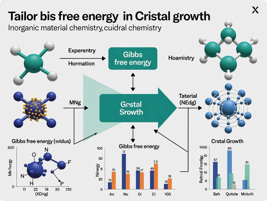
This article provides researchers, scientists, and drug development professionals with a comprehensive framework for understanding and manipulating Gibbs free energy in crystal growth processes.
Mastering Crystal Growth: A Comprehensive Guide to Tailoring Gibbs Free Energy for Pharmaceutical Development
Abstract
This article provides researchers, scientists, and drug development professionals with a comprehensive framework for understanding and manipulating Gibbs free energy in crystal growth processes. Covering fundamental thermodynamic principles through advanced computational methods and experimental techniques, we explore how controlled nucleation and growth directly impact critical pharmaceutical properties including polymorphism, solubility, and bioavailability. The content synthesizes current research on substrate temperature modulation, antisolvent treatment, solvent engineering, and computational prediction protocols, while addressing common optimization challenges and validation strategies for reliable crystal structure prediction and stability evaluation in drug development.
Gibbs Free Energy Fundamentals: The Thermodynamic Basis of Crystal Growth Control
Crystallization is a fundamental phase transition process critical in fields ranging from pharmaceutical development to materials science. This process occurs in two primary stages: nucleation, the formation of new, stable clusters (nuclei) from a supersaturated solution or melt, and crystal growth, the subsequent expansion of these nuclei into macroscopic crystals [1]. The driving force for both stages is supersaturation, a state where the concentration of a solute exceeds its equilibrium solubility [1] [2]. The control of crystallization is paramount in drug development, as it directly influences critical quality attributes of Active Pharmaceutical Ingredients (APIs), including purity, bioavailability, and stability [3]. Within the broader context of crystal growth research, a central theme is the deliberate tailoring of the Gibbs free energy landscape to guide these processes toward desired outcomes, such as specific polymorphs or crystal morphologies [4].
The Gibbs free energy change (( \Delta G )) for the formation of a spherical crystal nucleus can be described by the classical nucleation theory (CNT) as the sum of a volume term (negative, favoring crystallization) and a surface term (positive, representing an energy barrier) [5]: ( \Delta G = \frac{4}{3}\pi r^3 \Delta gv + 4\pi r^2 \sigma ) where ( r ) is the nucleus radius, ( \Delta gv ) is the Gibbs free energy change per unit volume, and ( \sigma ) is the surface free energy. This relationship gives rise to a critical nucleus radius (( rc )) and a corresponding free energy barrier (( \Delta G^* )) that must be overcome for a nucleus to become stable and grow [5]. The maximum barrier is defined as: ( \Delta G^* = \frac{16\pi \sigma^3}{3|\Delta gv|^2} ) Understanding and manipulating these thermodynamic parameters is the foundation of controlling crystallization processes in research and industry.
Theoretical Framework: Classical and Advanced Perspectives
Classical Nucleation Theory (CNT) and Kinetics
Classical Nucleation Theory (CNT) provides the principal theoretical framework for quantitatively describing nucleation kinetics. The central result of CNT is a prediction for the rate of nucleation, ( R ), which is the number of nuclei formed per unit volume per unit time [5]. This rate is given by: ( R = NS Z j \exp\left(-\frac{\Delta G^*}{kB T}\right) ) where:
- ( \Delta G^* ) is the free energy barrier to form a critical nucleus.
- ( k_B ) is the Boltzmann constant and ( T ) is the absolute temperature.
- ( N_S ) is the number of nucleation sites per unit volume.
- ( j ) is the rate at which molecules attach to the critical nucleus.
- ( Z ) is the Zeldovich factor, a dynamical correction factor.
The exponential term ( \exp(-\Delta G^/k_B T) ) represents the probability that a fluctuation will produce a critical nucleus, making the nucleation rate exquisitely sensitive to the barrier height ( \Delta G^ ) [5]. This kinetic formulation applies to both homogeneous and heterogeneous nucleation, with the latter occurring on surfaces or impurities and characterized by a lower effective energy barrier [5] [2]. For one-component systems, the thermodynamic driving force is often expressed as ( \Delta gv = \Delta \mu / v{\alpha} ), where ( \Delta \mu ) is the difference in chemical potential between the liquid and crystal phases, and ( v_{\alpha} ) is the volume per particle in the crystal phase [6].
The Role of Gibbs Free Energy and Driving Force
The Gibbs free energy provides the fundamental thermodynamic driving force for crystallization. In practical applications, the change in Gibbs free energy, ( \Delta G ), is directly related to the degree of supersaturation [7]. For a unit cell of a crystal, the Gibbs free energy, ( G{\text{unit}} ), is calculated as [4]: ( G{\text{unit}} = H{\text{unit}} + Uv - TS ) where ( H{\text{unit}} ) is the enthalpy, ( Uv ) is the zero-point vibrational energy, and ( S ) is the entropy. Under an applied pressure ( P ), the enthalpy is ( H{\text{unit}} = U{\text{int}} + P V{\text{unit}} ), where ( U{\text{int}} ) is the internal energy and ( V_{\text{unit}} ) is the unit cell volume [4]. The accurate computation of these energy terms, particularly for complex molecular crystals like active pharmaceutical ingredients (APIs), enables researchers to predict the most stable polymorphic form and rationally design crystallization processes to obtain it [4].
Table 1: Key Thermodynamic and Kinetic Parameters in Classical Nucleation Theory
| Parameter | Symbol | Description | Impact on Nucleation | ||
|---|---|---|---|---|---|
| Critical Radius | ( r_c ) | ( r_c = \frac{2\sigma}{ | \Delta g_v | } ) | Nuclei smaller than ( r_c ) dissolve; larger ones grow. |
| Free Energy Barrier | ( \Delta G^* ) | ( \Delta G^* = \frac{16\pi \sigma^3}{3 | \Delta g_v | ^2} ) | Determines the exponential term in the nucleation rate. |
| Nucleation Rate | ( R ) | ( R = NS Z j \exp\left(-\frac{\Delta G^*}{kB T}\right) ) | Number of nuclei formed per unit volume per unit time. | ||
| Interfacial Tension | ( \sigma ) | Energy per unit area of the nucleus-solution interface. | Lowering ( \sigma ) significantly reduces ( \Delta G^* ) and increases ( R ). | ||
| Driving Force | ( \Delta g_v ) | Gibbs free energy change per unit volume of crystal. | Increases with supersaturation, reducing ( r_c ) and ( \Delta G^* ). |
Beyond CNT: Non-Classical Pathways and Polymorphism
While CNT offers a foundational model, recent advances have revealed more complex, non-classical nucleation pathways. For instance, the softening of intermolecular interaction potentials, such as using a 7-6 potential instead of the standard 12-6 Lennard-Jones potential, can alter nucleation pathways without significantly changing the nucleation rate [8]. This softening can stabilize different polymorphic structures, such as the body-centered cubic (BCC) structure, introducing distinct nucleation pathways alongside the traditional face-centered cubic (FCC) pathway [8]. This demonstrates that polymorph selection can be achieved by modifying intermolecular interactions, a key aspect of tailoring the Gibbs free energy landscape.
Furthermore, the presence of interfaces (e.g., solid/liquid, air/liquid) can drastically alter nucleation behavior. Proteins and other macromolecules often exhibit preferential accumulation at interfaces, leading to increased local supersaturation and a reduced energy barrier for nucleation, making heterogeneous nucleation far more common than homogeneous nucleation in practical scenarios [2]. The application of external fields (electric, magnetic, ultrasonic) can also modify protein-protein interaction potentials and thus the thermodynamic and kinetic factors governing nucleation [2].
Quantitative Data and Energetics in Crystallization
Table 2: Comparative Nucleation and Growth Kinetics for Various Systems
| System | Relative Supersaturation | Nucleation Rate (J) | Growth Rate Prefactor | Key Finding |
|---|---|---|---|---|
| Lennard-Jones (12-6) [8] | Comparable driving force | Comparable to 7-6 system | N/A | Nucleation pathway predominantly leads to FCC structure. |
| Lennard-Jones (7-6) [8] | Comparable driving force | Comparable to 12-6 system | N/A | Two distinct pathways: one for BCC and one for FCC nuclei. |
| rAAV Capsids [9] | High | Similar tendency to nucleate as glycine | 7 orders smaller than lysozyme | Prolonged nucleation period; growth is transport-limited. |
| Ice (TIP4P/2005 model) [5] | At 19.5 °C supercooling | ( R = 10^{-83} \text{ s}^{-1} ) (calculated) | N/A | Highlights immense variation in nucleation rates. |
| Lysozyme [2] | High (typically ~100%) | Slow despite high S | Benchmark for growth rate | Slow kinetics due to limited patches for lattice bonds. |
The quantitative analysis of crystallization processes relies on measuring key parameters such as the nucleation induction time (the time between achieving supersaturation and the appearance of critical nuclei) and the metastable zone width (the region between the solubility and supersolubility curves where nucleation is kinetically unfavorable) [2]. For protein crystallization, the required supersaturation values are generally much higher (e.g., ~100%) compared to small molecules, yet the kinetics are often slower due to complex macromolecular configurations and a limited number of surface "patches" available for forming lattice bonds [2]. This is exemplified by recombinant adeno-associated virus (rAAV) capsids, which, despite their very high molecular weight (~3.6 MDa), have a similar nucleation tendency as small organic molecules like glycine (~75 Da) but exhibit a growth rate prefactor seven orders of magnitude smaller than that of lysozyme [9].
The thermodynamic stability of different polymorphs is determined by comparing their Gibbs free energies. For instance, a study on sulfathiazole polymorphs used density functional theory (DFT) calculations to determine the stability order of its five polymorphs (FI, FV, FIV, FII, FIII) based on their Gibbs free energy, confirming that form III (FIII) is the most stable structure at ambient conditions [4]. This approach moves beyond simple lattice energy calculations by incorporating the effects of entropy and temperature, providing a more accurate prediction for guiding experimental synthesis [4].
Experimental Protocols for Studying Nucleation and Growth
Protocol 1: Investigating Nucleation Pathways Using Modified Interaction Potentials in Silico
This protocol outlines a computational method to study how softening intermolecular potentials influences nucleation pathways and polymorph selection, based on the work by Minh et al. [8].
- Primary Objective: To characterize the effect of modified n-6 Lennard-Jones potentials on nucleation kinetics and mechanism under constant thermodynamic driving force.
- Reagents and Materials:
- Simulation software (e.g., LAMMPS, GROMACS).
- Parameter files for 12-6 and 7-6 potentials.
- High-performance computing (HPC) cluster resources.
- Methodology:
- System Setup: Create simulation boxes containing a sufficient number of particles (e.g., thousands to millions) for the system of interest.
- Potential Definition: Implement the standard 12-6 Lennard-Jones potential (Eq. 1 in [8]) and the softer 7-6 potential (Eq. 2 in [8]), ensuring the minima of both potentials are located at the same distance ( r = 2^{1/6}\sigma ).
- State Point Selection: Choose a thermodynamic state point (temperature and pressure) that provides the same level of supercooling and pressure for both systems to ensure an equivalent driving force for crystallization.
- Enhanced Sampling: Employ advanced sampling methods such as Replica Exchange Transition Interface Sampling (RETIS) [8] or seeding simulations [8] to overcome the nucleation free energy barrier and achieve sufficient sampling of the rare nucleation event.
- Trajectory Analysis: Use a suite of analysis tools:
- Free Energy Calculation: Compute the work of critical cluster formation, ( W_c ), using Classical Nucleation Theory or path sampling techniques.
- Pathway Analysis: Apply methodologies like the recently developed LeaPP method [8] to characterize the composition and structure (FCC, BCC, etc.) of nuclei along different trajectories.
- Rate Calculation: Determine the steady-state nucleation rate, ( J ), from the simulation data.
- Data Interpretation:
- Compare the critical nucleus composition and the prevalence of different nucleation pathways (e.g., FCC-dominant vs. BCC-dominant) between the 12-6 and 7-6 systems.
- Note that while nucleation rates may be comparable, the underlying mechanisms and resulting crystal structures can be significantly different [8].
Protocol 2: Extracting Nucleation and Growth Kinetics for a Complex Biotherapeutic
This protocol describes an integrated experimental and modeling approach to obtain kinetic constants for the crystallization of complex macromolecules, such as recombinant adeno-associated virus (rAAV) capsids, based on Bal et al. [9].
- Primary Objective: To extract nucleation and growth kinetics for a complex biotherapeutic from experimental data using coupled population balance and species balance equations.
- Reagents and Materials:
- Purified protein or macromolecule (e.g., rAAV capsids).
- Precipitating agents (e.g., salts, polymers).
- Hanging-drop vapor diffusion (HDVD) plates or batch crystallizers.
- In-line monitoring equipment (e.g., dynamic light scattering, microscopy, UV-vis spectroscopy).
- Methodology:
- Crystallization Setup: Perform crystallization trials, for example, in a hanging-drop vapor diffusion system. Here, a droplet containing the protein and precipitant is suspended over a reservoir of higher osmolyte concentration, leading to slow water removal and a dynamic increase in supersaturation [9].
- In-line Monitoring: Continuously monitor the crystallization process to track the evolution of crystal size and population. Techniques can include visual observation, laser-based methods, or spectroscopic techniques [9] [2].
- Model Development: Formulate coupled population balance equations (PBEs) and species balance equations to describe the system. The PBE accounts for the rates of nucleation and growth, while the species balance tracks the depletion of the solute from the solution.
- Parameter Estimation: Fit the developed model to the experimental data (e.g., crystal size distribution over time) to extract the kinetic parameters for nucleation and growth. This often involves determining the nucleation rate order and the growth rate expression.
- Kinetic Analysis: Analyze the extracted parameters. For rAAV capsids, this revealed a prolonged nucleation period and a growth rate that was weakly sensitive to supersaturation and limited by slow Brownian motion due to very high molecular weight [9].
- Data Interpretation:
- The model helps deconvolute the effects of vapor diffusion rate (which controls initial supersaturation) from the intrinsic nucleation and growth kinetics.
- Results can identify whether nucleation is homogeneous or heterogeneous, a key consideration for process control [9].
Protocol 3: Measuring Induction Times and Controlling Scaling in Membrane Crystallization
This protocol details a non-invasive method to measure induction times in different domains (bulk and membrane surface) to unify the understanding of nucleation and growth mechanisms in membrane systems, as per Karan et al. [10].
- Primary Objective: To relate boundary layer supersaturation to nucleation kinetics and identify a critical supersaturation threshold to avoid scaling.
- Reagents and Materials:
- Membrane distillation/crystallization setup.
- Temperature-controlled feed and permeate reservoirs.
- Non-invasive monitoring tools (e.g., video microscopy, image analysis).
- Aqueous salt solutions (e.g., NaCl, Na₂SO₄).
- Methodology:
- System Configuration: Set up a membrane crystallizer with controlled temperatures on the feed (( T ), between 45–60°C) and permeate sides to establish a temperature difference (( \Delta T ), between 15–30°C) [10].
- Induction Time Measurement: Use non-invasive techniques to independently measure the induction time for crystal formation in the bulk solution and at the membrane surface (scaling).
- Supersaturation Correlation: Relate the measured induction times to the calculated supersaturation level in the boundary layer adjacent to the membrane. A modified power law relation between supersaturation and induction time can be used to connect mass and heat transfer processes to Classical Nucleation Theory (CNT) [10].
- Crystal Size Distribution (CSD) Analysis: Analyze the final CSD to understand how ( \Delta T ) (which affects nucleation rate via supersaturation) and ( T ) (which affects growth rate) can be used collectively to control crystal morphology and population.
- Critical Supersaturation Identification: Determine the supersaturation threshold below which homogeneous nucleation (leading to scaling) is "switched off," and crystals form solely in the bulk solution with a preferred morphology [10].
- Data Interpretation:
- A log-linear relation between nucleation rate and boundary layer supersaturation confirms behavior characteristic of CNT.
- Discrimination of primary nucleation mechanisms reveals that scaling is formed homogeneously at extremely high supersaturation levels, while bulk crystals form at lower supersaturations.
The Scientist's Toolkit: Essential Research Reagents and Materials
Table 3: Key Research Reagent Solutions and Materials for Crystallization Studies
| Item | Function/Application | Example Use Case |
|---|---|---|
| Precipitating Agents | To induce supersaturation by reducing solute solubility. | Salts (e.g., ammonium sulfate), polymers (e.g., PEG), or organic solvents used in protein crystallization [2]. |
| Heteronucleants | Surfaces that lower the energy barrier for heterogeneous nucleation. | Functionalized surfaces or nanoparticles that provide a template for crystal formation [2]. |
| Modified Potentials | To computationally investigate the effect of interaction softness on nucleation pathways. | Using 7-6 vs. 12-6 Lennard-Jones potentials to study polymorph selection [8]. |
| Microreactors / Continuous Flow Systems | Process intensification for enhanced mixing, heat transfer, and control over nucleation-growth processes. | Manufacturing high-efficiency crystal particles with optimal form and structural stability [1]. |
| In-line Analytical Probes | For real-time monitoring of nucleation and growth kinetics. | Using dynamic light scattering (DLS) or microscopy to determine nucleation induction times and crystal size distributions [9] [2]. |
| Ionic Liquids | As a growth medium for potential-driven crystallization of metals. | Used in the growth of metal crystals from protic ionic liquids (PILs) and solvate ionic liquids (SILs) [1]. |
Workflow and Pathway Visualizations
The following diagrams illustrate key experimental workflows and conceptual pathways in crystallization research.
Diagram 1: Integrated Research Workflow for Crystallization Studies. This chart outlines parallel computational and experimental pathways for investigating nucleation and growth, which are integrated to inform strategies for tailoring the Gibbs free energy landscape.
Diagram 2: Nucleation Pathways and Polymorph Selection. This diagram visualizes the decision point in the nucleation pathway, influenced by factors like interaction potential softness, which can lead to different stable crystal structures (polymorphs) via distinct trajectories on the Gibbs free energy landscape [8].
A deep understanding of the two-step crystallization process, grounded in the principles of thermodynamics and kinetics, is indispensable for advancing research in drug development and materials science. The ability to tailor the Gibbs free energy landscape—whether through computational design of interaction potentials [8], the strategic use of heteronucleants and interfaces [2], or precise control over process parameters like temperature and supersaturation [10]—provides powerful levers for controlling nucleation mechanisms and crystal growth. The integrated application of advanced computational modeling, innovative experimental techniques, and robust process intensification strategies continues to deepen our fundamental understanding and enhance our control over these complex processes. This, in turn, enables the rational design and manufacturing of materials with tailored properties and enhanced functionality, ultimately accelerating the development of more effective therapeutics and advanced materials.
In crystal growth research, tailoring the Gibbs free energy of a system is paramount for controlling the formation, size, and purity of crystalline products. The chemical potential (μ), derived from the Gibbs free energy, represents the driving force for mass transfer and phase transitions, serving as a key parameter in predicting system equilibrium and spontaneous processes [11]. Supersaturation describes a non-equilibrium state where solute concentration exceeds its equilibrium solubility, providing the thermodynamic impetus for nucleation and crystal growth [12]. Together, these concepts form the foundational framework for understanding and manipulating crystallization processes across diverse fields, from pharmaceutical development to advanced material design and environmental technology.
The precise management of chemical potential and supersaturation enables researchers to navigate the complex energy landscape of crystallization, influencing critical outcomes including crystal size distribution, polymorph selection, and product purity. This application note examines the theoretical and practical aspects of these driving forces, providing structured protocols and data analysis techniques to advance crystal growth research within the broader context of Gibbs free energy optimization.
Theoretical Foundations
Chemical Potential and Gibbs Free Energy
Chemical potential (μ) is defined as the change in Gibbs free energy (G) of a system when a component is added or removed, while keeping temperature, pressure, and other component amounts constant: μᵢ = (∂G/∂nᵢ)ₜ,ₚ,ₙⱼ [11]. This intensive property represents the escaping tendency of a component and serves as the fundamental driving force for mass transfer in multicomponent systems. At equilibrium, the chemical potential of each component must be equal across all phases, as described by the Gibbs phase rule [13].
The relationship between Gibbs free energy and chemical potential extends to partial molar quantities, where the chemical potential of component i equals its partial molar Gibbs free energy: μᵢ = Ḡᵢ = Ḣᵢ - TṠᵢ + PṼᵢ [11]. This relationship connects the thermodynamic properties of individual components to the overall system energy, enabling prediction of phase behavior and equilibrium conditions.
Supersaturation as the Driving Force
Supersaturation describes a metastable state where the solute concentration exceeds its equilibrium solubility, creating a positive chemical potential difference (Δμ) between the solution and crystal phases [12] [14]. This chemical potential difference provides the thermodynamic driving force for both nucleation and crystal growth, with the system seeking to reduce Δμ by forming and growing solid particles.
The degree of supersaturation directly influences crystallization mechanisms: low supersaturation promotes diffusion-controlled crystal growth resulting in larger particles, while high supersaturation facilitates nucleation leading to smaller crystals [12]. This fundamental relationship enables researchers to control crystal size distribution by strategically managing supersaturation levels throughout the crystallization process.
Quantitative Relationships and Performance Data
Table 1: Quantitative Relationships Between Supersaturation, Chemical Potential, and Crystallization Outcomes
| System | Supersaturation Control Method | Chemical Potential Relationship | Key Performance Results | Reference |
|---|---|---|---|---|
| Photovoltaic Wastewater (CaF₂ Recovery) | Nucleation-Induced Crystallization Reflux Process (NCRP) with reflux ratios 5:1 to 10:1 | Lower supersaturation in reaction zone enhanced CaF₂ crystallization efficiency | Crystallization efficiency >90%; Effluent F⁻ <10 mg/L; F⁻ removal >98%; Crystal size D₅₀ = 1.62 mm | [12] |
| Protein Crystallization (Lysozyme) | Urea and NaCl additives to tune chemical potential difference (Δμ) | Δμ increases logarithmically with salt concentration and decreases linearly with urea content | Crystallization at lower supersaturations; Enhanced nucleation and growth rates at fixed Δμ | [14] |
| Pyramid Stepped Basin Solar Still (PSBSD) | Gibbs phase rule application to intensive state parameters | Chemical potentials establish relation between equilibrium of liquid and vapor mixture | System efficiency: 38.135%; Distillate yield: 4.280 l/m²day over 24h cycle | [13] |
| Sodium Halide Crystallization | Microdroplet evaporation across supersaturation range | Classical vs. nonclassical nucleation pathways based on supersaturation level | Identification of liquid crystal intermediate phases for NaBr and NaI at specific supersaturations | [15] |
Table 2: Crystallization Kinetics and Thermodynamic Parameters in Various Systems
| Parameter | Lysozyme with NaCl | Lysozyme with Urea | CaF₂ with NCRP | Sodium Halides |
|---|---|---|---|---|
| Solubility Trend | Decreases with increasing concentration | Increases with increasing concentration | N/A | N/A |
| Induction Time | Decreases with concentration | Increases with concentration | N/A | Varies by supersaturation |
| Crystal Growth Rate | Accelerates | Decelerates | Enhanced at lower supersaturation | Pathway-dependent |
| Nucleation Pathway | Classical | Classical | Classical | Classical (NaCl) vs. Nonclassical (NaBr, NaI) |
| Key Additive Effect | Reduces chemical potential difference | Enables crystallization at lower supersaturation | Reflux controls local supersaturation | Supersaturation determines intermediate phases |
Experimental Protocols
Protocol: Supersaturation-Controlled Fluoride Recovery via NCRP
Principle: This protocol describes the Nucleation-Induced Crystallization Reflux Process (NCRP) for recovering high-purity calcium fluoride from photovoltaic wastewater through precise supersaturation control [12].
Materials:
- Photovoltaic wastewater (Fluoride concentration: 450-2500 mg/L; pH: 2.1-9.4)
- Sodium hydroxide (NaOH) solution (For pH adjustment)
- Calcium chloride (CaCl₂) solution (Calcium source)
- NCRP reactor system (With controlled reflux capability)
- Analytical equipment (Ion-selective electrode for fluoride measurement, particle size analyzer)
Procedure:
- System Setup: Configure the NCRP reactor with direct recirculation of low-concentration effluent to the reactor base. Establish separate reaction zone (high-velocity, low-supersaturation) and clarification zone (low-velocity for crystal separation).
- Parameter Initialization: Set initial reflux ratio to 5:1 (effluent recycle:influent ratio). Maintain Ca/F molar ratio between 0.45-0.6 through controlled dosing of CaCl₂ solution.
- pH Adjustment: Co-dose NaOH solution to maintain system pH in the range of 6-8 throughout the operation. Monitor pH continuously with automatic controller.
- Supersaturation Control: Utilize dynamic reflux adjustment to maintain low supersaturation in the reaction zone. Higher reflux ratios (5:1 to 10:1) promote crystal growth over nucleation.
- Crystal Growth Phase: Operate system continuously for 25 days to allow development of crystalline particles with target median diameter (D₅₀) of 1.62 mm.
- Performance Monitoring: Sample effluent regularly to verify fluoride concentration remains below 10 mg/L. Analyze crystal products for purity (target CaF₂ content >85%) and surface characteristics.
Notes: The reflux mechanism is critical for mitigating influent water quality fluctuations and preventing excessive fine particle formation. System performance should be validated through characterization analyses including XPS, XRD, Raman, and zeta potential measurements [12].
Protocol: Protein Crystallization via Chemical Potential Tuning with Urea and Salt
Principle: This protocol employs urea and salt additives to independently tune thermodynamic and kinetic parameters of protein crystallization by modifying the chemical potential difference (Δμ) between solution and crystal phases [14].
Materials:
- Lysozyme protein (Sigma-Aldrich, prod. no. L6876)
- Sodium chloride (NaCl) (Fisher Scientific, prod. no. S/3160/60)
- Urea (Merck, prod. no. 1.08488; Sigma, prod. no. 33247)
- Sodium acetate (NaAc) buffer (50 mM, pH 4.5)
- Ultrafiltration cell (Amicon, Millipore, prod. no. 5121) with Omega 10 kDa membrane
- Video microscopy system (For crystallization kinetics monitoring)
Procedure:
- Sample Preparation: Dissolve lysozyme powder in 50 mM sodium acetate buffer (pH 4.5). Filter through 0.1 μm pore size syringe filter to remove aggregates. Concentrate using ultrafiltration cell to prepare stock solution (∼80 mg/mL).
- Solution Characterization: Determine protein concentration via UV absorbance. Prepare solutions with varying urea (0-2 M) and NaCl (0-5%) concentrations while maintaining constant protein concentration.
- Solubility Measurement: Determine solubility by equilibrating protein solutions at different additive concentrations until constant supernatant concentration is achieved. Plot solubility as function of second virial coefficient (B₂).
- Chemical Potential Calculation: Calculate chemical potential difference Δμ = kT ln(C/Csat), where C is protein concentration and Csat is solubility determined in step 3. Generate Δμ map across the phase diagram.
- Kinetics Monitoring: Use video microscopy to track crystallization at selected Δμ values. Measure induction time (delay between supersaturation achievement and first crystal detection) and crystal growth rates.
- Data Analysis: Fit nucleation data to classical nucleation theory and growth data to birth-and-spread growth model. Compare kinetics at constant Δμ with varying urea/salt ratios.
Notes: Urea increases protein solubility while salt decreases it, enabling independent control over thermodynamic and kinetic parameters. At fixed Δμ, urea enhances both nucleation and growth rates compared to salt alone, potentially by reducing energy barriers and suppressing non-productive binding [14].
Visualization of Concepts and Workflows
The Scientist's Toolkit: Essential Research Reagents and Materials
Table 3: Key Research Reagent Solutions for Crystallization Studies
| Reagent/Material | Function in Crystallization Research | Example Application | Key Considerations | |
|---|---|---|---|---|
| Urea | Modifies protein-protein interactions; increases protein solubility; alters dielectric properties of solution | Protein crystallization at sub-denaturing concentrations to tune nucleation and growth kinetics | Use at sub-denaturing concentrations (typically 0-2 M); enables crystallization at lower supersaturations | [14] |
| Sodium Chloride (NaCl) | Decreases protein solubility without salting-in effect; reduces induction time; accelerates crystal growth | Common precipitant for protein crystallization; enhances crystallization driving force | Concentration-dependent effect on chemical potential difference; combines effectively with urea | [14] |
| Calcium Chloride (CaCl₂) | Calcium source for fluoride crystallization; forms insoluble CaF₂ precipitate | Fluoride recovery from wastewater via crystallization; molar ratio control critical | Maintain Ca/F molar ratio 0.45-0.6 for optimal crystallization efficiency | [12] |
| Sodium Hydroxide (NaOH) | pH adjustment for crystallization systems; controls speciation and solubility | pH maintenance (6-8) in CaF₂ recovery; affects zeta potential and surface charge | Critical for optimizing crystallization in acidic wastewater streams | [12] |
| Crystal Seeds/Inducers | Provides surface for heterogeneous nucleation; reduces activation energy barrier | Fluidized-bed reactors; nucleation-induced crystallization processes | Surface characteristics and size distribution affect induction efficiency | [12] |
The strategic manipulation of chemical potential and supersaturation provides powerful leverage for controlling crystallization processes across diverse applications. Through the protocols and data presented, researchers can implement precise supersaturation control strategies—whether via reflux systems for inorganic crystallization or chemical potential tuning with additives for protein systems—to achieve target crystal size distributions, purity specifications, and process efficiencies. The fundamental relationship between Gibbs free energy, chemical potential differences, and supersaturation remains the unifying principle enabling rational design of crystallization processes, bridging theoretical thermodynamics with practical application in pharmaceutical development, materials science, and environmental technology.
In the pharmaceutical and materials sciences, the solid form is a critical quality attribute that can determine the success or failure of a product. Many organic compounds, including most active pharmaceutical ingredients (APIs), can crystallize in different three-dimensional arrangements, a phenomenon known as polymorphism. These different polymorphs, despite having identical chemical compositions, can exhibit dramatically different physical properties including solubility, dissolution rate, chemical stability, mechanical behavior, and bioavailability. The fundamental factor governing which polymorph dominates under specific conditions is the Gibbs free energy (G) of the crystalline form. At a given temperature and pressure, the polymorph with the lowest Gibbs free energy is thermodynamically stable, while other forms are metastable and will eventually transform to the stable form, though kinetic barriers may make this transformation impractically slow.
The Gibbs free energy of a crystal structure is defined by the relationship G = H - TS, where H is enthalpy, T is absolute temperature, and S is entropy. The relative stability between two polymorphs is determined by the difference in their Gibbs free energies (ΔG = ΔH - TΔS). While the enthalpy term (ΔH) primarily reflects differences in lattice energy from intermolecular interactions and packing efficiency, the entropy term (TΔS) accounts for vibrational freedom and disorder in the crystal lattice. This review explores how Gibbs free energy dictates polymorph stability, provides methodologies for its evaluation, and demonstrates its application in predicting and controlling crystalline forms in research and development.
Core Principles: Thermodynamic Foundations of Polymorph Stability
The Gibbs Free Energy Equation in Polymorphism
The competition between polymorphs is fundamentally governed by their relative Gibbs free energies. For any pair of polymorphs, the difference in their Gibbs free energy can be expressed as ΔG = G₂ - G₁ = (H₂ - H₁) - T(S₂ - S₁) = ΔH - TΔS. The stable polymorph under specific conditions of temperature and pressure is the one with the lowest Gibbs free energy. When ΔG < 0, polymorph 2 is more stable; when ΔG > 0, polymorph 1 is more stable. The temperature dependence of this relationship means that a polymorph with higher entropy (disorder) may become more stable at elevated temperatures even if it has higher enthalpy (less favorable intermolecular interactions).
The pressure dependence of polymorph stability is equally crucial, as described by the derivative (∂G/∂P)ₜ = V, where V is the molar volume. This relationship explains why denser polymorphs (with smaller molar volumes) typically become more stable at elevated pressures. As demonstrated in the case of benzophenone, the interplay between temperature and pressure creates a complex phase diagram where different polymorphs can dominate in different regions of pressure-temperature space [16].
Monotropic and Enantiotropic Relationships
Polymorphic systems are classified based on their thermodynamic relationships:
- Monotropic systems: One polymorph is always more stable than the other across all temperatures below melting. The higher-energy polymorph is always metastable and any transition between forms is irreversible.
- Enantiotropic systems: Each polymorph has a specific temperature range where it is thermodynamically stable. The transition between forms is reversible at a specific transition temperature where their Gibbs free energies are equal.
Large-scale computational studies have revealed that among organic molecular crystals, approximately 21% of polymorph pairs exhibit enantiotropic behavior, meaning temperature can reverse their relative stability [17]. This highlights the importance of considering entropy contributions and temperature effects in polymorph stability assessment.
Table 1: Thermodynamic Parameters Governing Polymorph Stability
| Parameter | Symbol | Definition | Impact on Polymorph Stability |
|---|---|---|---|
| Gibbs Free Energy | G | G = H - TS | Determines thermodynamic stability; lowest G is most stable |
| Enthalpy | H | Sum of internal energy and PV work | Reflects strength of intermolecular interactions and crystal packing efficiency |
| Entropy | S | Measure of disorder or vibrational freedom | Favors polymorphs with greater molecular mobility at higher temperatures |
| Volume | V | Molar volume of crystal | Determines pressure dependence; denser forms favored at high pressure |
| Heat Capacity | cₚ | Temperature derivative of enthalpy | Affects temperature dependence of enthalpy and entropy |
Experimental Determination of Gibbs Free Energy in Polymorph Stability
Thermodynamic Workflow for Amorphous Formation Risk Assessment
A practical thermodynamic workflow has been developed for pharmaceutical applications to evaluate the risk of amorphous formation during processing of either drug substances or drug products. This approach begins with understanding the thermodynamics of crystalline and amorphous phases through a three-step process:
First, thermodynamic equations are derived to calculate the enthalpy, Gibbs free energy, and solubility of each phase and their differences as a function of temperature. The enthalpy for each crystalline drug substance at its melting point is selected as the reference state (HcTm = 0, where Hc is the molar enthalpy and Tm is the melting temperature) to enable a consistent approach for all analyses [18].
Second, data from differential scanning calorimetry (DSC) measurements and the derived thermodynamic equations are used to construct enthalpy, Gibbs free energy, and solubility diagrams to compare the characteristics of the two phases. The Gibbs free energy difference between crystalline and amorphous phases (ΔGca) is calculated using the relationship: ΔGca(T) = ΔHca(T) - TΔSca(T), where ΔHca and ΔSca represent the differences in enthalpy and entropy between the crystalline and amorphous states [18].
Finally, the results of these calculations are used to evaluate the potential risk of crystalline-to-amorphous phase conversion during processing and the impact of amorphous formation on solubility. This workflow enables quantitative assessment of processing conditions that might inadvertently generate amorphous content, which could affect product stability and performance [18].
Experimental Protocol: Determining Thermodynamic Parameters via DSC
Principle: This protocol uses Differential Scanning Calorimetry (DSC) to obtain thermodynamic parameters needed to calculate Gibbs free energy differences between polymorphs or between crystalline and amorphous forms.
Materials and Equipment:
- Differential Scanning Calorimeter with high sensitivity and temperature calibration
- Hermetically sealed pans compatible with the DSC instrument
- Standard reference materials for temperature and enthalpy calibration (e.g., indium, tin)
- Powder samples of pure polymorphic forms
Procedure:
- Calibrate the DSC instrument using standard reference materials for both temperature and enthalpy flow.
- Precisely weigh 2-5 mg of each polymorphic sample and seal in DSC pans.
- Run DSC scans at controlled heating rates (typically 5-10°C/min) across the relevant temperature range.
- Identify and integrate thermal events: glass transitions (Tg), melting points (Tm), recrystallization exotherms, and solid-solid transitions.
- Calculate enthalpy changes (ΔH) from the area under endothermic or exothermic peaks.
- Determine heat capacity (cₚ) changes from step changes in the baseline at Tg.
Data Analysis:
- Calculate entropy of fusion: ΔSfus = ΔHfus/Tm
- Estimate entropy differences between polymorphs using heat capacity data and thermodynamic relationships
- Construct enthalpy and Gibbs free energy diagrams as functions of temperature using the reference state approach
- Calculate solubility differences between polymorphs using the Gibbs free energy data
Applications: This protocol enables quantitative risk assessment for polymorph conversion during processing, prediction of relative solubility of different forms, and identification of temperature ranges where enantiotropic transitions occur [18].
Computational Approaches for Gibbs Free Energy Prediction
Embedded Fragment QM Method for Pharmaceutical Molecules
Advanced computational methods have been developed to predict the relative stability of crystal structures from first principles. The embedded fragment quantum mechanical (QM) method has emerged as a powerful approach for calculating Gibbs free energies of molecular crystals, enabling stability evaluation without experimental input. This method is particularly valuable for pharmaceutical compounds like cabotegravir (GSK744), where crystal structure can significantly impact bioavailability and efficacy [19].
The protocol for Gibbs free energy-guided crystal structure prediction involves:
- Conformational Search: Identify flexible torsion angles and perform potential energy surface (PES) scans to find the lowest-energy molecular conformer.
- Crystal Structure Prediction: Use software packages like MOLPAK to generate plausible crystal packings in common space groups.
- Energy Ranking: Calculate lattice energies to identify low-energy candidates (typically within 10 kJ/mol of the global minimum).
- Gibbs Free Energy Calculation: Apply the embedded fragment QM method to calculate Gibbs free energies for the most promising candidates.
- Stability Determination: Identify the most stable structure at the bottom of the Gibbs free energy landscape.
The embedded fragment method calculates the internal energy (Uint) of the unit cell by dividing it into a proper combination of the energies of monomers and dimers that are embedded in the electrostatic field of the rest of the crystalline environment. This approach makes Gibbs free energy calculations feasible for large pharmaceutical molecules with practical computational resources [4] [19].
Computational Protocol: Gibbs Free Energy Calculation via Embedded Fragment Method
Principle: This protocol uses density functional theory (DFT) with the embedded fragment quantum mechanical approach to calculate Gibbs free energies of predicted crystal structures, enabling stability ranking of polymorphs.
Computational Requirements:
- Quantum chemistry software with periodic boundary condition capability
- High-performance computing cluster with parallel processing
- Crystal structure visualization software
Procedure:
- Initial Structure Generation:
- Perform conformational analysis to identify the most stable molecular conformer
- Generate crystal packing candidates using CSP methods (MOLPAK, PROM, GRACE)
Structure Optimization:
- Optimize lattice parameters using quasi-Newton algorithm
- Employ dispersion-corrected DFT functionals (e.g., ωB97XD/6-31G*)
- Set convergence criteria for geometry optimization (e.g., 0.001 Hartree/Bohr for maximum gradient)
Gibbs Free Energy Calculation:
- Calculate internal energy (Uint) using embedded fragment approach: Uint = ΣEμ + Σ(Eμν - Eμ - Eν) + ELR
- Compute zero-point vibrational energy (Uv) and entropy (S) using harmonic approximation
- Determine Gibbs free energy: G = H + Uv - TS = Uint + PV + Uv - TS
Stability Ranking:
- Compare Gibbs free energies of all candidate structures
- Identify the global minimum as the predicted most stable form
- Analyze energy differences to assess likelihood of polymorphism
Applications: This protocol enables ab initio prediction of the most stable polymorph for pharmaceutical compounds early in development, guides experimental polymorph screening, and provides understanding of structure-property relationships [4] [19].
Table 2: Computational Methods for Gibbs Free Energy Calculation
| Method | Key Features | Accuracy Considerations | Computational Cost |
|---|---|---|---|
| Embedded Fragment QM | Divides crystal into monomers/dimers in electrostatic field; uses DFT for energy calculations | Highly accurate when including entropy and temperature effects; accounts for polarization | High, but more efficient than full periodic QM |
| Lattice Energy Only | Considers only internal energy without entropy contributions | Limited accuracy; may misrank polymorph stability | Moderate |
| Force Field Methods | Uses parameterized atom-atom potentials | Speed vs. accuracy trade-off; may not capture subtle interactions | Low to Moderate |
| Quasi-Harmonic Approximation | Includes thermal expansion effects through volume dependence | Small effect on rankings but improves accuracy | High |
Case Studies in Pharmaceutical and Material Systems
Sulfathiazole: Resolving Polymorph Stability Through Gibbs Free Energy
Sulfathiazole, an antimicrobial drug, exists in five known polymorphs (FI, FII, FIII, FIV, FV) whose relative stability had been historically confusing. Researchers applied the embedded fragment QM method at the DFT level (ωB97XD/6-31G*) to calculate Gibbs free energies of all five forms at 300 K and atmospheric pressure. The results demonstrated that form III (FIII) is the most stable structure, with the overall stability order of FI < FV < FIV < FII < FIII. This computational ranking resolved longstanding confusion about sulfathiazole polymorphism and matched experimental observations [4].
The study highlighted the importance of using Gibbs free energy rather than lattice energy alone for stability evaluation. By including entropy and temperature effects, the calculations correctly identified the stability ordering that simple lattice energy calculations might have misranked. Additionally, the computed Raman spectra provided fingerprints to discriminate between the different polymorphs, offering both thermodynamic and spectroscopic validation of the computational approach [4].
Benzophenone: The Exception to the Density Rule
Benzophenone presents a fascinating case study where the less dense polymorph (form II) was found to possess a stable domain at high pressure and high temperature, despite historical classification as "totally unstable." This finding challenged the conventional wisdom that higher density polymorphs always become more stable at high pressure. The phase behavior of benzophenone demonstrates that both the volume term (VdP) and entropy term (-SdT) in the Gibbs free energy equation must be considered to understand polymorph stability [16].
The specific volumes of benzophenone forms I and II are very close (vI = 0.774 + 0.00016T cm³/g; vII = 0.781 + 0.00015T cm³/g), with virtually identical thermal expansivity. Despite form II having a slightly larger specific volume, it becomes stable at high pressure and temperature due to its higher entropic content. This case illustrates that small differences in both volume and entropy can lead to unexpected stability domains in the pressure-temperature phase diagram [16].
The Scientist's Toolkit: Essential Reagents and Materials
Table 3: Key Research Reagent Solutions for Polymorph Stability Studies
| Reagent/Material | Function/Application | Experimental Context |
|---|---|---|
| Differential Scanning Calorimeter (DSC) | Measures phase transitions, melting points, and enthalpy changes | Experimental determination of thermodynamic parameters for Gibbs free energy calculations |
| Hermetic Sealing pans | Encapsulates samples for DSC analysis | Prevents sample degradation or evaporation during thermal analysis |
| Temperature Calibration Standards (e.g., Indium) | Calibrates temperature scale of thermal analysis instruments | Ensures accuracy of melting point and enthalpy measurements |
| Powder X-ray Diffractometer | Identifies crystalline phases and determines unit cell parameters | Provides structural validation of polymorphic forms |
| Controlled Atmosphere Chambers | Maintains specific humidity and temperature conditions | Studies environmental effects on polymorph stability and transitions |
| ωB97XD/6-31G* Computational Method | Density functional theory with dispersion correction | Accurate calculation of intermolecular interactions in crystal lattice energy computations |
| Embedded Fragment QM Software | Implements fragment-based quantum mechanical approach | Enables Gibbs free energy calculations for large molecular crystals |
Workflow Diagram: Gibbs Free Energy Evaluation in Polymorph Stability
The following diagram illustrates the integrated experimental and computational workflow for evaluating polymorph stability through Gibbs free energy assessment:
Integrated Workflow for Polymorph Stability Assessment
This workflow illustrates the complementary experimental and computational paths for determining polymorph stability. The experimental path (green) begins with sample preparation and DSC analysis to measure thermal events and enthalpy changes, leading to thermodynamic parameter calculation. The computational path (red) starts with crystal structure prediction followed by DFT optimization and Gibbs free energy calculation. Both paths converge at stability ranking and phase diagram construction, ultimately enabling processing risk assessment for pharmaceutical development.
Gibbs free energy provides the fundamental thermodynamic criterion for understanding and predicting polymorph stability in crystalline materials. Through integrated experimental and computational approaches, researchers can now quantitatively evaluate the relative stability of polymorphs, predict stability domains across temperature and pressure ranges, and assess processing risks associated with polymorph conversion. The protocols and case studies presented demonstrate that accurate Gibbs free energy evaluation requires careful consideration of both enthalpy and entropy contributions, particularly for pharmaceutical systems where small energy differences can have significant implications for product performance and stability. As computational methods continue to advance, the ability to predict and control polymorphic outcomes through Gibbs free energy optimization will play an increasingly important role in materials design and drug development.
In the field of crystal engineering, nucleation is the critical first step that determines the final quality, morphology, and performance of crystalline materials. For researchers and drug development professionals, controlling nucleation is essential for producing materials with desired characteristics, from pharmaceutical actives to advanced materials. This process occurs through two primary pathways: homogeneous nucleation, which occurs spontaneously throughout the bulk solution, and heterogeneous nucleation, which is catalyzed by surfaces or impurities [20] [21]. The competition between these pathways directly influences critical quality attributes including crystal size distribution, polymorphism, purity, and stability [20] [22]. This application note examines the fundamental differences between these nucleation mechanisms within the overarching thesis that tailoring Gibbs free energy landscapes provides a powerful strategy for controlling crystal quality. We present quantitative comparisons, experimental protocols, and practical tools to guide nucleation control in research and development settings.
Theoretical Foundations: Gibbs Free Energy in Nucleation
Classical Nucleation Theory Framework
Classical Nucleation Theory (CNT) provides the fundamental framework for quantifying nucleation behavior. According to CNT, the formation of a new phase requires overcoming an energy barrier, known as the Gibbs free energy of nucleation (ΔG*) [5] [6]. This energy barrier arises from the competition between the energy penalty for creating a new interface and the energy gain from forming the more stable crystalline phase. For a spherical nucleus, the total Gibbs free energy change is described by:
ΔG = (4/3)πr³ΔGv + 4πr²γ
Where r is the nucleus radius, ΔGv is the Gibbs free energy change per unit volume (negative for stable phase formation), and γ is the interfacial tension [5] [21]. The critical radius (r) and critical nucleation barrier (ΔG) occur at the maximum of this function, where dΔG/dr = 0, yielding:
r* = -2γ/ΔGv and ΔG* = 16πγ³/(3ΔGv²)
The nucleation rate (J), which represents the number of nuclei formed per unit volume per unit time, depends exponentially on this energy barrier [5] [6]:
J = J₀exp(-ΔG*/kBT)
Where J₀ is a kinetic pre-exponential factor, kB is Boltzmann's constant, and T is absolute temperature [6].
The Role of Interfacial Energy
The crystal-liquid interfacial energy (σ) plays a decisive role in determining nucleation behavior [22]. This parameter contributes directly to the excess surface free energy required for nucleation and varies significantly between different compounds. Highly soluble salts typically exhibit low interfacial energy, resulting in lower nucleation barriers that favor heterogeneous mechanisms at limited supersaturations. In contrast, less soluble compounds possess higher interfacial energy, requiring greater supersaturation to overcome the nucleation barrier and often favoring homogeneous nucleation [22]. This relationship between solubility, interfacial energy, and nucleation mechanism has profound implications for membrane scaling in crystallization processes, with heterogeneous nucleation dominating for high-solubility compounds and homogeneous nucleation becoming significant for less soluble systems beyond supersaturation thresholds [22].
Comparative Analysis: Homogeneous vs. Heterogeneous Nucleation
Table 1: Fundamental characteristics of homogeneous and heterogeneous nucleation mechanisms
| Characteristic | Homogeneous Nucleation | Heterogeneous Nucleation |
|---|---|---|
| Nucleation Sites | Any monomer within the volume [20] | Foreign surfaces, impurities, structure defects, active centers [20] |
| Energy Barrier | Higher (ΔG*hom) [5] | Lower (ΔGhet = f(θ)ΔGhom) [5] |
| Contact Angle | Not applicable | 0° < θ < 180° [5] |
| Geometric Factor | f(θ) = 1 [5] | f(θ) = (2-3cosθ+cos³θ)/4 [5] |
| Critical Supersaturation | Higher [22] | Lower [22] |
| Typical Crystal Quality | Smaller crystals, uniform size distribution [20] | Larger crystals, potential defects from substrates [20] |
| Spatial Distribution | Random throughout volume [21] | Localized at active sites [20] |
| Probability in Practice | Rare [5] | Much more common [5] |
Table 2: Implications for crystal quality attributes in pharmaceutical development
| Quality Attribute | Homogeneous Nucleation Impact | Heterogeneous Nucleation Impact |
|---|---|---|
| Polymorphic Control | Potentially multiple polymorphs [20] | Substrate-directed polymorph selection [20] |
| Crystal Size Distribution | Narrower distribution [20] | Broader distribution [20] |
| Purity | Higher potential purity [21] | Risk of impurity incorporation [20] |
| Process Control | Challenging to initiate reliably [22] | More controllable via engineered substrates [20] |
| Scale Formation | Mitigates membrane scaling [22] | Promotes membrane scaling [22] |
| Reproducibility | Potentially variable between batches | More reproducible with controlled substrates |
The Geometric Factor in Heterogeneous Nucleation
The reduction of the nucleation barrier in heterogeneous nucleation is quantified by the geometric factor f(θ), which depends on the contact angle (θ) between the crystal nucleus and the substrate [5]. This factor ranges from 0 to 1, with lower values indicating greater catalytic effectiveness of the substrate. When θ = 90°, f(θ) = 0.5, meaning the nucleation barrier is halved compared to homogeneous nucleation. When θ = 180° (complete non-wetting), f(θ) = 1, and the system behaves as homogeneous nucleation. When θ = 0° (complete wetting), f(θ) = 0, and there is no nucleation barrier [5]. This relationship explains why surfaces with appropriate wettability can dramatically enhance nucleation rates at lower supersaturations.
Experimental Protocols for Nucleation Studies
Protocol: Determining Dominant Nucleation Mechanism
Purpose: To determine whether homogeneous or heterogeneous nucleation dominates in a given crystallizing system.
Materials:
- Pure solute compound
- High-purity solvent (HPLC grade)
- Nucleating agents/substrates (for controlled heterogeneous nucleation)
- Microdroplet array platform or small volume containers
- Temperature-controlled crystallization chamber
- Microscopy system for in-situ monitoring
Procedure:
- Prepare a series of solutions with varying supersaturation levels (β = 1.5-3.0 for pharmaceutical compounds).
- Divide each solution into multiple small droplets (1-100 μL) or use a microfluidic droplet generator for smaller volumes (10-100 nL) [20].
- For heterogeneous nucleation studies, introduce controlled substrates (functionalized surfaces, engineered particles, etc.) to selected droplets.
- Monitor nucleation events in real-time using optical microscopy or laser scattering detection.
- Record induction times (τ) for nucleation at each condition.
- Statistical analysis: For homogeneous nucleation, the probability of nucleation follows a Poisson distribution across multiple small droplets. For heterogeneous nucleation, the distribution will be skewed by the presence of active sites [20].
- Plot nucleation rate (J = 1/τ) versus supersaturation and fit with CNT models to extract energy barriers.
Data Interpretation:
- Homogeneous nucleation shows strong dependence on droplet volume and higher supersaturation thresholds
- Heterogeneous nucleation shows weaker volume dependence and occurs at lower supersaturations
- Linearized plots of ln(J) versus 1/(ΔT²) or 1/(lnS²) can distinguish mechanisms based on slope differences
Protocol: Tailoring Gibbs Free Energy Through Additives
Purpose: To modify nucleation barriers using selective additives to direct nucleation toward desired mechanisms.
Materials:
- Active pharmaceutical ingredient (API)
- Crystallization solvent
- Selected additives (polymers, surfactants, ions)
- Characterization tools (FTIR, Raman, XRD)
Procedure:
- Prepare stock solutions of API at constant supersaturation.
- Add varying concentrations of selected additives (0.01-1.0% w/w).
- Monitor nucleation induction times using focused beam reflectance measurement (FBRM) or particle vision microscope (PVM).
- Characterize resulting crystals for polymorphic form, crystal habit, and size distribution.
- Measure crystal-liquid interfacial energy through metastable zone width studies [22].
- Calculate effective changes to ΔG* using measured nucleation rates and the CNT equation.
Interpretation:
- Additives that significantly increase induction time are increasing the effective nucleation barrier
- Additives that selectively promote specific polymorphs are modifying the relative nucleation barriers between forms
- Correlation of interfacial energy changes with additive structure informs design of more effective modifiers
Visualization of Nucleation Pathways
The Scientist's Toolkit: Research Reagents and Materials
Table 3: Essential research reagents and materials for nucleation studies
| Reagent/Material | Function | Application Notes |
|---|---|---|
| Microdroplet Arrays | Confined volumes to study nucleation statistics [20] | Enables statistical analysis of nucleation events; silicon or PDMS platforms |
| Functionalized Surfaces | Engineered substrates for heterogeneous nucleation | Self-assembled monolayers with controlled wettability; contact angle critical |
| Nanoparticle Suspensions | Heterogeneous nucleation agents | Gold, silver, or functionalized nanoparticles; size and surface chemistry dependent effects |
| Molecular Additives | Modifiers of interfacial energy [22] | Polymers, surfactants, ionic additives; concentration typically 0.001-0.1% w/w |
| Seeds (Same Compound) | Controlled secondary nucleation | Size and characterization critical; typically 1-5% of final crystal mass |
| Seeds (Different Compounds) | Templated heteroepitaxial nucleation | Lattice matching important; potential regulatory considerations for pharmaceuticals |
| High-Purity Solvents | Minimize unintended heterogeneous sites [21] | HPLC grade or better; filtration through 0.2μm filters recommended |
Advanced Concepts: Beyond Classical Nucleation Theory
While CNT provides a valuable framework, recent research has revealed limitations in its application to crystal nucleation. The nucleation theorem provides a more general approach for analyzing experimental data without some of the restrictive assumptions of CNT [6]. This theorem relates the derivative of the work of critical cluster formation with respect to the thermodynamic driving force to the number of molecules in the critical cluster:
dWc/d(Δμ) = -nc
Where Wc is the work of critical cluster formation, Δμ is the difference in chemical potential, and nc is the number of molecules in the critical cluster [6]. This relationship allows researchers to extract critical cluster sizes from experimental nucleation rate data without assuming specific cluster properties.
Furthermore, the generalized Gibbs approach acknowledges that critical clusters may have properties different from the macroscopic crystal phase, particularly with respect to composition and structure [6]. This is especially relevant for polymorphic systems and multi-component crystals, where the pathway to the final crystal form may involve intermediate states not accounted for in standard CNT.
Understanding and controlling the competition between homogeneous and heterogeneous nucleation pathways provides powerful leverage for tailoring crystal quality in pharmaceutical and materials development. Through deliberate manipulation of Gibbs free energy landscapes—by controlling supersaturation, engineering substrates, modifying interfacial energy, or using selective additives—researchers can direct crystallization toward desired outcomes. The experimental protocols and analytical tools presented here offer practical approaches for investigating and controlling these fundamental processes. As crystallization science advances beyond classical nucleation theory toward more sophisticated models accounting for non-equilibrium clusters and complex multi-component systems, opportunities for precise crystal quality design continue to expand, promising enhanced control over critical quality attributes in pharmaceutical development.
Temperature and Pressure Effects on Thermodynamic Equilibrium
In crystal growth research, thermodynamic equilibrium represents a foundational concept where a system's properties remain constant over time, with no net flow of matter or energy. The Gibbs free energy (G) serves as the central thermodynamic potential determining phase stability and is defined as G = H - TS, where H is enthalpy, T is temperature, and S is entropy [23]. At equilibrium, a system achieves its minimum possible Gibbs free energy for given external conditions. For any process or reaction to occur spontaneously, the change in Gibbs free energy (ΔG) must be negative [23]. The driving force for crystallization is the difference in chemical potential (Δμ) between the liquid and solid phases, which relates directly to ΔG [24]. Understanding and manipulating how temperature and pressure affect Gibbs free energy enables researchers to tailor crystal growth processes for specific applications, from pharmaceutical development to advanced materials synthesis.
Theoretical Framework: Temperature and Pressure Dependence of Gibbs Free Energy
Fundamental Thermodynamic Relationships
The Gibbs free energy responds differently to changes in temperature and pressure, with these relationships quantified by fundamental thermodynamic equations:
Temperature Dependence: The dependence of G on temperature at constant pressure is given by (∂G/∂T)P = -S. This indicates that systems with higher entropy become more stable as temperature increases. For crystal growth, this relationship profoundly influences which polymorphic form dominates at different temperatures [25].
Pressure Dependence: The dependence of G on pressure at constant temperature is given by (∂G/∂P)T = V, where V is volume. This demonstrates that high-pressure conditions favor phases with smaller molar volumes, providing a pathway to access dense polymorphs [26].
Combined Effect: The complete differential dG = -SdT + VdP integrates both effects, enabling researchers to predict how simultaneous changes in temperature and pressure will impact phase stability [23].
Competition Between Enthalpy and Entropy
The balance between enthalpy (H) and entropy (S) in the Gibbs free energy equation G = H - TS underpins the temperature dependence of phase stability [25]. At low temperatures, the enthalpy term dominates, typically favoring crystalline forms with strong intermolecular bonds. As temperature increases, the -TS term becomes increasingly significant, potentially stabilizing disordered phases or liquids with higher entropy. This competition explains why some materials undergo polymorphic phase transitions with changing temperature and why crystals melt upon sufficient heating.
Quantitative Effects of Temperature and Pressure on Crystalline Systems
Temperature Effects on Crystal Structure and Stability
Experimental investigations consistently demonstrate significant temperature-dependent behavior in crystalline materials:
Table 1: Temperature Dependence of Piperidine-d11 Lattice Parameters [26]
| Temperature (K) | a (Å) | b (Å) | c (Å) | β (°) | Unit Cell Volume (ų) |
|---|---|---|---|---|---|
| 2 | 8.59695 | 5.21506 | 11.93271 | ~96.5 | ~532.1 |
| 255 | 8.6994 | 5.2552 | 11.9045 | ~96.5 | ~541.0 |
Analysis of the data reveals several key trends:
- Thermal Expansion: Piperidine exhibits anisotropic thermal expansion, with the a-axis expanding by approximately 1.19% from 2K to 255K, while the c-axis contracts slightly by 0.24% [26].
- Low-Temperature Behavior: At very low temperatures (below 100 K), thermal expansion is governed primarily by external lattice modes [26].
- High-Temperature Behavior: Above 100 K, intramolecular ring-flexing modes become significant contributors to thermal expansion [26].
Pressure Effects on Crystal Structure and Stability
High-pressure studies reveal how crystalline materials respond to confinement and compression:
Table 2: Pressure Dependence of Piperidine-d11 Lattice Parameters at Room Temperature [26]
| Pressure (GPa) | a (Å) | b (Å) | c (Å) | β (°) | Volume Change (%) |
|---|---|---|---|---|---|
| 0.22 | 8.6994 | 5.2552 | 11.9045 | 96.468 | 0.0 (reference) |
| 0.49 | 8.5969 | 5.2010 | 11.7936 | 96.507 | -3.2 |
| 0.80 | 8.5150 | 5.1577 | 11.6988 | 96.532 | -6.0 |
| 1.09 | 8.4452 | 5.1204 | 11.6181 | 96.560 | -8.4 |
Key observations from high-pressure data include:
- Anisotropic Compression: The b-axis compresses most significantly (approximately 2.6% at 1.09 GPa), while the a- and c-axes show lesser compression [26].
- Strain Orientation: The principal directions of strain under pressure align similarly to those in variable-temperature studies but exhibit more isotropic behavior due to the thermodynamic requirement to minimize volume and fill interstitial voids at elevated pressure [26].
- Hydrogen Bond Resilience: Strong intermolecular interactions like NH...N hydrogen bonds persist across the pressure range, while weaker van der Waals contacts compress more readily [26].
Experimental Protocols for Investigating Temperature and Pressure Effects
Protocol: Variable-Temperature Neutron Powder Diffraction
Purpose: To determine crystal structures and lattice parameters across a temperature range (2-255 K) [26].
Materials and Equipment:
- Perdeuterated compound (e.g., piperidine-d11)
- Cryostat capable of reaching 2 K
- Neutron powder diffractometer (e.g., HRPD instrument at ISIS)
- Rectangular aluminium sample can with heater
- Stainless steel mortar chilled with liquid nitrogen
Procedure:
- Sample Preparation:
- Cool the stainless steel mortar with liquid nitrogen.
- Cold-grind the crystalline sample to create a fine powder.
- Load the powder into the aluminium sample can.
Initial Measurement:
- Place the sample can in the cryostat at 100 K.
- Confirm the sample is in the known crystalline phase with a short diffraction collection.
Low-Temperature Data Collection:
- Reduce the temperature to 2 K.
- Collect neutron diffraction data at 2 K.
- Increase temperature in 5 K steps from 5 K to 250 K.
- Collect diffraction data at each temperature step.
High-Temperature Crystallization (for materials liquid at ambient):
- Melt the sample and transfer to a cylindrical vanadium can containing glass wool to promote random orientation.
- Place in cryostat held just below melting point (e.g., 245 K for piperidine).
- Monitor crystallization and collect data once crystallized.
Data Analysis:
- Perform Rietveld refinement against diffraction data.
- Extract lattice parameters and atomic coordinates at each temperature.
- Calculate thermal expansion coefficients from the temperature-dependent lattice parameters.
Protocol: High-Pressure Neutron Powder Diffraction
Purpose: To determine crystal structures and lattice parameters under hydrostatic pressure up to 2.77 GPa [26].
Materials and Equipment:
- Paris-Edinburgh press
- Null-scattering Ti-Zr alloy capsule gasket
- Powdered silica wool (to promote oriented powder)
- Lead pellet (as pressure marker)
- Neutron diffraction facility with high-pressure capabilities (e.g., PEARL beamline at ISIS)
Procedure:
- Sample Loading:
- Place the perdeuterated sample in the Ti-Zr alloy capsule gasket.
- Include powdered silica wool to aid formation of a well-oriented powder upon crystallization.
- Add a small lead pellet as a pressure marker.
Initial Crystallization:
- Increase pressure to approximately 0.22 GPa to crystallize the sample in situ.
- Confirm crystallization via diffraction.
Pressure-Dependent Data Collection:
- Collect neutron diffraction data at pressures of 0.22, 0.49, 0.80, 1.09, and 1.36 GPa.
- Collect shorter measurements at higher pressures (e.g., 2.06 and 2.77 GPa) if accessible.
- Note that peak broadening may become pronounced above 1.09 GPa without a hydrostatic medium.
Pressure Calibration:
- Refine the cell parameter of the lead pressure marker at each pressure point.
- Calculate actual pressure using a Birch-Murnaghan equation of state with known parameters (V₀ = 30.3128 ų, B = 41.92 GPa, B' = 5.72 for lead).
Data Analysis:
- Perform Rietveld refinement treating molecules as rigid groups using Z-matrix formalism.
- Include a fourth-order spherical harmonic preferred orientation correction.
- Refine common isotropic displacement parameters for non-hydrogen and deuterium atoms separately.
- Extract pressure-dependent lattice parameters and calculate compression tensors.
Protocol: Computational Assessment of Phase Stability
Purpose: To predict temperature-dependent phase stability using Gibbs free energy calculations [25].
Materials and Equipment:
- Density Functional Theory (DFT) software (e.g., VASP, Quantum ESPRESSO)
- Phonopy or similar phonon calculation package
- Computational resources for high-performance computing
Procedure:
- Structure Optimization:
- Optimize crystal structures of all competing phases using DFT.
- Ensure consistent computational parameters (exchange-correlation functional, pseudopotentials, energy cutoffs, k-point grids).
Phonon Calculations:
- Compute harmonic phonon frequencies for each structure using the finite displacement method.
- Confirm local stability by verifying all phonon frequencies are positive (no imaginary modes).
Free Energy Calculation:
- Calculate Helmholtz free energy F(T) = U - TS for each phase using the harmonic approximation or including anharmonic corrections if necessary.
- Compute Gibbs free energy G(T,P) = F(T) + PV, including the PV term for high-pressure studies.
Stability Assessment:
- Construct the temperature-dependent convex hull by comparing G(T) for all competing phases at each composition.
- Identify stable and metastable regions, noting phases that become stable at specific temperature ranges.
- Compare calculated stability with experimental observations.
The Scientist's Toolkit: Essential Research Reagents and Materials
Table 3: Essential Materials for Temperature and Pressure Crystallization Studies
| Item | Function | Application Notes |
|---|---|---|
| Perdeuterated Compounds | Reduces incoherent scattering in neutron diffraction; enables accurate hydrogen position determination | Essential for neutron studies; required for organic crystal structure determination under non-ambient conditions [26] |
| Null-Scattering Ti-Zr Alloy | Container material with minimal neutron scattering background; maintains integrity under high pressure | Critical for high-pressure neutron diffraction; minimizes background signal [26] |
| Paris-Edinburgh Press | Applies controlled high pressure to samples during neutron diffraction measurements | Enables studies up to several GPa; compatible with various neutron sources [26] |
| Lead Pressure Marker | Internal pressure standard with well-characterized equation of state | Allows accurate pressure determination in high-pressure experiments [26] |
| Silica Wool | Promotes formation of randomly-oriented powder when crystallizing liquids in situ | Reduces preferred orientation effects in powder diffraction patterns [26] |
| Density Functional Theory Codes | Computes electronic structure, phonon spectra, and thermodynamic properties | Enables prediction of phase stability and temperature-dependent behavior [25] |
Advanced Concepts: Beyond Equilibrium Thermodynamics
Interface-Induced Ordering Effects
Recent research reveals that the liquid adjacent to solid-liquid interfaces exhibits significant structural ordering, which affects crystal growth kinetics. This interface-induced ordering (IIO) reduces atomic mobility in the liquid near the interface, effectively slowing crystallization rates beyond predictions from classical models [24]. The extent of IIO varies with interface morphology, with atomically rough surfaces experiencing different ordering effects compared to flat low-index surfaces. Machine learning approaches can quantify this through parameters like "softness" (𝕊), which measures the propensity of liquid atoms to crystallize based on local structure [24].
Local Non-Equilibrium Effects at High Driving Forces
Under extreme undercooling or superheating conditions, crystal growth can enter regimes where local non-equilibrium effects dominate. Traditional models based on local thermodynamic equilibrium fail to predict the non-linear behavior of interface velocity at large driving forces, including velocity saturation or even maximum at fixed undercooling [27]. These effects become significant when interface velocities approach the diffusion speed in bulk phases (1-10 m/s for metallic alloys) [27]. Phase field models incorporating relaxation of gradient flow can quantitatively describe this non-linear crystal growth kinetics, matching molecular dynamics simulations for materials like pure iron [27].
Application to Pharmaceutical and Materials Development
The principles of temperature and pressure effects on thermodynamic equilibrium directly impact pharmaceutical and materials development:
Polymorph Control: Since different polymorphs have distinct Gibbs free energy temperature dependencies, controlled temperature profiles can selectively produce desired polymorphs [25]. Metastable polymorphs can be captured when they remain locally stable despite global instability.
Stability Assessment: The metastability window for crystalline phases is typically assessed relative to the amorphous state, with crystalline phases of lower energy than the amorphous "polymorph" considered potentially accessible [25].
Novel Phase Access: High pressure can produce polymorphs inaccessible through temperature variation alone, particularly denser forms with reduced molar volume [26]. This expands the solid form landscape for pharmaceutical development.
The tailored manipulation of Gibbs free energy through controlled temperature and pressure parameters provides a powerful strategy for accessing specific crystalline forms with optimized properties for research and development applications.
Advanced Techniques for Gibbs Free Energy Manipulation in Pharmaceutical Crystals
The deliberate engineering of substrate temperature is a powerful method to exert precise control over nucleation kinetics in crystalline materials. This process is fundamentally governed by the tailoring of the Gibbs free energy landscape, which dictates the thermodynamic driving force and kinetic pathways for nucleation. The formation of a stable nucleus from a supersaturated or undercooled parent phase requires overcoming an energy barrier, the nucleation barrier (W*), which is highly sensitive to thermal conditions [28]. Temperature influences all key parameters in the nucleation process: it modulates the supersaturation level, alters the interfacial energy between the new phase and its parent, and controls the atomic/molecular mobility for attachment processes [29]. Within a broader crystal growth research framework, mastering thermal control enables researchers to navigate the complex energy landscape to produce crystalline materials with targeted properties, whether for pharmaceutical polymorph control [29], advanced metallurgy [30] [28], or functional oxide layers [31].
Theoretical Foundations: Temperature-Modulated Nucleation Energetics
The Gibbs Free Energy Landscape
The work of formation (Wi) for a cluster of i molecules is given by the balance between volumetric and surface energy terms:
[ W_i = -i\Delta\mu + \Phi(i, \Delta\mu) ]
where (\Delta\mu = \mu{parent} - \mu{crystal}) is the difference in chemical potential (the thermodynamic driving force), and (\Phi) is the excess free energy associated with forming the interface [28]. The critical nucleation barrier, W, represents the maximum value of Wi at the critical cluster size i, where clusters become stable and tend to grow rather than dissolve.
Temperature influences this energy landscape through multiple mechanisms. Firstly, (\Delta\mu) is intrinsically temperature-dependent, typically increasing with higher undercooling or supersaturation. Secondly, the interfacial energy component of (\Phi) is also temperature-sensitive [29]. The nucleation rate (J), which quantifies the number of nuclei forming per unit volume per unit time, exhibits an exponential dependence on this nucleation barrier:
[ J \propto \exp\left(-\frac{W^*}{k_B T}\right) ]
where kB is the Boltzmann constant and T is absolute temperature [30] [28]. This profound mathematical relationship reveals why even small temperature variations can dramatically alter nucleation kinetics by orders of magnitude.
Heterogeneous Nucleation on Engineered Substrates
While homogeneous nucleation occurs within the bulk parent phase, most practical systems involve heterogeneous nucleation on substrates, impurities, or container walls [28]. The efficacy of a substrate in promoting nucleation is quantified by the net interfacial free energy ((γ{net})) at the crystal-substrate-liquid interface [32]. Effective substrates lower (γ{net}) below the crystal-liquid interfacial energy ((γ_{cl})), thereby reducing W* and increasing nucleation rates at a given temperature [32].
The geometry of the substrate further modulates this effect. Under identical thermodynamic conditions, concave substrates provide the most potent nucleation sites, followed by flat and then convex surfaces [28]. This geometric principle enables additional engineering strategies where substrate topography complements thermal control.
Table 1: Fundamental Parameters in Temperature-Controlled Nucleation
| Parameter | Symbol | Temperature Dependence | Impact on Nucleation |
|---|---|---|---|
| Chemical Potential Difference | (\Delta\mu) | Increases with undercooling/supersaturation | Enhances thermodynamic driving force |
| Nucleation Barrier | W* | Decreases as (\Delta\mu) increases | Exponential increase in nucleation rate |
| Interfacial Energy | (\sigma), (\gamma_{net}) | Generally decreases with temperature | Reduces energy barrier for stable nuclei |
| Critical Cluster Size | i, r | Decreases with increasing (\Delta\mu) | Smaller clusters become stable |
Figure 1: The conceptual framework of substrate temperature engineering, showing how temperature modulates both thermodynamic and kinetic parameters to control crystalline outcomes.
Quantitative Relationships: Experimental Data and Temperature Effects
Temperature-Dependent Nucleation Kinetics in Practical Systems
Experimental studies across material systems reveal consistent patterns of temperature influence on nucleation kinetics. In post-deposition crystallization of atomic layer deposited (ALD) TiO₂ thin films, the combined activation energy for nucleation and growth was measured between 1.40–1.58 eV atom⁻¹, with the critical Gibbs free energy for nucleation specifically calculated at ~1.3–1.4 eV atom⁻¹ [31]. This study confirmed nucleation as the rate-limiting step in the amorphous to anatase transformation, with the nucleation rate pre-exponential factor increasing at higher deposition temperatures, thereby enhancing nucleation likelihood [31].
For aluminum melts, research shows that the critical undercooling required for nucleation decreases with increasing cooling rate, leading to higher nucleation rates and finer microstructures [28] [30]. This principle forms the basis for controlling grain size in metallic alloys through thermal management.
Table 2: Experimentally Determined Kinetic Parameters in Various Material Systems
| Material System | Nucleation Type | Activation Energy | Key Temperature-Sensitive Parameter | Reference |
|---|---|---|---|---|
| ALD TiO₂ (TDMAT/H₂O) | Heterogeneous (PDA) | 1.40–1.58 eV atom⁻¹ (combined) | Nucleation rate pre-exponential factor | [31] |
| Aluminum Melt | Homogeneous/Heterogeneous | Not specified | Critical undercooling (ΔT) | [28] |
| Al–Cu Alloy | Liquid in Solid Solution | Not specified | Bimodal distribution transition | [30] |
| Calcite on Peptoid SAMs | Heterogeneous | Not specified | Net interfacial energy (γ_net) | [32] |
Metastable Zone Width and Polymorph Control
The metastable zone width (MZW) represents the temperature range where a system remains supersaturated but nucleation does not occur spontaneously. For compounds with temperature-independent solubility like sodium chloride (NaCl), evaporative crystallization at constant temperature demonstrates how nucleation times are probabilistic and strongly influenced by supersaturation levels controlled by solvent removal rather than cooling [33].
Temperature programming enables sophisticated polymorph control strategies. Competitive crystallization studies show that different polymorphs can be selectively promoted by designing specific cooling profiles that exploit differences in their nucleation and growth kinetics [29]. This approach is particularly valuable in pharmaceutical development where polymorph purity is critical.
Experimental Protocols: Thermal Control Methodologies
Protocol 1: Seeded Cooling Crystallization for Polymorph Control
This protocol describes a method to suppress homogeneous nucleation in favor of heterogeneous growth on seeded crystals, enabling the production of specific polymorphs through controlled thermal profiles [29].
Research Reagent Solutions:
- Active Pharmaceutical Ingredient (API): Target compound with known polymorphic forms.
- Crystalline Seeds: Pre-formed crystals of the desired polymorph (typically 0.1–2.0% w/w).
- Appropriate Solvent: Chosen based on solubility temperature coefficient.
Procedure:
- Prepare a saturated solution of the API at 5–10°C above the saturation temperature.
- Filter the solution through a 0.22 μm hydrophilic membrane to remove unintended particulates.
- Add precisely characterized seeds of the desired polymorph under stirring.
- Implement a controlled cooling ramp according to one of the following profiles:
- Linear Cooling: 0.1–0.5°C/min until 50–70% of solute is crystallized.
- Staged Cooling: Maintain at 2–5°C below saturation for 30–60 min, then cool at 0.2–0.8°C/min.
- Reverse Cooling: Initial rapid cooling to induce secondary nucleation, followed by slow heating for annealing.
- Monitor nucleation events and crystal growth using in-situ technologies (e.g., FBRM, PVM, or Raman spectroscopy).
- Hold the final temperature for 1–4 hours to allow for crystal maturation and complete Ostwald ripening.
Technical Notes:
- Seeding should occur <2°C above the saturation point to prevent seed dissolution.
- The optimal cooling rate depends on the specific polymorph kinetics and should be determined experimentally.
- Aggressive cooling promotes secondary nucleation but may risk amorphous precipitation.
Protocol 2: Evaporative Crystallization for Temperature-Insoluble Compounds
This protocol adapts thermal control for compounds like NaCl with temperature-independent solubility, where solvent evaporation at constant temperature controls supersaturation [33].
Research Reagent Solutions:
- Sodium Chloride (NaCl): Puriss. p.a., ≥99.5% purity.
- Ultrapure Water: Resistivity >18 MΩ·cm.
- Dry Air Supply: For controlled solvent evaporation.
Procedure:
- Prepare NaCl stock solutions with precise initial saturation ratios (S₀ = 0.8–0.9).
- Filter solutions through 0.22 μm hydrophilic PTFE syringe filters.
- Dispense 4 mL aliquots into 8 mL glass vials.
- Equilibrate vials at set-point temperature (20–60°C) for 10 minutes with stirring at 1250 rpm.
- Initiate evaporation by introducing dry air at controlled flow rates (1–2 Lₙ/min) through mass flow controllers.
- Monitor nucleation events via transmissivity laser sensors detecting sustained signal decreases.
- Record nucleation times for statistical analysis across multiple vials.
- Calculate nucleation rates by combining evaporation rate data with classical nucleation theory.
Technical Notes:
- Use overhead stirrers rather than magnetic bars to ensure crystal suspension.
- Constant evaporation rate is critical for reproducible results.
- Large data sets (multiple vials) are required due to the stochastic nature of nucleation.
Figure 2: Experimental workflow for controlled cooling crystallization demonstrating the key stages from solution preparation through temperature programming to final crystal harvesting.
The Scientist's Toolkit: Essential Research Reagents and Materials
Table 3: Key Research Reagent Solutions for Nucleation Kinetics Studies
| Reagent/Material | Function | Example Application | Technical Notes |
|---|---|---|---|
| Block-28 Peptoid | Organic template for heterogeneous nucleation | Calcite nucleation studies [32] | Self-assembles into nanosheets; contains carboxyl and amine functional blocks |
| Alkanethiol SAMs | Model substrates with controlled surface chemistry | Interface energy studies [32] | Terminations: carboxyl, amine, or mixed (1:1) functional groups |
| Crystalline Seeds | Provide controlled nucleation sites | Polymorph-specific crystallization [29] | Must be precisely characterized for polymorph identity and size distribution |
| TDMAT Precursor | Titanium source for ALD TiO₂ films | Thin film crystallization studies [31] | Avoids chlorine contamination; decomposes ~220°C |
| Sodium Chloride (High Purity) | Model compound for evaporative crystallization | Nucleation kinetics measurement [33] | ≥99.5% purity; temperature-independent solubility |
Implementation Strategies: Temperature Programming for Desired Outcomes
Optimizing Thermal Profiles for Microstructural Control
Effective temperature programming requires matching thermal profiles to specific material objectives. For microstructural control, consider these approaches:
Large Grain Production: Use high initial temperatures with slow cooling rates to minimize nucleation density while promoting growth of existing nuclei. In ALD TiO₂, lower deposition temperatures (120°C vs. 160°C) reduced nucleation rates and produced larger anatase grains during post-deposition annealing [31].
Fine Uniform Microstructures: Employ rapid cooling to high undercooling to maximize nucleation density, followed by isothermal holding for complete transformation. In Al-Cu alloys, a transition from monomodal to bimodal droplet distributions occurs less than 10K above the solidus temperature, indicating multiple nucleation site types [30].
Polymorph Selection: Design temperature trajectories that favor the nucleation kinetics of one polymorph over another. This often involves rapid cooling to a temperature where the desired polymorph has the highest nucleation probability, followed by controlled growth conditions [29].
Advanced Considerations: Non-Isothermal Kinetics
Many practical systems experience continuous temperature changes during processing. The maximum entropy production principle (MEPP) provides a thermodynamic framework for predicting system behavior under these non-isothermal conditions [34]. Systems tend to evolve along pathways that maximize entropy production, which can inform the design of temperature programs that guide nucleation along desired kinetic pathways.
For systems with multiple competing phases, temperature programs can be designed to sequentially activate different nucleation mechanisms. In solid solutions, two distinct types of nucleation sites for melting in the grain interior become active at different temperatures, producing bimodal size distributions [30]. Similar principles apply to crystalline polymorphs.
Substrate temperature engineering represents a sophisticated approach to controlling nucleation kinetics through deliberate manipulation of the Gibbs free energy landscape. By understanding the fundamental relationships between thermal conditions, interfacial energies, and kinetic barriers, researchers can design thermal protocols that produce crystalline materials with targeted characteristics. The experimental methodologies and theoretical frameworks presented here provide a foundation for advancing crystal growth research across diverse applications from pharmaceutical development to advanced materials synthesis. As in-situ characterization techniques continue to improve, real-time monitoring of nucleation events will further enhance our ability to implement precise thermal control strategies for complex crystallization scenarios.
In crystal growth research, the precise tailoring of Gibbs free energy is paramount for directing nucleation and growth mechanisms to achieve desired crystal characteristics. Antisolvent crystallization operates on the principle of inducing a state of supersaturation by introducing a miscible antisolvent into a solution of the solute in a good solvent. The antisolvent reduces the solute's solubility, thereby increasing the system's chemical potential and creating the driving force for crystallization. The core objective is to maneuver and maintain the system within the metastable zone—the region between the solubility curve and the spontaneous nucleation boundary—where crystal growth is favored over the formation of new nuclei. This control is directly linked to the manipulation of Gibbs free energy, which governs the thermodynamic likelihood of nucleation and the kinetics of crystal growth [35] [36] [37].
Theoretical Foundation: Linking Supersaturation to Gibbs Free Energy
The driving force for crystallization from solution is supersaturation. The degree of supersaturation (β) is quantitatively defined as the ratio of the compound concentration in the solvent-antisolvent mixture (C₀) to its equilibrium solubility (C) at the given conditions [37]: β = C₀ / C
Supersaturation is the key parameter that elevates the chemical potential of the system, dictating the change in Gibbs free energy (ΔG) for the formation of a solid nucleus. According to Classical Nucleation Theory (CNT), the energy barrier for nucleation (ΔG) and the critical radius (r) of a stable nucleus are derived from this fundamental relationship [37]:
ΔG* = (16πγ³Ω²) / (3(kBT)³(lnβ)²) r* = (2Ωγ) / (kBT lnβ)
Where:
- γ is the interfacial tension between the solid nucleus and the solution.
- Ω is the molecular volume within the nucleus.
- kB is the Boltzmann constant.
- T is the absolute temperature.
These equations demonstrate that an increase in supersaturation (β) leads to a lower energy barrier (ΔG) and a smaller critical nucleus size (r), thereby exponentially increasing the nucleation rate (J) [37]. Consequently, by controlling supersaturation, researchers can effectively tailor the Gibbs free energy landscape to steer the crystallization process toward either prolific nucleation (for small particles) or controlled growth (for large crystals).
Key Experimental Parameters and Quantitative Data
The following parameters are critical for designing an effective antisolvent crystallization experiment. The data below summarizes their effects and provides quantitative insights from recent research.
Table 1: Key Parameters in Antisolvent Crystallization and Their Effects
| Parameter | Effect on Process | Quantitative Impact |
|---|---|---|
| Supersaturation (β) | Primary driver for nucleation and growth. | Higher β decreases induction time and metastable zone width (MSZW), favoring nucleation over growth [35]. |
| Induction Time | Time between achieving supersaturation and the first detectable nucleation. | Decreases significantly as supersaturation approaches the metastable zone border [35]. |
| Stirring/Mixing Rate | Influences mass and heat transfer, ensuring uniformity. | Higher stirring speed shortens induction time [35]. |
| Solvent-to-Antisolvent (SAS) Ratio | Directly determines the final supersaturation level. | A higher antisolvent ratio increases β, generally leading to smaller particles [37]. |
| Antisolvent Addition Rate | Controls the rate at which supersaturation is generated. | Slower addition rates broaden the MSZW, promoting growth over nucleation [38]. |
| Interfacial Energy (γ) | Energy barrier at the solid-liquid interface. | Reductions in solvent polarity can increase interfacial energy, affecting nucleation kinetics [35]. |
Table 2: Exemplary Experimental Data from Caffeine Anti-Solvent Crystallization
| Initial Chloroform Mass Fraction | Metastable Zone Width (MSZW) | Induction Time | Interfacial Energy |
|---|---|---|---|
| Higher (e.g., 0.9) | Wider MSZW | Shorter at high supersaturation | Lower in more polar medium [35] |
| Lower (e.g., 0.1) | Narrower MSZW | Longer at low supersaturation | Higher in less polar medium [35] |
Detailed Experimental Protocols
Protocol 4.1: Determination of Metastable Zone Width (MSZW) and Induction Time
This protocol is adapted from studies on caffeine crystallization and provides a method to characterize the fundamental operating window for antisolvent processes [35].
I. Research Reagent Solutions
- Solute: Caffeine (≥0.99 mass fraction purity).
- Solvent: Chloroform (≥0.994 mass fraction purity).
- Antisolvent: Carbon Tetrachloride (≥0.996 mass fraction purity).
- Equipment: Jacketed crystallizer, online turbidimeter, temperature-controlled bath, precision balance, syringe pump.
II. Methodology
- Solubility Measurement: Add excess solute (∼1 g) to a known mass of solvent-antisolvent mixture (∼5 g) in a sealed vial. Place the vial in a thermostatted water bath and agitate for 24 hours. Analyze the supernatant to determine the equilibrium concentration (C*) [35].
- MSZW Determination:
- Prepare a saturated solution of the solute in the solvent at a specific known concentration.
- Place the solution in the crystallizer at a constant temperature with controlled stirring.
- Use a syringe pump to add the antisolvent at a constant, slow rate.
- Record the point at which a sustained change in turbidity is detected by the online turbidimeter. The amount of antisolvent added to reach this point defines the metastable zone limit [35].
- Induction Time Measurement:
- Prepare a solution with a specific concentration.
- Instantly achieve a desired supersaturation by rapidly adding a predetermined volume of antisolvent.
- Record the time interval between the addition of the antisolvent and the first detectable change in turbidity. This is the induction time (t_ind) [35].
- Variation of Parameters: Repeat steps 2 and 3 for different initial concentrations, temperatures, and stirring speeds to map the metastable zone and kinetic behavior under various conditions.
Protocol 4.2: Antisolvent Vapor-Assisted Crystallization (AVC) of Single Crystals
This protocol, inspired by the growth of CsPbBr₃ perovskite single crystals, outlines a strategy for growing large, high-quality crystals through controlled vapor diffusion, a method that gently elevates supersaturation [39].
I. Research Reagent Solutions
- Precursors: CsBr (≥99.8%), PbBr₂ (≥99.8%).
- Solvent: 9:1 (v/v) binary mixture of Dimethyl Sulfoxide (DMSO) and N,N-Dimethylformamide (DMF).
- Antisolvent: Ethanol (≥98%).
- Equipment: Glass vials, sealed growth container (e.g., desiccator), PTFE syringe filter (0.22 µm), magnetic stirrer.
II. Methodology
- Precursor Solution Preparation: Dissolve stoichiometric amounts of CsBr and PbBr₂ (e.g., with a 1.5x excess of PbBr₂ to suppress byproducts) in the DMSO/DMF binary solvent. Stir at 50°C for 2 hours until fully dissolved. Filter the solution through a 0.22 µm PTFE filter to remove any undissolved particles [39].
- Induction of a Metastable State (Optional Titration Step): To achieve a more controlled growth, pre-titrate the precursor solution by adding small volumes of ethanol antisolvent until the onset of turbidity. Re-filter this solution to obtain a clear, metastable precursor. This step preconditions the solution for slower, more controlled crystal growth [39].
- Crystal Growth Setup:
- Aliquot the (optionally pre-treated) precursor solution into several clean glass vials.
- Place these vials inside a larger, sealed growth container.
- Place a separate beaker containing the ethanol antisolvent (or an ethanol/DMSO mixture) in the same growth container. The antisolvent must not be poured into the precursor solution vials.
- Growth and Harvesting:
- Leave the setup undisturbed at room temperature.
- Over days, antisolvent vapor will slowly diffuse into the precursor solution, gradually increasing supersaturation and initiating crystallization.
- After a suitable growth period (e.g., one week), carefully extract the crystals from the vials.
- Wash the crystals gently with a solvent like DMF to remove residual mother liquor and air-dry [39].
<100: AVC Workflow>
The Scientist's Toolkit: Essential Reagents and Materials
Table 3: Key Research Reagent Solutions for Antisolvent Crystallization
| Reagent/Material | Function/Explanation | Exemplary Use Case |
|---|---|---|
| Good Solvent (e.g., DMSO, Chloroform) | Dissolves the target solute to a high concentration. Its miscibility with the antisolvent is crucial. | Chloroform for caffeine; DMSO/DMF mixture for perovskites [35] [39]. |
| Antisolvent (e.g., Ethanol, CCl₄) | Miscible with the solvent but reduces solute solubility, inducing supersaturation. | Carbon Tetrachloride for caffeine; Ethanol for perovskites [35] [39]. |
| Binary Solvent System | A mixture of solvents used to fine-tune solubility and kinetic parameters for optimal crystal growth. | 9:1 (v/v) DMSO/DMF for CsPbBr₃ to balance solubility and kinetics [39]. |
| Online Turbidimeter | Provides accurate, real-time detection of nucleation events for measuring induction time and MSZW. | Yields shorter, more accurate induction times compared to visual methods [35]. |
| Syringe Pump | Allows for precise and controlled addition of antisolvent, critical for reproducible supersaturation generation. | Used for constant-rate addition in MSZW determination [35]. |
| Seed Crystals | Pre-formed crystals used to provide a growth surface, suppressing primary nucleation and favoring controlled growth. | Added to precursor solution to promote seeded growth over spontaneous nucleation [39]. |
Discussion on Advanced Control Strategies
Advanced strategies involve the deliberate manipulation of process parameters to shape the final crystal product. For instance, in membrane distillation crystallization (MDC), using membrane area to adjust the concentration rate allows control over supersaturation without altering boundary layer dynamics. A higher concentration rate shortens induction time and raises supersaturation, favoring a homogeneous primary nucleation pathway for many small crystals. Conversely, modulating supersaturation to reposition the system within a specific region of the metastable zone can favor crystal growth over nucleation, leading to larger crystal sizes [38]. This is often achieved by maintaining a constant, low supersaturation after the initial nucleation event, allowing the system to desaturate through crystal growth, which in turn reduces the nucleation rate [38] [40].
The concept of seeding is a powerful strategy to bypass the stochastic nature of primary nucleation. Furthermore, dynamic seeding—where seed crystals are added not just initially but in a controlled profile over time—has been shown to achieve complex target crystal size distributions (CSDs) that are not possible with initial seeding alone [40]. This level of control, combined with supersaturation control, provides researchers with a robust toolkit for tailoring Gibbs free energy to meet specific crystallization objectives, from nanocrystals for enhanced drug bioavailability to large single crystals for high-performance optoelectronics [40] [37].
In the pursuit of advanced functional materials, the precise control of crystallization processes presents a fundamental challenge and opportunity. The paradigm of solvent engineering and precursor design has emerged as a powerful strategy for modulating the chemical potential landscapes that govern crystal nucleation, growth, and ultimate structural perfection. This approach is fundamentally rooted in the thermodynamic principle of Gibbs free energy (G = H - TS), where subtle manipulations of enthalpy (H) and entropy (S) through tailored chemical environments can dramatically alter material properties and stability [41].
The implications of controlling Gibbs free energy extend across multiple disciplines, from photovoltaics to pharmaceuticals. In perovskite solar cells, solvent engineering has enabled the stabilization of metastable phases with exceptional optoelectronic properties [42] [43]. In pharmaceutical development, precise prediction of sublimation Gibbs free energy allows researchers to anticipate polymorphism and stability issues that directly impact drug efficacy and shelf life [44]. This document provides a comprehensive set of application notes and experimental protocols for implementing these strategies within a research framework focused on tailoring Gibbs free energy in crystal growth.
Theoretical Foundation: Thermodynamic Principles of Chemical Potential Modulation
Chemical potential (μ) represents the change in Gibbs free energy when a component is added to a system while holding temperature, pressure, and other components constant. In crystallization processes, the difference in chemical potential between dissolved and solid states (Δμ) provides the driving force for nucleation and growth. Solvent engineering and precursor design modulate this driving force through several interconnected mechanisms:
Coordination Strength: Solvents with appropriate donor numbers (DN) form intermediate complexes with precursor ions, altering the enthalpy landscape of crystallization [45]. For instance, pyridine (DN = 33.1 kcal/mol) provides stronger coordination than DMSO (DN = 29.8 kcal/mol), leading to more stable intermediate phases that control crystallization kinetics [45].
Entropic Contributions: Solvent volatility influences the entropy change during crystallization, with high vapor pressure solvents like 2-methyltetrahydrofuran (MeTHF) enabling rapid solvent escape and reduced defect incorporation [42].
Surface Energy Modification: Additives selectively adsorb to specific crystal facets, altering their surface energies and enabling facet-driven performance optimization [46]. This facet engineering directly impacts the overall Gibbs free energy of the system by minimizing high-energy surfaces.
The relationship between these parameters and Gibbs free energy can be quantified through thermodynamic integration methods [47], which enable rigorous calculation of free energy differences along alchemical transformation pathways connecting different chemical states.
Table 1: Thermodynamic Parameters of Common Solvents in Crystal Engineering
| Solvent | Donor Number (kcal/mol) | Saturated Vapor Pressure (kPa) | Key Applications | Impact on ΔG |
|---|---|---|---|---|
| DMSO | 29.8 | 0.049 | Pb-based perovskites | Moderate stabilization of intermediates |
| Pyridine | 33.1 | 1.50 | Sn/Pb mixed perovskites | Strong intermediate formation, kinetic control |
| NMP | 27.3 | 0.037 | Two-step deposition | Slower crystallization, larger grains |
| MeTHF | ~20 | ~17.5 (at 20°C) | Green processing | Rapid removal, reduced defects |
| Butylamine | ~50 | ~2.5 (at 20°C) | 2D precursor phases | Alters reaction entropy pathway |
Application Notes: Solvent Engineering Strategies
Green Solvent Systems for Enhanced Phase Stability
The replacement of toxic, high-boiling-point solvents like DMF and DMSO with biorenewable alternatives represents a significant advance in sustainable materials processing. A MeTHF:butylamine (9:1) solvent system has demonstrated exceptional capability in producing formamidinium lead triiodide (α-FAPbI₃) perovskite films with remarkable stability, maintaining performance for >3000 hours under harsh environmental conditions [42].
Mechanistic Insight: Butylamine acts as both a coordinating solvent and a reactant, undergoing Brønsted-Lowry proton exchange with methylammonium iodide to produce n-butylammonium cations in situ. These cations template the formation of 2D Ruddlesden-Popper perovskite phases [(BA)₂(MA)ₙ₋₁PbₙI₃ₙ₊₁] that serve as precursor structures for subsequent transformation to the 3D α-FAPbI₃ phase. This pathway reduces the Gibbs free energy barrier for forming the metastable α-phase by providing a structurally compatible intermediate.
Performance Metrics: Solar cells fabricated using this green solvent approach showed exceptional stability, retaining >95% of initial performance after 2000 hours under operational conditions (85°C, 1-sun illumination) [42]. This represents a significant improvement over conventional processing methods.
High-Coordination-Strength Solvents for Narrow-Bandgap Perovskites
The fabrication of high-quality tin-lead (Sn/Pb) mixed narrow-bandgap (NBG) perovskite films for all-perovskite tandem solar cells presents unique challenges due to the disparate crystallization kinetics of Sn-based and Pb-based perovskite components. Introducing pyridine as a coordinating solvent with strong donor characteristics (DN = 33.1 kcal/mol) enables superior control over intermediate phase formation [45].
Coordination Chemistry: Pyridine demonstrates stronger coordination with both Pb²⁺ and Sn²⁺ ions compared to DMSO, forming distinct adducts (PbI₂·pyridine and SnI₂·pyridine) that serve as well-defined intermediate phases. This strong coordination suppresses heterogeneous nucleation and enables more homogeneous crystallization.
Process Advantages: The high saturated vapor pressure of pyridine (1.50 kPa vs. 0.049 kPa for DMSO) facilitates rapid solvent removal during vacuum annealing, minimizing solvent retention at buried interfaces that can lead to void formation and performance degradation [45].
Device Performance: All-perovskite tandem mini-modules fabricated using this pyridine-based solvent engineering approach achieved average efficiencies of 22.0 ± 0.4% with an aperture area of 10.4 cm², demonstrating excellent scalability potential [45].
Solvent-Additive Cascade Regulation for Facet Control
A sophisticated solvent-additive cascade regulation (SACR) strategy enables precise control over crystal facet orientation in perovskite films, providing a powerful tool for optimizing both efficiency and stability [46]. This approach sequentially couples solvent-driven intermediate assembly with additive-directed facet refinement.
Solvent Selection: Different solvent systems preferentially template specific orientations:
- DMF/DMSO solvents favor (111) orientation
- DMF/NMP solvents favor (100) orientation
Additive Engineering: The disordering effects of solvent systems are counteracted by specific additives:
- Cyclohexylamine (CHA) enhances (111) orientation in DMF/DMSO systems
- Cyclohexylamine iodide (CHAI) promotes (100) orientation in DMF/NMP systems
Performance Differentiation: Devices with controlled facet orientation show distinct characteristics:
- (100)-oriented devices achieve higher PCE (25.33%)
- (111)-oriented devices demonstrate superior long-term stability (>95% performance retention after 2000 hours)
Experimental Protocols
Protocol: Ambient-Air Processing of CsPbI₃ Perovskite Films Using PhDMAI₂ Additive
This protocol describes the fabrication of stable β-CsPbI₃ perovskite films under ambient air conditions (RH = 30-70%) using 1,4-phenyldimethylamine iodine (PhDMAI₂) as a stabilizing additive [43].
Table 2: Required Materials and Equipment
| Item | Specification | Purpose |
|---|---|---|
| PbI₂ | Ultrapure, ≥99.9% | Lead source |
| CsI | Ultrapure, ≥99.9% | Cesium source |
| PhDMAI₂ | Synthesized or commercial | Additive for phase stabilization |
| DMF | Anhydrous, ≥99.8% | Solvent |
| SnO₂ colloidal solution | 15% in H₂O | Electron transport layer |
| RbCl | Ultrapure, ≥99.9% | ETL dopant |
| Spin coater | Programmable | Film deposition |
| Hotplate | Temperature to 200°C | Annealing |
Step-by-Step Procedure:
Substrate Preparation: Clean FTO glass sequentially with Hellmanex solution, deionized water, acetone, and isopropanol under ultrasonication for 15 minutes each. Treat with UV-ozone for 15 minutes.
Electron Transport Layer (ETL) Fabrication:
- Prepare SnO₂ solution by diluting colloidal SnO₂ with deionized water (1:5 v/v).
- Add RbCl dopant at 0.1 M concentration.
- Spin-coat onto FTO at 3000 rpm for 30 seconds.
- Anneal at 150°C for 30 minutes.
Perovskite Precursor Solution Preparation:
- Dissolve PbI₂ (1.2 M) and CsI (1.2 M) in DMF.
- Add PhDMAI₂ additive at 5 mol% relative to PbI₂.
- Stir at 60°C for 2 hours until completely dissolved.
Film Deposition:
- Spin-coat perovskite solution in two steps: 1000 rpm for 10 seconds (spread) followed by 4000 rpm for 30 seconds (thin).
- During the second step, initiate anti-solvent treatment by dripping 200 μL chlorobenzene at the 20-second mark.
Two-Step Annealing:
- First step: 75°C for 2 minutes to remove residual solvent.
- Second step: 150°C for 10 minutes to complete crystallization.
Completion: Transfer samples to a nitrogen-filled glovebox for subsequent hole transport layer deposition and electrode evaporation.
Critical Parameters:
- Ambient conditions: 30-70% RH
- Solution temperature: Maintain at 60°C during spin-coating
- Anti-solvent timing: Precisely 20 seconds after initiating high-speed step
- Annealing temperature: Must not exceed 150°C to prevent phase degradation
Protocol: Solvent-Additive Cascade Regulation for Single-Orientation Perovskite Films
This protocol describes the SACR strategy for preparing perovskite films with homogeneous (111) or (100) orientation using a two-step method [46].
Table 3: Research Reagent Solutions for SACR
| Reagent | Function | Concentration | Solvent System |
|---|---|---|---|
| PbI₂ | Lead precursor | 1.5 M | DMF/DMSO or DMF/NMP |
| FAI/MABr/CsI | Cation sources | 1.5 M total | Isopropanol |
| CHA additive | (111) orientation promoter | 0.15 M | Isopropanol |
| CHAI additive | (100) orientation promoter | 0.15 M | Isopropanol |
Procedure:
PbI₂ Film Deposition (First Step):
- For (111) orientation: Dissolve PbI₂ in DMF/DMSO (4:1 v/v)
- For (100) orientation: Dissolve PbI₂ in DMF/NMP (4:1 v/v)
- Spin-coat at 3000 rpm for 30 seconds
- Anneal at 70°C for 1 minute
Additive Treatment (Second Step):
- Prepare cation solution (FAI/MABr/CsI in isopropanol)
- Add either CHA (for (111)) or CHAI (for (100)) at specified concentration
- Spin-coat additive-containing solution onto PbI₂ film at 4000 rpm for 30 seconds
Final Annealing: Heat at 150°C for 20 minutes to complete perovskite formation
Characterization:
- GIWAXS to confirm orientation homogeneity
- XRD to determine phase purity and crystallinity
- SEM to analyze morphology and coverage
Protocol: Machine Learning-Assisted Crystal Structure Prediction
The FastCSP workflow provides a high-throughput approach for predicting molecular crystal structures using machine learning interatomic potentials (MLIPs) [48]. This method enables rapid assessment of thermodynamic stability without extensive DFT calculations.
Workflow Implementation:
Input Preparation:
- Generate molecular conformers using conformational analysis
- Optimize geometry at DFT level (e.g., M08-HX-D3/aug-cc-pVTZ)
- Calculate partial charges and electrostatic potential
Random Structure Generation (Genarris 3.0):
- Specify molecular geometry and composition
- Generate packing arrangements across multiple space groups
- Apply Rigid Press compression with hard-sphere potential
- Remove duplicates using StructureMatcher
Structure Relaxation with UMA Potential:
- Relax all generated structures using UMA-S-1.1 model
- Discard non-converged or chemically altered structures
- Remove duplicates post-relaxation
Stability Ranking:
- Calculate lattice energies for all unique structures
- Apply energy window filter (e.g., 15 kJ/mol above global minimum)
- Compute free energy corrections (quasi-harmonic approximation)
Polymorph Analysis:
- Identify experimentally observed structures
- Assess ranking performance (target: within 5 kJ/mol of global minimum)
Computational Requirements:
- Modern NVIDIA H100 80 GB GPU
- ~15 seconds per relaxation
- Total workflow: hours on tens of GPUs
Data Analysis and Characterization Methods
Thermodynamic Parameter Quantification
Accurate determination of Gibbs free energy changes requires multiple complementary approaches:
Experimental Determination:
- Temperature-dependent XRD for thermal expansion coefficients
- Differential scanning calorimetry for phase transition enthalpies
- Vapor pressure measurements for sublimation thermodynamics
Computational Methods:
- Machine learning interatomic potentials for high-throughput screening [48]
- Thermodynamic integration for absolute free energies [47]
- Quasi-harmonic approximation for vibrational contributions [41]
Table 4: Comparison of Solvent Models for Thermodynamic Predictions
| Solvent Model | Theoretical Basis | Best Application | ΔG Prediction Accuracy |
|---|---|---|---|
| SMD | Electron density-based | Charged species, cyclodextrin complexes | Highest agreement with experiment [49] |
| PCM | Polarizable continuum | General solvation energy | Moderate for neutral compounds |
| CPCM | Conductor-like PCM | Electrostatic-dominated processes | Good for polar solvents |
| Onsager | Spherical cavity model | Quick estimates | Limited accuracy |
Structural Characterization Techniques
- GIWAXS: Determine crystal orientation and texture [46]
- XRD: Phase identification and crystallinity assessment [43]
- SEM: Morphology analysis and film quality evaluation [45]
Workflow Visualization
Diagram 1: Chemical Potential Modulation Workflow. This diagram illustrates the sequential process of modulating chemical potential through solvent engineering and precursor design, ultimately leading to controlled crystallization through Gibbs free energy minimization.
Diagram 2: Solvent Parameters to Gibbs Free Energy Relationship. This diagram shows how key solvent parameters influence thermodynamic components that collectively determine the Gibbs free energy, which ultimately drives crystallization outcomes.
Solvent engineering and precursor design represent a sophisticated approach to chemical potential modulation that enables unprecedented control over material properties and stability. By systematically manipulating the thermodynamic parameters that govern crystallization through tailored solvent systems, additives, and processing conditions, researchers can direct materials toward desired structural outcomes with precision. The protocols and analysis methods presented here provide a framework for implementing these strategies across diverse materials systems, from photovoltaics to pharmaceuticals. As computational methods continue to advance, particularly through machine learning potentials [48] and high-throughput screening, the rational design of crystallization pathways through chemical potential engineering will become increasingly powerful and accessible.
Crystal Structure Prediction (CSP) represents a cornerstone of modern materials science and pharmaceutical development, aiming to determine the stable crystalline forms of a compound from its molecular structure alone. The thermodynamic stability of any crystal structure is governed by its Gibbs free energy (G), which integrates both enthalpy and entropy contributions at a given temperature and pressure [50]. The fundamental challenge in CSP lies in the fact that different polymorphs—distinct crystal structures of the same compound—are often separated by mere few kJ/mol in free energy, yet exhibit significantly different physical, chemical, and mechanical properties [48] [51]. This narrow free energy window necessitates highly accurate computational methods to reliably rank the stability of predicted structures.
The process of crystallization itself is understood through nucleation and growth theory, where the system moves through a supersaturated state before forming stable nuclei that grow into macroscopic crystals [50]. The chemical potential (μ), derived from the partial derivative of the Gibbs free energy with respect to particle number at constant temperature and pressure, drives this process forward [50]. In computational approaches, tailoring the Gibbs free energy landscape has become a central strategy for guiding search algorithms toward thermodynamically stable structures and for understanding polymorphic stability under different environmental conditions.
Current Methodologies in Crystal Structure Prediction
Methodological Spectrum and Workflows
The field of CSP has evolved into several distinct methodological approaches, each with unique strengths and limitations. Global search algorithms like CALYPSO and USPEX combine exploration of the configuration space with Density Functional Theory (DFT) energy calculations [52] [53]. Template-based methods leverage known crystal structures as templates for new compounds through element substitution [53]. Recently, machine learning (ML) approaches have emerged, utilizing either ML potentials to accelerate energy evaluations or complete end-to-end prediction pipelines [54] [48] [55].
The following diagram illustrates a modern ML-enhanced CSP workflow that integrates multiple methodological approaches:
Quantitative Performance Comparison of CSP Methods
Recent benchmarking studies on diverse crystal structures have provided quantitative insights into the performance of various CSP methodologies. The table below summarizes key performance indicators for major CSP approaches:
Table 1: Performance Benchmarking of CSP Algorithms (adapted from CSPBench [52] [53])
| Algorithm Category | Representative Methods | Success Rate* | Computational Cost | Key Strengths |
|---|---|---|---|---|
| Global Search + DFT | CALYPSO, USPEX, CrySPY | 30-60% | Very High | High accuracy, proven reliability |
| ML Potential + Search | GN-OA, AGOX, GOFEE | 40-70% | Medium | Good speed/accuracy balance |
| Template-Based | TCSP, CSPML | 50-80% | Low | High efficiency for similar compounds |
| End-to-End ML | SPaDe-CSP, FastCSP | 70-80% | Low | Very fast, high throughput |
| Ab Initio Random | AIRSS | 20-40% | High | No prior knowledge required |
Success rate measured as percentage of cases where experimentally known structure is found within top 10 predictions. *Highly dependent on availability of appropriate templates in database.
The integration of machine learning has particularly transformed CSP workflows. For instance, the SPaDe-CSP approach uses ML models to predict the most probable space groups and crystal densities, effectively filtering out unstable candidates before computationally intensive relaxation steps [55]. This preprocessing enables a more direct path to identifying experimentally observed crystal arrangements, doubling the success rate compared to conventional random CSP for organic molecules [55].
Detailed Experimental Protocols
Protocol 1: FastCSP with Machine Learning Interatomic Potentials
Purpose: To predict stable crystal structures of organic molecules using universal machine learning interatomic potentials (MLIPs) for high-throughput screening.
Background: The FastCSP workflow leverages the Universal Model for Atoms (UMA) MLIP, trained on the Open Molecular Crystals (OMC25) dataset containing over 25 million configurations from thousands of putative molecular crystal structures [48].
Table 2: Research Reagent Solutions for FastCSP Protocol
| Component | Specification | Function | Source/Reference |
|---|---|---|---|
| Genarris 3.0 | Random structure generator | Generates initial packing arrangements across space groups | [48] |
| UMA-S-1.1 Model | Small version 1.1 Universal Model for Atoms | MLIP for geometry relaxation and energy evaluation | [48] |
| Pymatgen | Python materials genomics | Structure analysis and duplicate removal | [48] |
| OMC25 Dataset | 25M+ DFT-labeled configurations | Training data for UMA model | [48] |
Procedure:
- Input Preparation: Begin with a 3D molecular structure of the compound of interest, typically generated from a 2D diagram using structure generation software.
- Structure Generation: Use Genarris 3.0 to construct molecular packing arrangements across compatible space groups. Apply the "Rigid Press" feature with a regularized hard-sphere potential to achieve close-packing.
- Initial Deduplication: Remove duplicate structures using Pymatgen's StructureMatcher with default tolerance settings.
- Structure Relaxation: Perform full geometry relaxation of all candidate structures using the UMA-S-1.1 model. This typically takes approximately 15 seconds per structure on a modern NVIDIA H100 80 GB GPU.
- Quality Control: Discard any non-converged structures or those with altered molecular connectivity during relaxation.
- Final Deduplication: Reapply StructureMatcher to eliminate redundant structures after relaxation.
- Energy Ranking: Calculate the lattice energy for each unique relaxed structure and retain those within 5 kJ/mol per molecule of the global minimum for the final energy landscape.
Validation: The method has been validated on 28 mostly rigid molecules, consistently generating known experimental structures and ranking them within 5 kJ/mol per molecule of the global minimum [48].
Protocol 2: Hierarchical CSP with Free Energy Calculations
Purpose: To predict crystal structures with thermodynamic stability rankings across temperature ranges using a hierarchical approach combining machine learning force fields and DFT.
Background: This protocol, validated on 66 molecules with 137 experimentally known polymorphic forms, combines systematic crystal packing search with hierarchical energy ranking [54].
Procedure:
- Crystal Packing Search: Employ a novel systematic algorithm that divides the parameter space into subspaces based on space group symmetries, searching each consecutively.
- Initial Ranking with Molecular Dynamics: Perform MD simulations using a classical force field for initial structure optimization and ranking.
- MLFF Refinement: Optimize structures and rerank using a machine learning force field (MLFF) with long-range electrostatic and dispersion interactions.
- Final DFT Ranking: Apply periodic Density Functional Theory calculations with the r2SCAN-D3 functional for final ranking of the shortlisted structures.
- Free Energy Calculations: Evaluate temperature-dependent stability of different polymorphs using established methods for Gibbs free energy computation.
- Cluster Analysis: Group similar structures (RMSD₁₅ < 1.2 Å) into representative structures to address over-prediction, selecting the lowest energy structure from each cluster.
Validation: This method achieved reproduction of all experimentally known polymorphs for the 66-molecule test set, with known experimental structures ranked among the top candidates [54].
Thermodynamic Framework and Data Analysis
Gibbs Free Energy in Crystal Structure Stability
The ranking of predicted crystal structures relies fundamentally on accurate calculation of Gibbs free energy, which for crystalline solids includes multiple contributions:
G(T) = E₀ + Fvib(T) + Fconf(T) + pV
Where E₀ is the internal energy at 0 K, Fvib is the vibrational free energy, Fconf is the configurational free energy, and pV is the pressure-volume work term [41]. For molecular crystals, the vibrational contributions are particularly significant and require careful treatment within the harmonic or quasi-harmonic approximation.
Recent assessments of high-throughput Gibbs free energy predictions have revealed both progress and limitations. While machine learning interatomic potentials show promising performance, much of the calculated and experimental data for G still lack the accuracy and precision required for robust thermodynamic modeling of polymorphic systems [41].
Performance Metrics and Validation Standards
The evaluation of CSP algorithms has evolved toward quantitative metrics that enable objective comparison between methods. Key metrics include:
- RMSD₁₅: Root-mean-square deviation of a spherical cluster of 15 molecules following the CSD standard [54]
- Success Rate: Percentage of cases where experimentally known structures are found within a specified energy window (typically 5-10 kJ/mol) of the global minimum [48]
- Ranking Accuracy: The position of experimentally observed structures in the energy-ranked list of predictions [54]
The following diagram illustrates the critical relationship between thermodynamic properties and CSP methodology:
The development of standardized benchmarks like CSPBench with 180 test structures enables systematic evaluation of CSP algorithms across diverse chemical space [52]. Current results indicate that ML potential-based CSP algorithms are achieving competitive performance compared to established DFT-based methods, with their performance strongly determined by the quality of the neural potentials as well as the global optimization algorithms employed [52].
Applications in Pharmaceutical and Materials Science
CSP methods have found particularly critical applications in pharmaceutical development, where late-appearing polymorphs have caused significant issues, including patent disputes, regulatory challenges, and market recalls [54]. The case of ritonavir exemplifies the substantial risks associated with incomplete polymorph screening [54]. Computational CSP can complement experimental approaches to de-risk polymorphic changes during drug development by identifying low-energy polymorphs that may not be accessible through conventional experimental methods [54].
In functional materials design, CSP enables the exploration of structure-property relationships for organic electronics, pigments, and energetic materials [48]. The ability to predict crystal structures from molecular diagrams allows researchers to computationally screen for materials with optimal electronic, optical, or mechanical properties before undertaking synthetic efforts.
The continuing development of more accurate and efficient CSP protocols, particularly those leveraging machine learning and advanced free energy calculations, promises to accelerate the discovery of new materials and pharmaceutical forms while reducing the risks associated with polymorphic uncertainty in industrial applications.
Fragment-Based Quantum Mechanical Approaches for Large Pharmaceutical Molecules
Fragment-based quantum mechanical (QM) approaches represent a breakthrough in computational chemistry, enabling accurate quantum mechanical calculations for large molecular systems that are intractable for conventional ab initio methods. These methods operate on a divide-and-conquer principle, where a large molecular system is partitioned into smaller, manageable fragments. The properties of the entire system are then reconstructed through a proper combination of individual fragment calculations [56]. This strategy circumvents the steep computational cost scaling of traditional QM methods, making it feasible to study pharmaceutically relevant systems such as protein-ligand complexes, molecular clusters, and crystalline materials at quantum mechanical levels of theory.
The electrostatically embedded generalized molecular fractionation with conjugate caps (EE-GMFCC) method exemplifies this approach. In EE-GMFCC, the large system is divided into fragments with conjugate caps to treat boundary effects, while an electrostatic embedding scheme accounts for the long-range interactions from the remaining parts of the system [56]. This methodology has demonstrated remarkable efficiency in applications ranging from total energy calculations and binding affinity predictions to geometry optimization and excited-state properties for biomolecular systems. For pharmaceutical researchers, these advances enable unprecedented accuracy in modeling molecular interactions that underlie drug action and material properties.
Key Methodological Frameworks
Fundamental Principles of System Fragmentation
The theoretical foundation of fragment-based QM approaches lies in the systematic decomposition of large molecular systems. The molecular fractionation process typically involves cutting through covalent bonds, with capping atoms (such as hydrogen atoms) added to satisfy valence requirements. More sophisticated methods employ conjugate caps that preserve the local electronic environment [56]. The electrostatic embedding component represents a critical advancement, where each fragment calculation is performed in the presence of point charges representing the electrostatic potential of the entire molecular system. This embedding accounts for polarization effects and long-range electrostatic interactions that are crucial for accurate energy evaluations.
The general workflow involves: (1) System decomposition into logical fragments based on chemical motifs or functional groups; (2) Fragment cap with appropriate atoms or groups to maintain valence; (3) Electrostatic embedding using partial atomic charges; (4) QM calculation on individual fragments; and (5) Data recombination to reconstruct total system properties. The accuracy of these methods depends critically on fragmentation strategy and embedding scheme, with careful attention to boundary effects and long-range interactions.
EE-GMFCC Methodology
The EE-GMFCC method extends basic fragmentation approaches through several key features. It employs generalized conjugate caps that more accurately represent the electronic structure at fragmentation boundaries. The method incorporates a systematic electrostatic embedding where the electron density of each fragment is polarized by the point charge representation of the entire environment [56]. For a system with N fragments, the total energy is expressed as:
[E{\text{total}} = \sum{i} E{i} - \sum{i>j} E_{ij} + \cdots]
where (E{i}) represents the energy of individual fragments in the electrostatic environment, and (E{ij}) corrects for overcounting of interactions between fragments. The approach has been implemented at various ab initio levels, including Hartree-Fock, density functional theory (DFT), and post-Hartree-Fock methods, providing a balance between accuracy and computational feasibility for pharmaceutical applications.
Applications in Pharmaceutical Research
Protein-Ligand Binding Affinity Prediction
Accurate prediction of protein-ligand binding affinities remains a cornerstone of structure-based drug design. Fragment-based QM approaches have demonstrated particular value in this domain by enabling quantum mechanical treatment of binding interactions at feasible computational cost. The EE-GMFCC method, for instance, allows for precise calculation of interaction energies between drug candidates and their protein targets, incorporating electronic effects such as charge transfer, polarization, and many-body interactions that are poorly described by classical force fields [56].
In practical applications, the binding site is fragmented into chemically relevant units, with the ligand typically treated as a separate fragment. QM calculations are performed on the ligand and each protein fragment in the electrostatic environment of the entire complex. The binding energy is then reconstructed from these calculations, often achieving accuracy within 1-2 kcal/mol of experimental values [56]. This precision is crucial for lead optimization in drug discovery, where small energy differences can determine compound efficacy. The methodology has been successfully applied to various pharmaceutical targets, including kinases, GPCRs, and viral proteases, providing insights that guide medicinal chemistry efforts.
Fragment-Based Drug Design (FBDD)
Fragment-based quantum mechanical approaches synergize powerfully with experimental fragment-based drug discovery (FBDD). In FBDD, initial low-molecular-weight fragment hits are identified through biophysical screening and subsequently optimized into lead compounds through fragment growing, linking, or merging strategies [57]. X-ray crystallography of protein-fragment complexes provides crucial structural information for this optimization process, though structural information is not always available [58].
Computational approaches complement experimental FBDD by enabling in silico fragment screening and optimization. The COVID-19 pandemic showcased this synergy when researchers used crystallographic data of fragments bound to the SARS-CoV-2 main protease (Mpro) to design potent inhibitors [57]. Through fragment merging strategies, three fragments (JFM, U0P, and HWH) binding adjacent subsites of the Mpro active site were combined into a single molecule (B19) with predicted binding affinity comparable to native protein ligands [57]. Molecular dynamics simulations confirmed the stability of these designed compounds within the target active site, demonstrating the value of integrated computational and experimental approaches for rapid therapeutic development.
Table 1: Key Applications of Fragment-Based QM in Pharmaceutical Research
| Application Area | Methodological Approach | Key Performance Metrics | Pharmaceutical Relevance |
|---|---|---|---|
| Binding Affinity Prediction | EE-GMFCC with electrostatic embedding | Accuracy within 1-2 kcal/mol of experimental values | Lead optimization for kinase inhibitors, antiviral drugs |
| Fragment-Based Drug Design | Fragment merging/linking with QM optimization | Improved binding affinity from mM-μM range to nM-pM | SARS-CoV-2 Mpro inhibitors, cancer therapeutics |
| Crystal Structure Prediction | QM treatment of intermolecular interactions | Lattice energy prediction for polymorph screening | API formulation stability, intellectual property |
| Solvation and Tautomerism | QM/MM with explicit solvation models | pKa prediction, tautomer population ratios | Bioavailability optimization, salt selection |
Gibbs Free Energy in Crystal Engineering
Thermodynamic Fundamentals
Gibbs free energy (G) represents a fundamental thermodynamic quantity governing the stability and phase behavior of molecular crystals. The Gibbs free energy of a system is defined as G = H - TS, where H is enthalpy, T is temperature, and S is entropy. In crystalline solids, G determines thermodynamic stability relative to other polymorphs or disordered phases [44]. For pharmaceutical materials, the Gibbs free energy difference between polymorphs dictates their relative stability and interconversion tendencies, with direct implications for drug formulation, shelf life, and intellectual property.
The sublimation Gibbs free energy (ΔGsub) specifically describes the transition from crystalline solid to gas phase and relates to crystal lattice stability. Recent research has demonstrated that the entropy contribution to ΔGsub increases with decreasing molecular weight, indicating greater vibrational freedom in crystals of smaller molecules [44]. Understanding these thermodynamic relationships enables rational design of crystalline materials with desired physical properties, such as enhanced solubility or stability.
Prediction Methods for Gibbs Free Energy
Predicting Gibbs free energy for crystalline solids remains challenging due to the complex interplay of intermolecular interactions and thermal effects. Recent advances include clusterization approaches that group structurally related compounds using Tanimoto similarity coefficients, then develop correlation models within each cluster [44]. These models employ descriptors such as melting temperature and HYBOT parameters describing specific and non-specific intermolecular interactions, achieving standard deviations of 2.85-3.12 kJ/mol in ΔGsub prediction [44].
Machine learning interatomic potentials (MLIPs) represent another promising approach, though benchmark studies reveal limitations in accuracy and precision required for some pharmaceutical applications [41]. For nucleation processes, classical nucleation theory provides a framework for determining Gibbs free energy of nucleation (ΔGnuc) from metastable zone width (MSZW) data [59]. This approach has been validated across diverse compounds, revealing ΔGnuc values from 4-49 kJ/mol for most small molecules, increasing to 87 kJ/mol for larger biomolecules like lysozyme [59].
Table 2: Experimental Gibbs Free Energy Values for Various Compound Classes
| Compound Category | Specific Compound | Gibbs Free Energy (kJ/mol) | Experimental Context |
|---|---|---|---|
| APIs | Paracetamol (in water) | 14.2 | Nucleation free energy [59] |
| APIs | Sulfamethizole (in methanol) | 17.8 | Nucleation free energy [59] |
| Amino Acids | Glycine (in water) | 26.7 | Nucleation free energy [59] |
| Biomolecules | Lysozyme (in NaCl solution) | 87.1 | Nucleation free energy [59] |
| Molecular Crystals | Diverse organic crystals | 40-180 | Sublimation free energy [44] |
Experimental Protocols
Protocol: Binding Affinity Calculation Using EE-GMFCC
Purpose: Predict protein-ligand binding affinity using fragment-based QM approach.
Materials and Software:
- Protein-ligand complex structure (PDB format)
- Quantum chemistry software (Gaussian, ORCA, or Q-Chem)
- EE-GMFCC implementation (in-house or commercial)
- Molecular visualization software (PyMOL, VMD)
Procedure:
- System Preparation:
- Obtain crystal structure of protein-ligand complex or generate reasonable model
- Add hydrogen atoms appropriate for physiological pH
- Optimize hydrogen bonding network
Fragmentation:
- Divide protein into amino acid residues or functional groups
- Treat ligand as separate fragment
- Apply conjugate caps at fragmentation boundaries
Electrostatic Embedding:
- Calculate partial atomic charges for entire system
- Generate point charge field for electrostatic embedding
QM Calculations:
- Perform geometry optimization on individual fragments in electrostatic environment
- Conduct single-point energy calculations at higher theory level
- Calculate interaction energies between fragments
Energy Reconstruction:
- Combine fragment energies using generalized molecular fractionation formula
- Apply corrections for overcounting
- Calculate binding energy as difference between complex and separated components
Validation:
- Compare with experimental binding constants where available
- Perform sensitivity analysis on fragmentation scheme
- Calculate energy components to identify key interactions
Protocol: Nucleation Rate and Gibbs Free Energy Determination
Purpose: Determine nucleation kinetics and thermodynamics from metastable zone width data.
Materials and Equipment:
- Pure compound for crystallization
- Appropriate solvent system
- Crystallization workstation with temperature control
- Turbidity probe or visualization system for nucleation detection
- Data analysis software (Python, R, or MATLAB)
Procedure:
- Solubility Determination:
- Prepare saturated solution at known temperature
- Determine concentration by gravimetric or spectroscopic methods
- Establish solubility curve across temperature range of interest
Metastable Zone Width Measurement:
- Start with undersaturated solution at 5°C above saturation temperature
- Apply constant cooling rate (0.1-2.0 K/min)
- Record temperature at nucleation onset (Tnuc)
- Repeat at different cooling rates and initial saturation temperatures
Data Analysis:
- Calculate ΔTmax = Tsat - Tnuc for each experiment
- Determine corresponding ΔCmax from solubility curve
- Plot ln(ΔCmax/ΔTmax) versus 1/Tnuc
Parameter Extraction:
- Obtain nucleation rate constant (kn) from intercept
- Calculate Gibbs free energy of nucleation (ΔG) from slope
- Compute surface energy and critical nucleus size using classical nucleation theory
Applications:
- Polymorph screening and control
- Crystallization process optimization
- Prediction of crystal size distribution
Essential Research Reagent Solutions
Successful implementation of fragment-based QM approaches requires specialized computational tools and resources. The following table outlines key components of the computational infrastructure needed for these studies.
Table 3: Essential Research Reagents and Computational Tools
| Resource Category | Specific Tools/Software | Function in Research | Implementation Considerations |
|---|---|---|---|
| Quantum Chemistry Software | Gaussian, ORCA, Q-Chem, PSI4 | Perform QM calculations on fragments | Support for electrostatic embedding; MPI parallelism for large systems |
| Fragment-Based Methods | EE-GMFCC, FMO, MFCC | Implement fragmentation protocols | Custom scripting often required; integration with QM software |
| Molecular Dynamics | AMBER, GROMACS, NAMD | System equilibration; binding stability assessment | GPU acceleration for millisecond timescales |
| Molecular Visualization | PyMOL, VMD, Chimera | System preparation; results analysis | Integration with analysis scripts; publication-quality rendering |
| Force Field Parameters | GAFF, CGenFF, AMBER FF | Classical MD simulations; system preparation | Parameterization for novel fragments; validation against QM |
| Crystallization Data Analysis | MATLAB, Python, R | MSZW data processing; nucleation modeling | Custom scripts for classical nucleation theory analysis |
Data Management and Validation
Robust data management practices are essential for reliable fragment-based QM studies. Researchers should implement version control for computational scripts and maintain comprehensive records of all calculation parameters. Validation against experimental data remains crucial, particularly for novel systems where method performance may be uncertain. For pharmaceutical applications, validation should include comparison with available binding affinity data, crystal structures, and thermodynamic measurements.
Access to high-performance computing resources is typically necessary, with fragment-based methods benefiting from parallelization across multiple compute nodes. Cloud computing platforms offer flexible alternatives to traditional computing clusters, particularly for exploratory studies or resource-constrained research groups.
Workflow Visualization
Diagram 1: Fragment-Based QM Workflow. The diagram illustrates the sequential process for applying fragment-based quantum mechanical approaches to pharmaceutical systems, from initial system preparation through to various application domains.
Fragment-based quantum mechanical approaches have emerged as powerful tools for addressing complex challenges in pharmaceutical research and crystal engineering. By enabling accurate quantum mechanical treatment of large molecular systems, these methods provide insights into molecular interactions, binding energetics, and material properties that were previously inaccessible through computational approaches. The integration of these methods with experimental fragment-based drug discovery and crystallization studies creates synergistic opportunities for accelerating pharmaceutical development.
The connection to Gibbs free energy concepts provides a unifying theoretical framework bridging molecular interactions and macroscopic material properties. As methodological advances continue to improve the accuracy and efficiency of these approaches, and as computational resources grow more powerful, fragment-based QM methods are poised to become increasingly central to pharmaceutical research and development efforts. Future directions include more sophisticated embedding schemes, integration with machine learning approaches, and expanded applications to complex pharmaceutical formulations and delivery systems.
Overcoming Crystal Growth Challenges: Defect Mitigation and Polymorph Control
Reducing Defect Density and Ion Migration in Perovskite and Pharmaceutical Crystals
Controlling crystal growth to minimize defects and enhance stability is a fundamental challenge in materials science and pharmaceutical development. This application note details advanced protocols, framed within the broader thesis of tailoring Gibbs free energy (ΔG), for growing high-quality perovskite and pharmaceutical crystals. The principles outlined here enable researchers to systematically reduce crystal defect density and suppress deleterious ion migration, leading to significant improvements in optoelectronic device performance and pharmaceutical product stability.
Theoretical Foundation: Gibbs Free Energy in Crystal Growth
The nucleation and growth of crystals are governed by thermodynamic and kinetic parameters, with Gibbs free energy serving as the central driving force. The process initiates when a supersaturated solution provides sufficient chemical potential (μ) for stable nuclei formation, where μ is defined as (∂G/∂Nᵢ) at constant temperature (T) and pressure (P) [60]. According to classical nucleation theory, the nucleation rate (J) exhibits an exponential dependence on the Gibbs free energy barrier: J = kₙexp(-ΔG/RT), where kₙ is the nucleation rate kinetic constant, R is the gas constant, and T is temperature [59]. Tailoring the ΔG of nucleation, typically ranging from 4 to 87 kJ mol⁻¹ across various compounds, provides a powerful lever to control crystal quality [59].
Table 1: Key Thermodynamic Parameters in Crystal Nucleation
| Parameter | Symbol | Relationship | Experimental Range |
|---|---|---|---|
| Nucleation Rate | J | J = kₙexp(-ΔG/RT) | 10²⁰ to 10³⁴ molecules m⁻³ s⁻¹ [59] |
| Gibbs Free Energy of Nucleation | ΔG | Derived from MSZW data | 4-49 kJ mol⁻¹ (most compounds); up to 87 kJ mol⁻¹ (lysozyme) [59] |
| Surface Free Energy | γ | γ = [ΔG/(16πVₘ²/3)]¹/³ | System-dependent [59] |
| Critical Nucleus Radius | r* | r* = 2γVₘ/(RT·lnS) | System-dependent [59] |
Advanced Crystallization Control Strategies
Ligand-Assisted Solution Growth for Perovskite Single Crystals
Background: Low defect density in metal halide perovskite single crystals is critical for high-performance optoelectronic devices. The ligand-assisted solution process using 3‐(decyldimethylammonio)‐propane‐sulfonate inner salt (DPSI) as an additive significantly enhances crystal quality by reducing defect density and suppressing ion migration [61].
Experimental Protocol:
- Solution Preparation: Prepare MAPbI₃ precursor solution in appropriate solvent system (typically GBL/DMSO mixtures). Add DPSI at 10-16.3% molar ratio to Pb²⁺ [61].
- Nucleation Control: Heat the precursor solution to nucleation temperature (approximately 18-20°C higher than control solutions without DPSI) [61].
- Crystal Growth: Maintain growth temperature approximately 8°C higher than control conditions after pre-seeding to maintain optimal growth rate [61].
- Growth Duration: Continue crystal growth for 50 hours to obtain crystals with dimensions up to 12.9 mm × 12.6 mm × 3.7 mm [61].
- Characterization: Analyze crystal quality through X-ray diffraction (XRD), Fourier transform infrared spectroscopy (FTIR), and trap density measurements [61].
Mechanism Insight: DPSI ligands anchor with lead ions on perovskite crystal surfaces through ionic bonding of the -SO₃⁻ group with Pb²⁺, as confirmed by FTIR peak shifts from 1030 cm⁻¹ to 1018 cm⁻¹ and 1024 cm⁻¹ [61]. This interaction suppresses random nucleation in solution and regulates proper ion addition to growing surfaces. The long alkyl chain of DPSI creates steric hindrance (1-2 nm thickness) that hinders ion diffusion to crystal surfaces, effectively slowing growth rates of specific facets and promoting superior crystallinity [61].
Dual-Stage Crystallization Regulation for Perovskite Films
Background: Perovskite crystallization involves distinct nucleation and grain growth stages during solution-based processing. Most additive strategies treat crystallization as a single process, lacking stage-specific control. Betahistine mesylate enables differential regulation of both stages, accelerating nucleation while extending grain growth for superior film quality [62].
Experimental Protocol:
- Precursor Modification: Add betahistine mesylate to perovskite precursor solution (typical composition: FA/MA/Cs/PbI₂/PbBr₂ in DMF/DMSO) [62].
- Spin Coating: Deposit precursor solution via one-step spin coating with antisolvent quenching.
- Nucleation Stage: Observe accelerated nucleation during initial film formation due to colloidal complexes providing nucleation sites [62].
- Growth Stage: Experience prolonged crystal growth during annealing process due to surface coordination between betahistine mesylate and perovskite [62].
- Film Characterization: Analyze resulting films through SEM, XPS, and photovoltaic performance testing [62].
Performance Outcomes: This dual-stage regulation produces perovskite grains with larger size, smoother surfaces, higher symmetry, and smoother boundaries, reducing defect density and improving device performance. Photovoltaic parameters show improvement: efficiency increases from 22.84% to 24.19%, open-circuit voltage from 1.148V to 1.162V, fill factor from 77.15% to 79.77%, and short-circuit current from 25.80 mA cm⁻² to 26.10 mA cm⁻² [62].
Metastable Zone Width Optimization for Pharmaceutical Crystals
Background: The metastable zone width (MSZW) defines the supersaturation range where spontaneous nucleation does not occur but crystal growth is possible. Operating within this zone enables controlled crystal growth with consistent size and quality [59].
Experimental Protocol:
- Solubility Determination: Establish precise solubility temperature (T*) for the active pharmaceutical ingredient (API) in selected solvent system.
- Polythermal Method: Cool solutions from approximately +5°C above saturation temperature at fixed cooling rates (dT*/dt) while monitoring for nucleation onset (Tₙᵤc) [59].
- MSZW Calculation: Determine metastable zone width as ΔTₘₐₓ = T* - Tₙᵤc [59].
- Data Analysis: Apply the model ln(ΔCₘₐₓ/ΔTₘₐₓ) = ln(kₙ) - ΔG/(RTₙᵤc) to extract nucleation parameters from MSZW data at different cooling rates [59].
- Parameter Extraction: Calculate nucleation rate kinetic constant (kₙ), Gibbs free energy of nucleation (ΔG), surface energy (γ), and critical nucleus radius (r*) from linear regression [59].
Mathematical Framework: The model enables direct estimation of nucleation rates from MSZW data:
- Linear relationship: ln(ΔCₘₐₓ/ΔTₘₐₓ) vs. 1/Tₙᵤc
- Slope = -ΔG/R
- Intercept = ln(kₙ)
- Surface energy: γ = [ΔG/(16πVₘ²/3)]¹/³
- Critical nucleus radius: r* = 2γVₘ/(RT·lnS) [59]
Table 2: Quantitative Improvements from Advanced Crystallization Methods
| Method | System | Key Parameter | Improvement | Reference |
|---|---|---|---|---|
| Ligand-Assisted Growth | MAPbI₃ SC | Trap Density | 23-fold reduction (7 × 10¹⁰ cm⁻³ vs. control) [61] | |
| Ligand-Assisted Growth | MAPbI₃ SC | X-ray Sensitivity | (2.6 ± 0.4) × 10⁶ µC Gy⁻¹ₐᵢᵣ cm⁻² [61] | |
| Dual-Stage Regulation | PSCs | PCE | 22.84% → 24.19% [62] | |
| Dual-Stage Regulation | PSCs | Open-Circuit Voltage | 1.148V → 1.162V [62] | |
| Cation Engineering | FA-rich vs. MA-rich | Carrier Lifetime | FA₀.₆MA₀.₄PbI₃ > FA₀.₄MA₀.₆PbI₃ [63] |
Cation Engineering and Ion Migration Suppression
Background: In mixed-cation perovskite single crystals, the selection of organic cations (formamidinium [FA⁺] vs. methylammonium [MA⁺]) significantly influences surface defect formation and ion migration behavior, which critically determines charge carrier dynamics and operational stability [63].
Experimental Protocol:
- Crystal Growth: Prepare mixed-cation single crystal perovskites (FA₀.₆MA₀.₄PbI₃ and FA₀.₄MA₀.₆PbI₃) using inverse temperature crystallization (ITC) method [63].
- Surface Characterization: Employ 4D ultrafast scanning electron microscopy (4D-USEM) to probe photogenerated carrier transport at the first few nanometers of crystal surfaces [63].
- Theoretical Modeling: Complement experimental data with density functional theory (DFT) calculations to track defect centers and ion migration pathways [63].
- Lifetime Analysis: Measure charge carrier recovery dynamics, observing longer lifetimes in FA-rich compositions (FA₀.₆MA₀.₄PbI₃) compared to MA-rich counterparts [63].
Mechanistic Insights: DFT calculations reveal that iodide ions in FA-rich perovskites have a lower energy barrier for migration from bulk to surface (passivating surface vacancies) and a higher energy diffusion barrier to escape from surface to vacuum. This results in fewer surface vacancies and longer-lived hole-electron pairs compared to MA-rich compositions where iodide ions more readily escape the outermost layer, creating higher defect densities [63].
Research Reagent Solutions
Table 3: Essential Research Reagents for Advanced Crystallization
| Reagent | Function | Application | Key Mechanism |
|---|---|---|---|
| DPSI (3‐(decyldimethylammonio)‐propane‐sulfonate inner salt) | Ligand additive | Perovskite single crystals | Coordinates with Pb²⁺, reduces nucleation, regulates ion addition [61] |
| Betahistine Mesylate | Dual-stage crystallization regulator | Perovskite thin films | Accelerates nucleation, prolongs grain growth [62] |
| Mixed Cation Systems (FA/MA) | Cation engineering | Perovskite single crystals & films | Modulates ion migration barriers, reduces surface vacancies [63] |
| Chlorine Additives | Mixed-halide crystallization | Perovskite single crystals | Alters surface and edge free energies, enhances growth rate [64] |
The strategic tailoring of Gibbs free energy through advanced crystallization control methods provides a powerful framework for reducing defect density and suppressing ion migration in both perovskite and pharmaceutical crystals. The protocols detailed in this application note—including ligand-assisted growth, dual-stage crystallization regulation, MSZW optimization, and cation engineering—enable researchers to precisely manipulate nucleation and growth processes. Implementation of these approaches yields substantial improvements in critical material properties, including 23-fold reductions in trap density, significant enhancements in optoelectronic device performance, and controlled pharmaceutical crystal formation. These methodologies establish a foundation for developing next-generation materials with tailored properties for specific applications across multiple scientific and industrial domains.
In the solid-state development of active pharmaceutical ingredients (APIs) and other molecular crystals, controlling polymorphic outcomes is a critical challenge with direct implications for product efficacy, safety, and manufacturability. Polymorphism—the ability of a compound to exist in multiple crystalline forms with different spatial arrangements—can dramatically alter key physicochemical properties including solubility, dissolution rate, chemical stability, and bioavailability [65]. The phenomenon represents a significant manifestation of the thermodynamic–kinetic dualism in crystalline materials, where the final polymorphic form is determined by the delicate interplay between thermodynamic stability and kinetic factors during crystallization [65].
The strategic tailoring of Gibbs free energy landscapes during crystal growth represents the fundamental approach to controlling polymorphic outcomes. As defined by the Gibbs free energy equation (ΔG = ΔH - TΔS), the relative stability of polymorphs is governed by enthalpic (ΔH) and entropic (TΔS) contributions that can be manipulated through careful control of crystallization parameters [66]. This application note provides a structured framework for controlling polymorphic outcomes through thermodynamic and kinetic strategies, supported by experimental protocols and mechanistic insights relevant to researchers and drug development professionals.
Thermodynamic Fundamentals of Polymorphic Systems
Stability Relationships in Polymorphic Systems
The thermodynamic relationship between polymorphs falls into two distinct categories with significant practical implications. In enantiotropic systems, two polymorphs exhibit a reversible solid-phase transition at a specific temperature below their melting points, with a transition point where ΔG = 0 [65]. In monotropic systems, one polymorph is always thermodynamically stable relative to the other across the temperature range below melting, with no solid-phase transition point [65]. These relationships determine the temperature-dependent stability and transformation behavior of polymorphic systems.
Table 1: Thermodynamic Stability Relationships in Polymorphic Systems
| System Type | Transition Behavior | Free Energy Relationship | Practical Implications |
|---|---|---|---|
| Enantiotropic | Reversible transition below melting | ΔGtrans = 0 at transition temperature | Stability depends on temperature; phase transitions possible during processing or storage |
| Monotropic | Irreversible transition | No transition point below melting; one form always stable | "Disappearing polymorph" risk; spontaneous conversion to stable form |
| Virtual Transition | No actual transition | ΔG curves cross in liquid phase stability field | Metastable forms cannot interconvert in solid state |
The thermodynamic stability relationship between polymorphs can be inferred from melting data through the derivation of Gibbs free energy differences (ΔG) between forms [67]. This approach enables estimation of relative stability at different temperatures and identification of transition temperatures through extrapolation, providing critical data for polymorph control strategies.
Energetic Landscape of Polymorphic Systems
The free-energy landscape of polymorphic systems is typically rugged with multiple low-lying metastable states, while exceedingly slow solid-state kinetics prevents full ergodic relaxation [68]. For organic molecular crystals, the energy difference between polymorphic forms is usually small—on the order of a few kJ/mol—primarily due to entropic contributions to the free energy [65]. This narrow energy window makes polymorphic outcomes highly sensitive to processing conditions and explains why kinetic forms often crystallize preferentially despite thermodynamic instability.
Large-scale studies of temperature-dependent properties of polymorphic organic crystals have revealed that vibrational contributions to free energies significantly impact thermodynamic stability, while thermal expansion generally has minimal effect on polymorph free energy differences [69]. Computational approaches including coarse-grained metadynamics simulations and density functional theory (DFT) calculations have emerged as powerful tools for mapping these complex free-energy landscapes and predicting polymorphic stability [68].
Mechanisms of Polymorphic Transformation
Transformation Pathways
Unwanted polymorphic transformations occur through two primary mechanisms with distinct kinetic profiles and controlling factors. Understanding these pathways is essential for designing effective prevention strategies.
Table 2: Characteristics of Polymorphic Transformation Mechanisms
| Transformation Type | Mechanism | Rate-Determining Factors | Typical Timescale |
|---|---|---|---|
| Solvent-Mediated Polymorphic Transformation (SMPT) | Dissolution of metastable form followed by crystallization of stable form from solution | Nucleation and crystal growth of stable form; solvent-solute interactions | Hours to days |
| Solid-State Polymorphic Transformation (SSPT) | Direct molecular rearrangement in solid state without dissolution | Molecular mobility; crystal defects; temperature | Seconds to thousands of years |
The solvent-mediated polymorphic transformation (SMPT) process involves dissolution of the metastable form and crystallization of the stable form from solution, with the crystal growth of the stable form often being rate-determining [70]. In contrast, solid-state polymorphic transformation (SSPT) occurs through direct molecular rearrangement in the solid phase, initiated at crystal surfaces and defects before propagating through the crystal lattice [70].
The diagram above illustrates the competitive pathways leading to either desired or unwanted crystal forms. The kinetic pathway (red) typically dominates under high supersaturation and fast cooling conditions, favoring metastable forms that may subsequently transform to the stable form through SMPT or SSPT mechanisms. The thermodynamic pathway (green) prevails under conditions of low supersaturation and slow cooling, directly yielding the stable polymorph.
Case Study: Azelaic Acid Polymorphic Transformations
Research on azelaic acid polymorphs provides compelling evidence of both SMPT and SSPT mechanisms. The crystal growth of the stable β form was identified as the rate-determining step for SMPT, with ethanol proving more effective than other solvents due to lower adsorption energy [70]. For SSPT, elevated temperatures (increasing molecular mobility) and seeding with the stable form (3% β form doping) significantly reduced the energy barrier and accelerated the transformation [70]. Quantum chemical and molecular dynamics calculations revealed that SSPT initiates at the crystal surface of the metastable α form and progressively propagates through the crystal lattice [70].
Experimental Protocols for Polymorphic Control
Comprehensive Polymorph Screening and Characterization
Objective: Identify all potentially crystallizable forms of an API and characterize their relative stability.
Materials:
- API compound (≥99% purity)
- Pharmaceutical-grade solvents covering diverse properties (polar, non-polar, protic, aprotic)
- Temperature-controlled crystallization platforms
- Analytical instrumentation (PXRD, DSC, Raman spectroscopy, HPLC)
Procedure:
- Solvent Selection: Prepare a solvent library representing diverse chemical properties (polarity, hydrogen bonding capability, dielectric constant).
- Crystallization Experiments:
- Perform cooling crystallizations from 5°C above saturation temperature to 5°C at 0.1°C/min, 0.5°C/min, and 1.0°C/min rates.
- Conduct solvent evaporation experiments at 25°C and 40°C with controlled evaporation rates.
- Perform antisolvent addition experiments with varying addition rates.
- Solid Form Characterization:
- Analyze all solid products by PXRD to identify distinct crystalline forms.
- Characterize thermal behavior by DSC from 25°C to melting point.
- Confirm chemical identity and purity by HPLC and Raman spectroscopy.
- Stability Assessment:
- Store each form under accelerated conditions (40°C/75% RH) for 4-8 weeks.
- Monitor for form conversion weekly by PXRD.
- Perform slurry bridging experiments to determine relative stability.
Thermodynamic Stability Relationship Determination
Objective: Determine the thermodynamic stability relationship between polymorphs and identify transition temperatures.
Materials:
- Pure samples of each polymorph
- High-purity solvents for solubility studies
- Precision temperature control equipment
- Calorimetry instrumentation
Procedure:
- Solubility Measurement:
- Prepare saturated solutions of each polymorph in selected solvents.
- Equilibrate with excess solid at controlled temperatures (5°C increments from 5°C to 50°C).
- Sample clear solution after equilibrium (24-72 hours).
- Determine concentration by UV-Vis or HPLC.
- Thermal Analysis:
- Determine melting temperature and enthalpy of fusion for each form by DSC.
- Measure heat capacity (Cp) for each polymorph by modulated DSC.
- Data Analysis:
- Plot solubility vs. temperature for each polymorph.
- Calculate free energy differences from solubility data: ΔG = -RTln(K), where K is the solubility ratio.
- Apply the Heat of Fusion Rule to classify the system as monotropic or enantiotropic.
- Estimate transition temperature from intersection of solubility curves or free energy extrapolation.
Transformation Kinetics Monitoring
Objective: Quantify transformation kinetics between polymorphic forms under various conditions.
Materials:
- Pure polymorph samples
- Suitable solvent systems
- In-situ monitoring tools (FTIR, FBRM, PVM)
- Powder X-ray diffractometer
Procedure:
- SMPT Monitoring:
- Prepare slurry of metastable form in selected solvent at controlled temperature.
- Monitor transformation in real-time using in-situ tools (FTIR, FBRM).
- Sample periodically for ex-situ analysis by PXRD.
- Model kinetics using Kolmogorov–Johnson–Mehl–Avrami (KJMA) equation.
- SSPT Monitoring:
- Store pure metastable form under controlled temperature and humidity.
- Monitor for transformation by PXRD at predetermined intervals.
- Assess impact of mechanical stress (grinding, compression).
- Accelerated Transformation Studies:
- Introduce seeds of stable form (0.1-5% w/w) to metastable form.
- Apply thermal cycling to enhance molecular mobility.
- Analyze transformation rates as function of temperature and seed concentration.
The Scientist's Toolkit: Essential Research Reagents and Materials
Table 3: Research Reagent Solutions for Polymorph Control Studies
| Category | Specific Materials | Function/Application | Key Considerations |
|---|---|---|---|
| Solvent Systems | Methanol, acetone, water, ethyl acetate, acetonitrile, ethanol | Polymorph screening; SMPT studies | Varying polarity, hydrogen bonding, and solvation power to access different polymorphs |
| Analytical Standards | High-purity polymorph references (>99%) | Form identification and quantification | Essential for calibration of analytical methods; critical for patent protection |
| Crystallization Platforms | Crystal16 parallel crystallizer; Crystalline with particle visualization | High-throughput solubility and metastable zone width determination | Enables rapid screening of crystallization conditions and in-situ monitoring |
| Computational Tools | Schrödinger MacroModel; DFT-D (wB97X-D3(BJ)/def2-TZVPP) | Conformational analysis; dimer energy calculations; crystal structure prediction | Identifies solution-phase conformational preferences guiding polymorph selection |
| Temperature/Humidity Control | Stability chambers; hygrothermal control systems | Accelerated stability studies; SSPT monitoring | ICH guidelines: 25°C/60% RH; 30°C/65% RH; 40°C/75% RH |
Strategic Approaches to Polymorphic Control
Thermodynamic Strategy: Targeting the Stable Form
The most robust approach to polymorphic control involves identifying and consistently producing the thermodynamically stable form. For Tegoprazan (TPZ), comprehensive investigation revealed Polymorph A as thermodynamically stable across all conditions, with both amorphous TPZ and Polymorph B converting to A through solvent-mediated processes [71]. The crystallization outcome was governed by solution-phase conformational preferences and hydrogen bonding, with protic solvents favoring direct crystallization of stable Polymorph A while aprotic solvents promoted transient formation of metastable Polymorph B [71].
Kinetic Strategy: Metastable Form Stabilization
When the metastable form offers superior properties, kinetic stabilization becomes necessary. For p-aminobenzoic acid, all nucleation experiments resulted in crystallization of the high-temperature stable α-polymorph, which was kinetically favored across all evaluated conditions despite β being more stable at lower temperatures [72]. This demonstrates how kinetic control can consistently deliver a specific polymorph, even when it is not the thermodynamic stable form at the crystallization temperature.
Formulation strategies can further stabilize metastable forms. For lorazepam infusion solutions, crystallization was prevented by optimizing the co-solvent composition to maintain concentrations in the stable solution region throughout the dilution process [73]. Phase diagram construction identified critical concentration thresholds where crystallization occurred, enabling formulation redesign to avoid supersaturation during administration.
Mitigating "Disappearing Polymorph" Risks
The phenomenon of "disappearing polymorphs"—where a previously accessible form becomes irreproducible—presents significant challenges in pharmaceutical development [71]. This typically occurs when spontaneous transformation to a more stable form is triggered by trace contamination with seed crystals or partial dissolution-recrystallization during storage [71]. Prevention strategies include:
- Rigorous isolation of crystallization processes for different polymorphs
- Implementation of dedicated equipment for each form
- Controlled seeding with desired form to dominate crystallization landscape
- Storage conditions that minimize molecular mobility and transformation risk
Controlling polymorphic outcomes requires integrated application of thermodynamic principles and kinetic strategies within a comprehensive crystal engineering framework. The systematic approach outlined in this application note—encompassing thorough polymorph screening, stability relationship determination, transformation kinetics monitoring, and strategic crystallization control—provides a robust methodology for avoiding unwanted crystal forms. By deliberately tailoring the Gibbs free energy landscape through careful manipulation of crystallization parameters, researchers can reliably produce desired polymorphic forms with optimal properties for pharmaceutical development and other industrial applications.
Optimizing Crystal Morphology for Drug Solubility and Bioavailability
In the pursuit of developing orally administered drugs, solubility and bioavailability present significant challenges. A critical but often overlooked factor governing these properties is crystal morphology—the external shape of a crystal—which is fundamentally dictated by the thermodynamic principle of minimizing Gibbs free energy. During crystal growth, molecules arrange themselves into a structure that minimizes the total surface energy of the system, resulting in an equilibrium morphology known as the Wulff shape [74]. This morphology determines which crystal facets are exposed and their respective surface energies, which in turn directly influences key pharmaceutical properties including dissolution rate, filtration, and compaction behavior [75].
The ability to rationally tailor crystal morphology by modulating Gibbs free energy represents a powerful strategy in pharmaceutical development. As crystal habit modification is an economically viable approach to mitigate manufacturing challenges, understanding and controlling these thermodynamic drivers enables scientists to design crystal forms with optimized performance characteristics without altering the chemical structure of the active pharmaceutical ingredient (API) [75]. This application note details the theoretical frameworks, experimental protocols, and characterization methods for controlling crystal morphology to enhance drug solubility and bioavailability, framed within the context of Gibbs free energy optimization.
Computational Prediction of Crystal Morphology
Predicting the equilibrium morphology of a crystal structure is the crucial first step in any morphology optimization workflow. Several computational models have been developed for this purpose, each with distinct theoretical foundations and applications.
Table 1: Computational Models for Crystal Morphology Prediction
| Model Name | Theoretical Basis | Key Input Parameters | Predicted Output | Primary Applications |
|---|---|---|---|---|
| Gibbs-Curie-Wulff Principle [74] | Minimum Total Surface Energy | Surface energy (γi) of each crystal face (hkl) | Equilibrium crystal shape (Wulff construction) | Determining theoretical equilibrium morphology |
| BFDH Model [74] | Crystal Geometry & Symmetry | Lattice parameters, crystal symmetry, face spacing (dhkl) | List of likely growth faces and relative growth rates | Initial morphology screening based on internal structure |
| Attachment Energy (AE) Model [74] | Intermolecular Interactions & Periodic Bond Chains (PBC) | Crystal structure, intermolecular force fields | Growth rate of crystal faces (Ghkl proportional to Eatt) | Modeling crystal morphology under vacuum conditions |
| Machine Learning Potentials (e.g., FastCSP) [48] | Machine-learned interatomic potentials trained on DFT data | Single molecule conformer | Low-energy crystal polymorphs and their structures | High-throughput polymorph screening for diverse compounds |
The Gibbs-Curie-Wulff principle provides the fundamental thermodynamic basis for morphology prediction, stating that a crystal in equilibrium will form a shape that minimizes its total surface energy for a given volume [74]. This principle is operationalized through the Wulff construction, where the distance from the crystal's center to a specific face (hkl) is proportional to its surface energy (γhkl). The BFDH model offers a simpler, geometry-based approach, predicting that the growth rate of a crystal face is inversely proportional to its interplanar spacing (dhkl) [74]. In contrast, the more advanced Attachment Energy (AE) model calculates the energy released when a new growth layer attaches to a crystal face, with the central premise that faces with lower attachment energies grow more slowly and therefore become more prominent in the final morphology [74].
Recent advancements have introduced machine learning interatomic potentials (MLIPs) into crystal structure prediction. Frameworks like FastCSP leverage universal models trained on diverse quantum mechanical data to rapidly predict stable crystal polymorphs and their structures without requiring system-specific tuning [48]. This approach accelerates the identification of potential morphologies with accuracy comparable to dispersion-inclusive density functional theory (DFT) but at a fraction of the computational cost, making high-throughput crystal structure prediction feasible for pharmaceutical applications [48].
Experimental Control of Crystal Morphology
While computational models predict equilibrium morphologies, experimental conditions during crystallization profoundly influence the final crystal habit by modulating surface energies and growth kinetics. The following factors represent primary control levers for morphological engineering.
Solvent Selection
The choice of solvent significantly impacts crystal morphology through solute-solvent interactions at specific crystal faces. Different solvents can stabilize or destabilize crystal faces by forming specific interactions with functional groups exposed on those faces, thereby altering their relative growth rates and the final crystal habit [75] [74]. This solvent-induced morphological change is fundamentally a modulation of the effective surface energy of different crystal facets.
Additives and Impurities
The intentional introduction of growth-modifying additives represents a highly targeted approach to morphology control. These additives, which can be structurally related to the API or specifically designed impurities, selectively adsorb to particular crystal faces through molecular recognition. This adsorption reduces the surface energy of those faces and inhibits their growth, resulting in morphological changes [74]. For instance, in Cu₂O systems, surfactants can selectively bind to specific surfaces and dramatically alter particle morphology from cubic to octahedral by changing the relative stability of (100) versus (111) facets [76].
Supersaturation Control
Supersaturation level during crystallization serves as a powerful kinetic control parameter for morphology. At high supersaturation levels, crystal growth tends to be diffusion-controlled rather than energy-minimizing, often resulting in elongated, needle-like habits. Conversely, low supersaturation favors equilibrium morphology development by allowing sufficient time for molecules to adopt the lowest-energy configuration [75] [74]. The cooling rate directly influences supersaturation in cooling crystallizations, with faster cooling typically generating higher supersaturation and potentially different morphologies [59].
Advanced Crystallization Techniques
Several advanced techniques offer enhanced control over crystallization parameters:
- Membrane-Assisted Crystallization: Provides precise control over supersaturation generation by regulating solvent removal or antisolvent addition rates [74].
- External Physical Fields: Application of ultrasound, magnetic, or electric fields can influence molecular orientation and assembly during nucleation and growth [74].
- Ball Milling: Mechanochemical approaches can induce polymorphic transformations and create crystals with unique morphology and surface characteristics [74].
Thermodynamics of Nucleation and Growth
The crystallization process initiates with nucleation, where molecules assemble into stable clusters that can grow into crystals. According to classical nucleation theory, the nucleation rate (J) is governed by the Gibbs free energy of nucleation (ΔG), which represents the energy barrier that must be overcome for a stable nucleus to form [59]:
J = knexp(-ΔG/RT)
where kn is the nucleation rate constant, R is the gas constant, and T is temperature. The Gibbs free energy of nucleation comprises two competing terms: a volume term that promotes nucleation (negative) and a surface term that inhibits it (positive). This relationship explains why operating within the metastable zone width (MSZW)—where sufficient supersaturation exists for growth but not for spontaneous nucleation—is crucial for controlling crystal size and morphology [59].
A recent study established a mathematical model to predict nucleation rates and Gibbs free energy of nucleation using MSZW data obtained at different cooling rates [59]. This approach enables direct estimation of key thermodynamic parameters, including surface free energy and critical nucleus size, from experimental crystallization data. The Gibbs free energy of nucleation for various APIs typically ranges from 4 to 49 kJ mol⁻¹, reaching up to 87 kJ mol⁻¹ for larger molecules like lysozyme [59].
Table 2: Experimentally Determined Thermodynamic Parameters for Various Compounds [59]
| Compound | Solvent System | Gibbs Free Energy of Nucleation, ΔG (kJ mol⁻¹) | Nucleation Rate Kinetic Constant, kn |
|---|---|---|---|
| Glycine | Aqueous | 14.1 | 3.98 × 10²² |
| Lysozyme | NaNO₃ / Water | 87.0 | 3.98 × 10³⁴ |
| Paracetamol | Ethanol/Water | 18.2 | 1.58 × 10²³ |
| L-Arabinose | Aqueous | 12.6 | 6.31 × 10²¹ |
| Sodium Nitrate | NaCl / Water | 4.4 | 3.16 × 10²⁰ |
Protocol: Crystal Habit Modification through Additive-Based Crystallization
Scope and Application
This protocol describes a method for modifying crystal habit using growth-modifying additives to enhance solubility and dissolution rate. The procedure is applicable to a wide range of organic crystalline APIs and involves identifying additives that selectively inhibit growth of specific crystal faces.
Principle
Crystal growth modifiers adsorb preferentially to specific crystal faces through molecular recognition, reducing their surface energy and growth rate. This selective inhibition changes the relative face growth rates, resulting in altered crystal morphology [75] [74].
Experimental Procedure
Materials and Equipment
Table 3: Research Reagent Solutions and Materials
| Item | Specification | Function/Purpose |
|---|---|---|
| Active Pharmaceutical Ingredient (API) | High purity (>98%) | Target compound for crystallization |
| Crystallization solvent | Appropriate for API, pharma grade | Medium for crystal growth |
| Growth modifiers | Additives, impurities, structurally related compounds | Selective face inhibition |
| Heating/cooling crystallization apparatus | Programmable temperature control | Controlled supersaturation generation |
| Particle imaging system | Microscopy with image analysis | Morphology characterization |
Procedure
Saturated Solution Preparation
- Prepare a saturated solution of the API in the selected solvent at 5-10°C above the saturation temperature.
- Filter the solution through a 0.45 μm membrane to remove undissolved particles or dust.
Additive Screening
- Prepare crystallization solutions with different additives at concentrations ranging from 0.01-1.0% w/w.
- Use structurally diverse additives to identify effective growth modifiers.
Crystallization Execution
- Transfer 50 mL of filtered solution to each crystallization vessel.
- Cool the solutions linearly at a controlled rate (0.1-0.5°C/min) through the metastable zone.
- Maintain gentle agitation (100-200 rpm) to ensure uniform mixing.
Crystal Harvesting
- Terminate crystallization when crystals reach 50-200 μm.
- Separate crystals by filtration or centrifugation.
- Wash crystals with a small volume of cold solvent to remove surface-adhered additive.
- Dry crystals under vacuum at ambient temperature.
Protocol: Cocrystal Engineering for Solubility Enhancement
Scope and Application
This protocol describes the development of pharmaceutical cocrystals to enhance aqueous solubility and bioavailability of poorly soluble APIs, using valsartan as a model compound [77].
Principle
Pharmaceutical cocrystals consist of an API and a pharmaceutically acceptable coformer in the same crystal lattice. By modifying the crystal packing and reducing lattice energy, cocrystals can significantly improve solubility and dissolution rate while maintaining thermodynamic stability [78] [77].
Experimental Procedure
Materials and Equipment
Table 4: Cocrystallization Materials and Reagents
| Item | Specification | Function/Purpose |
|---|---|---|
| Valsartan (API) | Pharmaceutical grade | Poorly soluble model drug |
| Saccharin | Pharmaceutical grade | Hydrogen bond acceptor coformer |
| Glutaric acid | Pharmaceutical grade | Dicarboxylic acid coformer |
| Solvent system | Ethanol, methanol, or mixtures | Cocrystallization medium |
| Central composite design | Statistical software | Formulation optimization |
Procedure
Coformer Selection
- Select GRAS (Generally Recognized As Safe) coformers with complementary hydrogen bond functionality to the API.
- Screen multiple coformer candidates at stoichiometric ratios.
Solvent Evaporation Method
- Dissolve API and coformer in minimal appropriate solvent.
- Use stoichiometric ratios of API:coformer (typically 1:1 or 2:1).
- Agitate solution at ambient temperature for 2-4 hours to ensure molecular interaction.
- Allow solvent to evaporate slowly at controlled temperature (25-30°C).
Formulation Optimization
- Employ experimental design (e.g., central composite design) to optimize critical process parameters.
- Variables may include API:coformer ratio, solvent composition, and temperature.
Characterization and Evaluation
- Confirm cocrystal formation using PXRD and DSC.
- Evaluate solubility in aqueous buffer (pH 7.4) and compare to pure API.
- Measure dissolution rate using USP apparatus.
Expected Results
Valsartan-saccharin (VAL-SAC) cocrystals typically demonstrate significantly enhanced aqueous solubility (0.7710 ± 0.012 mg/mL) compared to the plain drug (0.0201 ± 0.001 mg/mL), representing approximately 38-fold improvement [77]. This solubility enhancement translates to improved in vivo antihypertensive efficacy.
Analytical Techniques for Morphology and Solubility Characterization
Comprehensive characterization of modified crystals is essential to correlate morphological changes with solubility and performance enhancements.
Morphological Characterization
- Scanning Electron Microscopy (SEM): Provides high-resolution images of crystal habit and surface topography [77].
- Optical Microscopy with Image Analysis: Quantifies crystal size distribution and aspect ratio [75].
Solid-State Characterization
- Powder X-ray Diffraction (PXRD): Confirms crystallinity and identifies polymorphic forms [77].
- Differential Scanning Calorimetry (DSC): Determines thermal properties and identifies phase transitions [77].
Solubility and Dissolution Assessment
- Equilibrium Solubility Measurement: Quantifies aqueous solubility in physiologically relevant buffers [78] [77].
- Dissolution Testing: Evaluates drug release kinetics using USP apparatus [77].
- Surface Energy Analysis: Determines interfacial properties affecting dissolution [75].
Application Case Studies
Valsartan Cocrystals for Hypertension Treatment
Valsartan cocrystals with saccharin and glutaric acid coformers demonstrated markedly improved solubility and antihypertensive efficacy compared to the plain drug. The cocrystal formulations showed in vitro release profiles characterized by an initial burst followed by sustained release, potentially optimizing therapeutic performance [77].
Needle-to-Isometric Morphology Transition
For APIs with inherently needle-like morphology, which causes poor flow and handling, crystal habit modification can transform crystals to more isometric forms. This transformation improves filtration, drying, and bulk density, directly addressing manufacturing challenges while potentially enhancing dissolution through increased specific surface area [75] [74].
The strategic optimization of crystal morphology through thermodynamic control represents a powerful approach to enhancing drug solubility and bioavailability. By understanding and manipulating Gibbs free energy during crystal nucleation and growth, pharmaceutical scientists can design crystal forms with tailored properties that address both biopharmaceutical and manufacturing challenges. The integration of computational prediction, experimental control strategies, and comprehensive characterization enables rational crystal engineering that can significantly improve drug product performance.
Addressing Phase Segregation and Spectral Instability Issues
Phase segregation and spectral instability present significant challenges in the development of advanced materials, from organic semiconductors and metal-halide perovskites to pharmaceutical compounds. These phenomena, wherein a mixed-composition system separates into distinct domains or exhibits unstable properties under operational conditions, directly undermine material performance and longevity. At its core, the propensity for a system to undergo such detrimental transformations is governed by its Gibbs free energy (G). The fundamental thermodynamic relationship G = H - TS dictates that a system will evolve toward states of lower free energy, making the tailoring of Gibbs free energy a central strategy for stabilizing desired crystal phases and compositions.
This Application Note frames the issues of phase segregation and spectral instability within the context of Gibbs free energy landscapes. It provides researchers with structured experimental protocols and quantitative data to guide the development of stable materials for applications in photovoltaics, light-emitting diodes (LEDs), and pharmaceutical solid forms.
Theoretical Background: Gibbs Free Energy and Stability
The stability of a crystal phase or a mixed-composition system is determined by its Gibbs free energy. For a system at constant temperature and pressure, the state with the lowest Gibbs free energy is the most thermodynamically stable. Phase segregation often occurs because a homogeneous mixture is metastable, and the system can lower its overall free energy by separating into distinct phases with different compositions.
In crystal growth and stabilization, the goal is to engineer the enthalpy (H) and entropy (S) components of the Gibbs free energy to create a deep, single minimum corresponding to the desired phase. Computational methods, particularly Density Functional Theory (DFT), allow for the prediction of this landscape. For instance, the relative stability of five polymorphs of the antibiotic drug Sulfathiazole was successfully predicted by calculating and comparing their Gibbs free energies, establishing the stability order as FI < FV < FIV < FII < FIII at 300 K [4]. This demonstrates the power of a first-principles approach in guiding experimental synthesis toward the most stable polymorph.
Diagram 1: Thermodynamic and Kinetic Pathways in Phase Stability. A homogeneous system can lower its Gibbs free energy (G) via phase segregation. Kinetic interventions can trap the system in a metastable, homogeneous state.
Quantitative Data: Stability and Segregation in Material Systems
The following tables summarize key quantitative findings from recent investigations into phase stability and segregation, highlighting the materials, observed issues, and thermodynamic insights.
Table 1: Observed Phase Segregation and Stability Issues in Material Systems
| Material System | Observed Issue | Key Quantitative Finding | Reference |
|---|---|---|---|
| Mixed-Halide Perovskites (for Pure-Blue/Red PeLEDs) | Halide Phase Segregation under electrical bias, leading to spectral instability (unstable EL emission). | Spectral shift due to segregation deteriorates color purity and generates trap centers, reducing EQE and operational stability. | [79] |
| Sulfathiazole (Pharmaceutical Compound) | Existence of Five Polymorphs with different stabilities. | Gibbs free energy calculation established stability order: FI < FV < FIV < FII < FIII at 300 K. | [4] |
| Highly Doped InP (Semiconductor) | Uneven impurity distribution (striped inhomogeneity) after Czochralski growth. | Electrochemical etching revealed segregation correlated with dislocation densities >10⁴ cm⁻², acting as recombination centers. | [80] |
| Ice Crystal from Melt | Step-Bunching Instability (SBI) on vicinal faces during growth. | SBI self-organizes elementary steps, transiently induces screw dislocations, and governs late-stage spiral growth. | [81] |
Table 2: Computational Parameters for Gibbs Free Energy and Stability Analysis
| Computational Method | System Studied | Key Parameters & Functionals | Output and Application | |
|---|---|---|---|---|
| Embedded Fragment QM/DFT | Sulfathiazole Polymorphs | ωB97XD/6-31G*; dcut = 4 Å; 3x3x3 supercell for QM, 11x11x11 for background charges. | Gibbs free energy per unit cell; Prediction of most stable polymorph (Form III). | [4] |
| VIP (Volume Integral of Pressure) Method | fcc Al, bcc/hcp Ti, ZrO₂ | Based on Grüneisen parameter and Birch-Murnaghan EOS; uses constant-volume phonon calculations. | Efficient evaluation of Gibbs free energy including anharmonic effects and thermal expansion. | [82] |
| Density Functional Theory (DFT) | Pyridine-1-ium-2-carboxylate-hydrogenbromide (PHBr) | B3LYP/6-31+G(d,p) basis set for geometry optimization, vibrational, and HOMO-LUMO analysis. | Molecular optimized geometry, vibrational wavenumbers, and energy gap (4.745 eV) for NLO applications. | [83] |
Experimental Protocols
This section provides detailed methodologies for key experiments cited in this note, from computational analysis to empirical characterization.
Protocol: Calculating Relative Polymorph Stability via Embedded Fragment DFT
This protocol is adapted from the study on Sulfathiazole polymorphs to determine the most stable crystal form using Gibbs free energy calculations [4].
- Objective: To computationally predict the relative stability of molecular crystal polymorphs at a specific temperature and pressure.
Materials & Software:
- Initial Crystal Structures: From databases such as the Cambridge Structural Database (CSD).
- Computational Code: Software capable of performing Density Functional Theory (DFT) and phonon calculations (e.g., Gaussian, VASP, Quantum ESPRESSO).
- Functional and Basis Set: ωB97XD functional with 6-31G* basis set is recommended.
Procedure:
- Structure Optimization: Obtain experimental crystal structures (e.g., CCDC reference codes). Optimize all atom positions and lattice parameters of the polymorphs using a quasi-Newton algorithm (e.g., BFGS) to minimize the enthalpy of the unit cell. Convergence criterion for the maximum gradient should be set to 0.001 Hartree/Bohr.
- Calculate Internal Energy (Uint): Use the embedded fragment QM method to compute the internal energy of the unit cell.
- Calculate the energies of monomers and dimers (for intermolecular distances ≤ 4 Å) embedded in the electrostatic field of the rest of the crystal.
- Use a 3x3x3 supercell for the QM calculation, an 11x11x11 supercell for the background charges, and a 41x41x41 supercell for long-range electrostatic interactions.
- Compute Gibbs Free Energy (Gunit): Calculate the Gibbs free energy of the unit cell using the formula:
Gunit = Hunit + Uv - TSHunitis the enthalpy (Uint + PVunit). At atmospheric pressure, thePVterm is often negligible.Uvis the zero-point vibrational energy, calculated from phonon frequencies using a 21x21x21 k-point grid.Sis the entropy, also derived from the phonon calculations.
- Compare Stability: The polymorph with the lowest Gibbs free energy per unit cell at the target temperature (e.g., 300 K) is the most thermodynamically stable.
Diagram 2: Workflow for Computational Prediction of Polymorph Stability.
Protocol: Investigating Impurity Segregation via Selective Electrochemical Etching
This protocol outlines the use of selective electrochemical etching to visualize phase and impurity segregation in semiconductor crystals like InP [80].
- Objective: To detect and observe uneven impurity distribution (striped inhomogeneity) in highly doped single crystals.
Materials:
- Samples: Single-crystalline n-type or p-type InP plates.
- Electrolytes: Aqueous or aqueous-alcohol solutions of HF, HNO₃, or HCl (analytical grade).
- Cell Setup: Three-electrode electrochemical cell with a PTFE body.
- Electrodes: InP sample (Working Electrode), Pt plate (Counter Electrode), Ag/AgCl reference electrode.
- Instrumentation: Potentiostat/Galvanostat, Scanning Electron Microscope (SEM).
Procedure:
- Sample Preparation: Mechanically and chemically polish the InP plates. Wash thoroughly with distilled water and degrease in an alcohol solution.
- Electrochemical Cell Setup: Place the InP sample as the working electrode in the three-electrode cell. Connect the reference electrode via a Luggin capillary.
- Anodic Etching:
- Use a potentiostatic or galvanostatic regime. For p-InP, continuous front illumination with white light is typically required.
- For a potentiostatic run, initially hold at 0 V for 30-60 s, then stepwise shift the potential to the working region (several volts relative to Ag/AgCl) and hold for tens to hundreds of seconds.
- Monitor the anodic current transient
I(t). The etching duration should be adjusted based on the shape of this transient, which shows an induction stage, a current jump (breakdown), and a quasi-steady dissolution stage.
- Post-Treatment: Upon completion, immediately rinse the sample in deionized water and then alcohol. Dry with a stream of high-purity nitrogen.
- Morphology Examination: Image the surface morphology using SEM (e.g., at 10 kV accelerating voltage) to observe etch pits, terraces, tracks, and crystallites that reveal the underlying impurity segregation.
Protocol: Mitigating Halide Segregation in Mixed-Halide Perovskites
This protocol synthesizes strategies reported to suppress halide phase separation in perovskite light-emitting diodes (PeLEDs) [79].
- Objective: To enhance the spectral stability of mixed-halide perovskite films, particularly for pure-blue and pure-red emission.
Materials:
- Precursors: Lead halide (e.g., PbBr₂, PbI₂) and organic halide salts (e.g., CH₃NH₃Br, CH₃NH₃I, CsI).
- Additives/Engineering Agents: Compositional engineering additives (e.g., alkali cations), dimensional engineering agents (e.g., long-chain alkylammonium cations for 2D/3D heterostructures).
- Solvents: Dimethylformamide (DMF), Dimethyl sulfoxide (DMSO), etc.
Procedure:
- Compositional Engineering:
- Prefer cation engineering (e.g., mixing formamidinium (FA⁺), cesium (Cs⁺), and methylammonium (MA⁺)) over anion mixing to target emission wavelengths.
- If mixed halides are necessary, incorporate small amounts of additives like Rb⁺ or K⁺ to suppress halide migration.
- Dimensional Engineering:
- Form a 2D/3D heterostructure by introducing a large cation (e.g., phenylethylammonium bromide, PEA-Br) into the 3D perovskite precursor solution.
- The 2D perovskite phases act as passivating barriers at grain boundaries, inhibiting ion migration and halide segregation.
- Film Formation & Crystallization:
- Use antisolvent dripping during spin-coating to control crystallization.
- Perform post-deposition annealing at an optimized temperature and time to create uniform, highly crystalline films with low defect density.
- Characterization: Monitor spectral stability by measuring electroluminescence (EL) spectra of devices under continuous electrical bias over time. A stable device will show minimal shift in its EL peak wavelength.
- Compositional Engineering:
Diagram 3: Strategies for Mitigating Halide Segregation in Perovskites.
The Scientist's Toolkit: Essential Reagents and Materials
Table 3: Key Research Reagents and Materials for Investigating Phase Stability
| Reagent/Material | Function and Application | Example Use Case |
|---|---|---|
| ωB97XD/6-31G* (Computational) | Density Functional Theory (DFT) functional and basis set for accurate computation of intermolecular interactions and lattice energies in molecular crystals. | Calculating Gibbs free energy of pharmaceutical polymorphs (e.g., Sulfathiazole) [4]. |
| B3LYP/6-31+G(d,p) (Computational) | DFT functional and basis set for geometry optimization, vibrational analysis, and HOMO-LUMO energy gap calculation of organic crystals. | Characterizing nonlinear optical (NLO) organic crystals like PHBr [83]. |
| Hydrofluoric Acid (HF) / Hydrochloric Acid (HCl) Electrolytes | Selective electrochemical etchants for revealing defect and impurity segregation patterns in semiconductor crystals. | Detecting striped inhomogeneity in highly doped InP [80]. |
| Phenylethylammonium Bromide (PEA-Br) | A bulky organic ammonium salt used to form 2D perovskite phases or 2D/3D heterostructures, which inhibit ion migration. | Suppressing halide segregation in mixed-halide perovskites for stable pure-blue PeLEDs [79]. |
| Cesium Bromide (CsBr) | Inorganic cation source for mixed-cation perovskite engineering. Enhances formation energy and phase stability. | Improving the thermal and spectral stability of perovskite films in solar cells and LEDs [79] [84]. |
Balancing Computational Cost and Accuracy in Free Energy Predictions
In crystal growth research, the precise prediction of Gibbs free energy of nucleation (ΔG) is a cornerstone for controlling polymorphism, crystal morphology, and ultimate product performance in industries ranging from pharmaceuticals to organic electronics. The central challenge lies in navigating the trade-off between computational cost and predictive accuracy when applying free energy calculation methods. Accurately capturing the thermodynamic stability of different molecular configurations requires highly precise methods, yet the computational expense of these approaches often limits their practical application in high-throughput discovery workflows. This application note examines contemporary computational strategies for free energy prediction, evaluating their respective strengths and limitations within the context of crystal engineering and design. We provide a structured comparison of quantitative performance across methods, detailed experimental protocols for implementation, and visual workflows to guide researchers in selecting appropriate methodologies for their specific crystal growth challenges.
Computational Methodologies: Quantitative Comparison
Modern computational approaches for free energy prediction span from physics-based simulations to machine learning-powered methods, each offering distinct accuracy and computational cost profiles. Table 1 summarizes the key performance characteristics of these methodologies, while Table 2 presents quantitative free energy data across diverse material systems.
Table 1: Performance Comparison of Free Energy Prediction Methods
| Methodology | Accuracy Range | Computational Cost | Throughput | Key Applications in Crystal Growth |
|---|---|---|---|---|
| Classical Nucleation Theory (CNT) Models [59] | ΔG: 4–87 kJ/mol for APIs/inorganics | Low | High | Metastable Zone Width (MSZW) analysis, nucleation rate prediction |
| Machine Learning Interatomic Potentials (MLIP/MM) [85] | Hydration Free Energy: MAE ~1.0 kcal/mol | Medium | Medium | Hydration free energy, protein-ligand binding, conformational sampling |
| Nonequilibrium Switching (NES) [86] | Comparable to FEP/TI | Medium-High | High (5-10X FEP/TI) | Relative binding free energy (RBFE) for drug candidate ranking |
| Universal MLIPs (e.g., UMA) [48] | Lattice energy ranking within 5 kJ/mol of global minimum | High (but much lower than DFT) | Very High (vs. DFT) | Crystal Structure Prediction (CSP), polymorph ranking |
| Thermodynamic Integration (TI) [87] | High (but system-dependent) | Very High | Low | Absolute binding free energy, solvation free energy |
Table 2: Experimental Gibbs Free Energy of Nucleation and Key Parameters Across Various Systems [59]
| Compound Category | Example Compounds | Gibbs Free Energy of Nucleation, ΔG (kJ/mol) | Nucleation Rate, J (molecules/m³s) | Critical Nucleus Radius (nm) |
|---|---|---|---|---|
| APIs & Intermediate | 10 different APIs, L-arabinose | 4 – 49 | 10²⁰ – 10²⁴ | System-dependent |
| Large Biomolecule | Lysozyme | ~87 | Up to 10³⁴ | System-dependent |
| Amino Acid | Glycine | Within range for APIs | Within range for APIs | System-dependent |
| Inorganic Compounds | 8 different compounds | 4 – 49 | Not Reported | System-dependent |
The data demonstrates that CNT-based models applied to MSZW experiments provide a experimentally accessible route to estimate ΔG and nucleation rates across diverse compounds [59]. For more computationally intensive predictions, MLIPs now enable high-throughput CSP with sufficient accuracy to rank polymorph stability, a task that previously required expensive DFT calculations [48].
Detailed Experimental Protocols
Protocol 1: Determining Nucleation Kinetics from Metastable Zone Width (MSZW) Data
This protocol outlines the procedure for extracting nucleation rates and Gibbs free energy of nucleation from experimental MSZW measurements at different cooling rates, based on a model rooted in Classical Nucleation Theory [59].
Step 1: Experimental Data Collection
- Prepare a saturated solution of the compound of interest at a known solubility temperature (T*).
- Using a polythermal method, cool the solution from approximately 5°C above T* at a fixed, predefined cooling rate (dT*/dt).
- Detect the temperature at which nucleation is first observed (T_nuc) using an appropriate in-situ tool (e.g., FBRM, PVM, or turbidity probe).
- Repeat the experiment for multiple cooling rates.
- For each experiment, record the MSZW (ΔTmax = T* - Tnuc) and the corresponding supersaturation at the nucleation point (ΔC_max). The relationship between these parameters is illustrated in Figure 1.
Step 2: Data Processing and Linear Regression
- For each cooling rate, calculate the natural logarithm of the ratio ΔCmax/ΔTmax.
- Plot ln(ΔCmax/ΔTmax) against the inverse of the nucleation temperature (1/T_nuc).
- Perform a linear regression analysis on the data points. The slope of the resulting line is equal to -ΔG/R, and the y-intercept is equal to ln(kn), where R is the universal gas constant, ΔG is the Gibbs free energy of nucleation, and kn is the nucleation rate kinetic constant.
Step 3: Calculation of Nucleation Parameters
- Calculate the Gibbs free energy of nucleation: ΔG = -slope × R.
- Calculate the nucleation rate kinetic constant: k_n = exp(intercept).
- Determine the nucleation rate (J) at a specific cooling rate using the equation: J = kn × exp(-ΔG/(R × Tnuc)).
- Optionally, calculate the surface free energy (γ) and the critical nucleus radius (rc) using the following equations, where k is a constant that incorporates molecular volume and other parameters [59]:
- γ = k × (ΔG)^{2/3}
- rc = 2γ / (ΔGv) (where ΔGv is the free energy change per unit volume)
Protocol 2: High-Throughput Crystal Structure Prediction with a Universal MLIP
This protocol describes the use of the open-source FastCSP workflow, which leverages a universal Machine Learning Interatomic Potential (MLIP) for the rapid prediction and ranking of molecular crystal polymorphs [48].
Step 1: Input Preparation and Initial Structure Generation
- Provide a 3D molecular structure file (e.g., in .mol or .sdf format) of the target compound in a specified conformer.
- Use Genarris 3.0 (integrated into FastCSP) to generate a large number of initial crystal packing arrangements. The software automatically creates random structures across a broad set of compatible space groups.
- Subject the initial structures to a compression step using the "Rigid Press" feature in Genarris 3.0, which employs a regularized hard-sphere potential to achieve physically realistic close-packing.
- Perform an initial deduplication to remove identical structures using a tool like Pymatgen's
StructureMatcher.
Step 2: MLIP-Driven Geometry Relaxation and Filtering
- Relax the entire set of unique candidate structures using the Universal Model for Atoms (UMA) MLIP. This is a key step where the MLIP evaluates and adjusts the geometry of each crystal structure with near-DFT accuracy but at a fraction of the computational cost.
- Discard any structures that did not converge during relaxation or whose molecular connectivity changed.
- Perform a final deduplication using
StructureMatcherto eliminate redundant relaxed structures.
Step 3: Free Energy Calculation and Stability Ranking
- Calculate the lattice energy for each unique relaxed structure using the UMA potential.
- For higher accuracy, especially for closely ranked polymorphs, calculate the vibrational free energy contribution at the desired temperature. The FastCSP workflow supports these calculations using the UMA potential.
- Rank all structures by their total free energy (lattice energy + vibrational contribution).
- The final output is a crystal structure landscape, typically retaining structures within an energy window of 5 kJ/mol per molecule above the global minimum. The experimentally observed polymorph(s) are expected to be found within this window.
Workflow Visualization
The following diagram illustrates the logical relationship and decision pathway for selecting an appropriate free energy calculation method based on research goals and constraints, integrating the methodologies discussed in this note.
The subsequent diagram outlines the specific high-throughput workflow for crystal structure prediction using a universal machine learning potential.
The Scientist's Toolkit: Essential Research Reagents and Computational Solutions
Successful implementation of free energy prediction protocols requires both software tools and conceptual frameworks. The following table details key resources for researchers in this field.
Table 3: Key Research Reagent Solutions for Free Energy Calculations
| Tool/Solution Name | Type | Primary Function | Relevance to Free Energy Prediction |
|---|---|---|---|
| MSZW Experimental Setup [59] | Laboratory Instrumentation | Measures metastable zone width via polythermal cooling. | Provides experimental data (ΔTmax, Tnuc) to feed into CNT models for determining ΔG and nucleation kinetics. |
| FastCSP Workflow [48] | Software Workflow | Open-source platform for Crystal Structure Prediction. | Integrates Genarris 3.0 for structure generation and UMA MLIP for relaxation/ranking, enabling high-throughput polymorph screening. |
| Universal Model for Atoms (UMA) [48] | Machine Learning Interatomic Potential | Provides energies and forces for diverse molecular crystals. | Serves as a fast, accurate engine for geometry relaxation and free energy calculations in CSP, avoiding costly DFT computations. |
| pmx & GROMACS [88] [87] | Molecular Dynamics Software Suite | Performs MD simulations and free energy calculations (e.g., TI). | Implements rigorous, physics-based alchemical free energy methods for relative binding affinities and solvation energies. |
| Cadence NES Tools [86] | Computational Chemistry Software | Implements Nonequilibrium Switching for free energy calculations. | Offers a highly parallelizable method for RBFE calculations, providing 5-10x higher throughput than traditional FEP/TI. |
| ML/MM Framework in AMBER [85] | Multiscale Simulation Interface | Combines MLIP accuracy with MM scalability in MD simulations. | Enables highly accurate free energy calculations for solvation and binding using a hybrid ML/MM potential. |
The strategic selection of free energy calculation methods is paramount for advancing crystal growth research. For rapid estimation of nucleation parameters from standard laboratory crystallization experiments, Classical Nucleation Theory models applied to MSZW data provide an accessible and validated path. For de novo prediction of crystal structures and polymorph stability, universal Machine Learning Interatomic Potentials, as demonstrated by the FastCSP workflow, now offer a transformative balance of speed and accuracy, achieving reliable rankings without final DFT re-evaluation. Meanwhile, for drug discovery applications requiring precise relative binding affinities, advanced molecular simulation methods like Nonequilibrium Switching and ML/MM hybrids provide scalable, high-fidelity options. By understanding the quantitative performance and implementation requirements of these complementary approaches, researchers can effectively tailor their computational strategy to efficiently navigate the complex free energy landscapes that govern crystal formation and stability.
Validating Crystal Growth Strategies: From Computational Predictions to Experimental Verification
In crystal growth research, particularly in the pharmaceutical industry, predicting the stability and synthesisability of solid forms is a fundamental challenge. The overarching goal of tailoring Gibbs free energy for specific crystallization outcomes requires reliable computational methods to guide experimental efforts. Two primary thermodynamic approaches dominate this field: lattice energy calculations and Gibbs free energy calculations. While both are used to assess crystal stability, their applications, underlying assumptions, and reliability differ significantly. Lattice energy represents the energy change upon formation of a crystalline compound from its infinitely separated gaseous ions, essentially measuring cohesive forces in ionic solids [89]. In contrast, Gibbs free energy incorporates temperature and entropy effects through the relationship (G = U + PV - TS), providing a more complete thermodynamic description relevant to real experimental conditions [25]. This application note provides a systematic benchmarking framework for these methods, enabling researchers to select appropriate computational tools for predicting crystal stability and polymorphism.
Comparative Analysis: Lattice Energy vs. Gibbs Free Energy
The table below summarizes the fundamental characteristics, applications, and limitations of these two computational approaches.
Table 1: Benchmarking Lattice Energy and Gibbs Free Energy Calculations for Crystal Stability Prediction
| Aspect | Lattice Energy | Gibbs Free Energy |
|---|---|---|
| Definition | Energy change when gaseous ions form a crystal [89]. | (G = U + PV - TS); Free energy accounting for enthalpy and entropy [25]. |
| Primary Application | Explaining stability and high melting points of ionic solids [90] [89]. | Assessing temperature-dependent polymorphism and global crystal stability [25] [4]. |
| Theoretical Basis | Born-Haber cycle, Born-Landé equation, Born-Mayer equation [89]. | Density Functional Theory (DFT) with vibrational contributions [4]. |
| Treatment of Entropy (S) | Typically neglected. | Explicitly included via vibrational entropy [25]. |
| Temperature Dependence | Athermal (constant with temperature). | Explicitly temperature-dependent. |
| Strengths | Simple, intuitive, good for trends in ionic compounds. | More complete thermodynamic picture, essential for polymorph ranking. |
| Limitations | Limited to ionic solids; ignores temperature and entropy [4]. | Computationally expensive; requires phonon calculations. |
Experimental Protocols
Protocol for Lattice Energy Calculation via the Born-Haber Cycle
The Born-Haber cycle provides an indirect, experimental pathway to determine lattice energy by applying Hess's Law to a series of thermochemical steps [90] [89].
- Step 1: Obtain Formation Enthalpy - Determine the standard enthalpy of formation ((ΔHf)) for the target ionic compound (M{a}L_{b}(s)) from its elements in their standard states. This value is typically available from experimental thermochemical databases.
- Step 2: Sublimation Enthalpy - Measure or obtain the sublimation enthalpy ((ΔH_{sub})) for the metal (M(s) \rightarrow M(g)). This converts the solid metal into gaseous atoms.
- Step 3: Dissociation Enthalpy - Measure or obtain the bond dissociation enthalpy ((ΔH{diss})) for the diatomic non-metal, e.g., ( \frac{1}{2}L{2}(g) \rightarrow L(g) ).
- Step 4: Ionization Energy - Obtain the ionization energy ((IE)) for the process (M(g) \rightarrow M^{b+}(g) + b e^{-}), where (b) is the charge of the cation.
- Step 5: Electron Affinity - Obtain the electron affinity ((EA)) for the process (L(g) + a e^{-} \rightarrow L^{a-}(g)), where (a) is the charge of the anion. Note: By convention, the electron affinity is often reported as a positive value for exothermic processes, but it must be subtracted in the cycle calculation [90].
- Step 6: Calculate Lattice Energy - The lattice energy ((U{lattice})) is the unknown that closes the thermodynamic cycle. Calculate it using the following relationship, ensuring careful attention to the signs of all terms: (U{lattice} = ΔHf - [ΔH{sub} + \frac{1}{2}ΔH_{diss} + IE + (-EA)]) [91] [90].
Protocol for Gibbs Free Energy Calculation via the Embedded Fragment QM Method
For molecular crystals like active pharmaceutical ingredients (APIs), a first-principles approach using quantum mechanics (QM) is required to compute the Gibbs free energy, which includes entropy [4].
- Step 1: Initial Structure Preparation - Obtain the crystal structure of the target molecule (e.g., a drug molecule like Sulfathiazole) from databases like the Cambridge Structural Database (CSD). This includes space group, unit cell parameters, and atomic coordinates [4].
- Step 2: Crystal Structure Optimization - Use a quantum chemical method (e.g., Density Functional Theory with the ωB97XD functional and 6-31G* basis set) to optimize the crystal geometry by minimizing the enthalpy of the unit cell. The quasi-Newton (BFGS) algorithm is effective for this, with a convergence criterion of 0.001 Hartree/Bohr on the maximum gradient [4].
- Step 3: Internal Energy Calculation with Embedded Fragments - Calculate the internal energy ((U_{int})) of the unit cell using an embedded fragment QM approach to make the calculation tractable for large systems. The total energy is computed by summing the energies of monomers and dimers embedded in the electrostatic field of the rest of the crystal, with a distance cutoff (e.g., 4 Å) for explicit QM treatment of interactions [4].
- Step 4: Enthalpy Calculation - Calculate the enthalpy ((H{unit})) of the unit cell at the desired pressure (usually atmospheric) using (H{unit} = U{int} + PV{unit}).
- Step 5: Vibrational Free Energy Contribution - Compute the phonon (vibrational) frequencies of the crystal. Use these frequencies within the harmonic approximation to calculate the zero-point vibrational energy ((Uv)) and the vibrational entropy ((Sv)) [4].
- Step 6: Final Gibbs Free Energy - Compute the final Gibbs free energy of the unit cell ((G{unit})) using the equation: (G{unit} = H{unit} + Uv - TSv). Compare the (G{unit}) values for different polymorphs at the target temperature (e.g., 300 K) to determine their relative stability, with the lowest (G_{unit}) indicating the most stable form [4].
Workflow Visualization
The following diagram illustrates the logical relationship and decision process for selecting between these two computational methods.
Method Selection Workflow
The Scientist's Toolkit: Research Reagent Solutions
This section details the essential computational tools and "reagents" required to perform the benchmarked calculations.
Table 2: Essential Research Reagents and Software for Computational Crystal Stability Assessment
| Tool / Reagent | Type | Primary Function | Relevance |
|---|---|---|---|
| Born-Haber Cycle | Thermodynamic Protocol | Indirect experimental determination of lattice energy for ionic solids. | Foundational method for validating theoretical lattice energy calculations [90] [89]. |
| DFT Software (e.g., ORCA, Gaussian) | Quantum Chemistry Software | Performs electronic structure calculations to obtain total energies for Gibbs free energy. | Core engine for QM-based Gibbs free energy and lattice energy calculations [4] [92]. |
| Embedded Fragment Method | Computational Algorithm | Approximates the energy of a large crystal by combining calculations on smaller fragments. | Makes Gibbs free energy calculation for molecular crystals computationally feasible [4]. |
| Phonon Calculation Code | Computational Module | Calculates vibrational frequencies from the second derivatives of the energy. | Essential for obtaining the vibrational entropy (S) term in the Gibbs free energy [4]. |
| Hirshfeld Atom Refinement (HAR) | Refinement Method | Improves the accuracy of crystal structures (especially H-atom positions) from XRD data. | Provides high-quality initial structural models, which are critical for accurate energy calculations [92]. |
| Cambridge Structural Database (CSD) | Data Repository | Source of experimental crystal structures for initial coordinates and validation. | Provides essential input structures for optimization and benchmarking [4]. |
Within the broader context of tailoring Gibbs free energy in crystal growth research, the prediction and verification of polymorph stability represent a fundamental challenge in pharmaceutical development. Sulfathiazole (STZ), an antimicrobial agent, serves as a classic model system for polymorphic studies due to its complex structural landscape featuring five distinct polymorphic forms. The ability to accurately predict the most stable polymorph under ambient conditions is crucial for ensuring drug product stability, bioavailability, and manufacturing consistency [93]. This application note provides a comprehensive framework combining computational prediction of polymorph stability via Gibbs free energy calculation with experimental verification protocols, establishing a robust methodology for crystal engineering research.
Computational Prediction of Polymorph Stability
Theoretical Framework
The relative stability of molecular crystals at finite temperatures is governed by Gibbs free energy, which incorporates both enthalpy and entropy contributions, rather than lattice energy alone which neglects temperature effects. For sulfathiazole polymorphs, the Gibbs free energy per unit cell (G_unit) can be calculated using the equation:
Gunit = Hunit + U_v - TS
Where Hunit represents the enthalpy of the unit cell, Uv is the zero-point vibrational energy, T is temperature, and S denotes entropy [4]. The enthalpy term incorporates both internal energy (Uint) and pressure-volume work (PVunit), providing the thermodynamic connection to experimental conditions.
Embedded Fragment QM Methodology
To manage the computational complexity of periodic crystal systems, the embedded fragment quantum mechanical (QM) method provides an accurate yet efficient approach for evaluating intermolecular interactions:
- Energy Calculations: The internal energy (U_int) is computed through a combination of monomer, dimer, and long-range interaction terms evaluated at the ωB97XD/6-31G* level of theory
- Electrostatic Embedding: Molecular fragments are embedded in the electrostatic field of the surrounding crystal environment represented by fitted atomic charges
- Cutoff Criteria: Quantum mechanical treatment of dimer interactions with interatomic distances ≤ 4 Å, with longer-range interactions handled via Coulombic approximations [4]
- Thermodynamic Integration: Vibrational frequencies derived from harmonic approximation enable calculation of zero-point vibrational energies and entropy contributions across a 21×21×21 k-point grid [4]
Computational Protocol
Figure 1: Computational workflow for predicting sulfathiazole polymorph stability.
Initial Structure Acquisition: Obtain experimental crystal structures for all five sulfathiazole polymorphs from the Cambridge Structural Database (reference codes: FI, FII, FIII, FIV, FV) [4]
Crystal Structure Optimization:
- Employ quasi-Newton algorithm for geometry optimization
- Utilize BFGS procedure to update Hessian matrix
- Set convergence criterion to 0.001 Hartree/Bohr for maximum gradient
- Perform optimization at ωB97XD/6-31G* level of theory
Gibbs Free Energy Calculation:
- Calculate internal energy via embedded fragment approach using 3×3×3 supercell for QM computation
- Employ 11×11×11 supercell for background charges
- Use 41×41×41 supercell for long-range electrostatic interactions
- Compute vibrational frequencies and thermal corrections at 300K
Stability Ranking: Compare Gibbs free energy values across all five polymorphs to establish stability order [4]
Computational Results
Table 1: Predicted relative stability of sulfathiazole polymorphs from computational analysis
| Polymorph Form | Space Group | Molecules per Unit Cell | Relative Gibbs Free Energy (300K) | Stability Rank |
|---|---|---|---|---|
| Form I | P2₁/c | 8 | Highest | 5 (Least stable) |
| Form II | P2₁/c | 4 | Low | 2 |
| Form III | P2₁/c | 8 | Lowest | 1 (Most stable) |
| Form IV | P2₁/n | 4 | Intermediate | 3 |
| Form V | P2₁/n | 8 | High | 4 |
The computational prediction establishes that Form III is the most thermodynamically stable polymorph under ambient conditions (300K, atmospheric pressure), with the overall stability order of FI < FV < FIV < FII < FIII [4]. This stability ranking derives from the comprehensive Gibbs free energy calculation that incorporates both enthalpic and entropic contributions, providing superior accuracy compared to lattice energy-based predictions.
Experimental Verification Protocols
Polymorph Generation and Characterization
Experimental verification of computational predictions requires careful isolation and characterization of pure polymorphic forms. The following protocols adapt established methodologies from literature with specific modifications to ensure polymorphic purity [93].
Crystallization Protocols
Protocol A: Form I Crystallization
- Prepare saturated solution of sulfathiazole in sec-butanol at 65°C
- Cool rapidly to 25°C at a rate of 10°C/minute under continuous stirring
- Maintain at 25°C for 24 hours with agitation
- Collect crystals by vacuum filtration
- Wash with cold sec-butanol and air-dry [93]
Protocol B: Form II Crystallization
- Dissolve sulfathiazole in acetonitrile at 50°C
- Cool slowly to 5°C at 0.5°C/minute
- Age the suspension at 5°C for 48 hours
- Filter and wash with cold acetonitrile
- Dry under nitrogen atmosphere [93]
Protocol C: Form III Crystallization
- Prepare supersaturated solution in isopropanol at 60°C
- Cool gradually to 20°C at 1°C/minute
- Allow slow evaporation at 20°C for 72 hours
- Collect formed crystals [93]
Protocol D: Form IV Crystallization
- Dissolve sulfathiazole in 1:1 water:isopropanol mixture at 70°C
- Cool rapidly to 4°C in ice bath
- Age for 24 hours at 4°C
- Filter and wash with cold solvent mixture [93]
Protocol E: Form V Crystallization
- Use solvent evaporation technique with acetone as solvent
- Evaporate slowly at room temperature over 7 days
- Ensure minimal disturbance during crystal growth [93]
Characterization Techniques
Figure 2: Experimental workflow for polymorph characterization.
X-ray Powder Diffraction (XRPD):
Thermal Analysis:
- Differential Scanning Calorimetry (DSC): Mettler Toledo module, 5°C/minute heating rate, 26-420°C range, nitrogen purge at 15 mL/min
- Thermogravimetric Analysis (TGA): 10°C/minute heating rate, 23-950°C range
- Hot-Stage Microscopy (HSM): Linkam hot stage, 10°C/minute heating, visual observation of phase transitions [93] [95]
Spectroscopic Characterization:
Microscopy:
- Scanning Electron Microscopy (SEM): Phenom G2 pro, 5 kV voltage, backscattered electron detector
- Sample Preparation: Mount on double-sided carbon adhesive tape [93]
Solubility and Stability Assessment
Solubility Measurement Protocol:
- Prepare supersaturated solutions by adding excess solid (75 mg drug equivalent) to 10 mL water
- Stir continuously at 26°C for 72 hours in thermostatic bath
- Centrifuge suspensions and filter supernatant through 0.45 μm nitrocellulose membranes
- Quantify dissolved drug concentration by UV spectroscopy at 284 nm
- Perform five replicates for statistical significance [94]
Stability Assessment:
- Monitor polymorphic purity over time under accelerated storage conditions (40°C/75% RH)
- Assess phase transformations using in situ XRPD and DSC
- Compare dissolution profiles of fresh and aged samples [93]
Experimental Results and Validation
Table 2: Experimental characterization data for sulfathiazole polymorphs
| Characterization Method | Form I | Form II | Form III | Form IV | Form V |
|---|---|---|---|---|---|
| Melting Point (°C) [93] | 173-175 | 198-200 | 200-202 | 196-198 | 174-176 |
| FTIR N-H Stretch (cm⁻¹) [96] | 3325, 3260 | 3320, 3255 | 3315, 3250 | 3322, 3258 | 3328, 3262 |
| XRPD Characteristic Peaks (°2θ) [96] | 9.8, 15.2, 26.1 | 10.2, 16.8, 27.3 | 11.5, 17.2, 28.4 | 10.8, 16.3, 27.9 | 9.5, 15.8, 26.7 |
| Solubility in Water (mg/mL) [94] | - | - | Lowest | - | - |
| Relative Density [93] | Lowest | High | Highest | Intermediate | Low |
Experimental verification confirms that Form III exhibits the highest melting point, greatest crystal density, and lowest aqueous solubility, consistent with its designation as the most stable polymorph [93]. The characteristic XRPD patterns and vibrational spectra provide distinct fingerprints for each polymorph, enabling unambiguous identification [96]. These experimental observations align precisely with the computational predictions establishing the stability order FI < FV < FIV < FII < FIII [4].
Advanced Applications: Solubility Enhancement Strategies
The stability prediction framework enables rational design of enhanced drug formulations. For sulfathiazole, the low aqueous solubility of the most stable polymorph presents biopharmaceutical challenges that can be addressed through composite material design.
Montmorillonite Clay Composite Protocol
Objective: Enhance sulfathiazole solubility through intercalation with montmorillonite (MMT) clay [94]
Procedure:
- Dissolve 0.5 g sulfathiazole in 100 mL acetone
- Disperse 2.5 g MMT (Veegum HS) in drug solution
- Maintain under magnetic stirring at room temperature for 24 hours
- Remove solvent by rotary evaporation at 40°C
- Dry resultant solid interaction product at 37°C
- Characterize by XRPD, TGA, and FTIR [94]
Results: The MMT-sulfathiazole interaction product demonstrates a 220% increase in aqueous solubility compared to pristine drug substance, significantly enhancing dissolution characteristics while maintaining polymorphic stability [94].
The Scientist's Toolkit: Research Reagents and Materials
Table 3: Essential research reagents and materials for sulfathiazole polymorph studies
| Reagent/Material | Specification | Function/Application |
|---|---|---|
| Sulfathiazole API | 98% purity, Sigma-Aldrich | Primary compound for polymorph generation |
| Montmorillonite (Veegum HS) | Pharmaceutical grade, Vanderbilt Company | Clay excipient for solubility enhancement |
| sec-Butanol | Analytical reagent grade, Fisher Scientific | Solvent for Form I crystallization |
| Acetonitrile | Analytical reagent grade, Fisher Scientific | Solvent for Form II crystallization |
| Isopropanol | Analytical reagent grade, Fisher Scientific | Solvent for Form III crystallization |
| Acetone | Pure grade, Sigma-Aldrich | Solvent for Form V crystallization |
| Silicon Zero-Background Plates | XRPD grade | Sample mounting for diffraction analysis |
| Nitrocellulose Membranes | 0.45 μm pore size, Merck Millipore | Filtration for solubility studies |
This application note demonstrates a comprehensive methodology integrating computational prediction and experimental verification of sulfathiazole polymorph stability. The embedded fragment QM approach for Gibbs free energy calculation successfully predicts Form III as the most stable polymorph, with the stability order FI < FV < FIV < FII < FIII, subsequently validated through rigorous experimental characterization. The protocols outlined provide researchers with a robust framework for polymorph stability assessment that can be extended to other pharmaceutical systems. Furthermore, the montmorillonite clay composite strategy demonstrates how stability knowledge enables rational design of enhanced drug formulations with improved biopharmaceutical properties. This integrated approach represents a significant advancement in the tailoring of Gibbs free energy for crystal growth research and pharmaceutical development.
Comparing Alchemical and Path-Based Free Energy Calculation Methods
The accurate prediction of Gibbs free energy is a cornerstone of modern computational chemistry, with profound implications for tailoring crystal growth, optimizing drug-target interactions, and understanding biomolecular folding. Among the various computational strategies developed, alchemical and path-based methods represent two fundamentally different philosophical and technical approaches for calculating these critical thermodynamic quantities. Alchemical methods, such as Free Energy Perturbation (FEP) and Thermodynamic Integration (TI), utilize non-physical intermediate states to compute free energy differences between two thermodynamic end states [97]. In contrast, path-based approaches focus on constructing a physical, often optimized, transition pathway between initial and final states, as demonstrated in methods that build transition paths for peptides by harmonically restraining dihedral angles [98]. This article provides a detailed comparison of these methodologies, framed within the context of crystal growth research and drug development, offering application notes and experimental protocols to guide researchers in selecting and implementing the appropriate technique for their specific scientific challenges.
Theoretical Foundations and Methodological Comparison
Core Principles and Underlying Physics
Alchemical Methods derive their name from the non-physical "alchemical" transformations they employ to compute free energy differences. These methods connect two physical states of interest (e.g., a ligand bound to a receptor and the same ligand in solvent) through a series of artificial intermediate states where the chemical identity of molecules is progressively changed [97]. The fundamental theoretical basis lies in statistical mechanics, where the free energy difference is calculated as a ratio of partition functions between these states. Popular implementations include Free Energy Perturbation (FEP), which uses the Zwanzig relation; Thermodynamic Integration (TI), which numerically integrates the derivative of the Hamiltonian with respect to the coupling parameter; and the more statistically efficient Bennett Acceptance Ratio (BAR) [97].
Path-Based Methods, conversely, focus on physical pathways between initial and final states. These approaches aim to identify and characterize realistic transition mechanisms, such as the geometrical optimization path for peptide transitions described by Chen et al., where harmonic potentials restrain non-hydrogen atom dihedrals in the initial state, with equilibrium angles gradually shifted to those of the final state through a series of optimization steps [98]. This creates a "smooth and short path" that can be used for free energy calculation, demonstrating self-convergence and cross-convergence in helix-helix and helix-hairpin transitions [98]. Other path-based approaches include string methods and nudged elastic band, which explicitly map the minimum free energy path between states.
Comparative Analysis of Methodological Characteristics
Table 1: Fundamental Characteristics of Free Energy Calculation Methods
| Characteristic | Alchemical Methods | Path-Based Methods |
|---|---|---|
| Fundamental Approach | Non-physical intermediates connecting thermodynamic states | Physical pathway along configurational coordinates |
| Theoretical Basis | Statistical mechanics (partition function ratios) | Pathway optimization and integration |
| Typical Applications | Relative binding affinities, solvation free energies, mutation effects | Conformational transitions, reaction pathways, nucleation events |
| Computational Efficiency | Highly efficient for small structural changes; scales with number of intermediates | Efficiency depends on path optimization; can be superior for large conformational changes |
| Convergence Behavior | Depends on sufficient phase space overlap between adjacent states | Can exhibit self-convergence and cross-convergence when paths are optimized [98] |
| Implementation Complexity | Moderate to high (requires careful selection of λ schedule) | Moderate (requires definition of collective variables or path parameters) |
Performance and Accuracy Considerations
Alchemical methods typically excel when calculating free energy differences between similar molecular structures, as in relative binding free energy calculations where they can achieve high precision with proper implementation [97]. However, they face challenges when the end states have poor phase space overlap, requiring many intermediate states and consequently greater computational resources. Path-based methods can be more effective for systems with large conformational changes, as demonstrated by their application to peptide transitions where they proved "more efficient than conventional molecular dynamics method in accurate free energy calculation" [98]. The accuracy of both methods depends heavily on proper implementation, force field selection, and adequate sampling of relevant degrees of freedom.
Application in Crystal Growth and Drug Development
Crystal Growth Research Applications
In crystal growth research, accurate prediction of Gibbs free energy of nucleation (ΔG) is essential for controlling crystallization processes, polymorphism, and crystal morphology. The recent work by Vashishtha and Kumar demonstrates a path-based approach within classical nucleation theory, developing a mathematical model that uses metastable zone width (MSZW) data at different cooling rates to predict nucleation rates and Gibbs free energy of nucleation [59]. Their model successfully applied to 22 solute-solvent systems including APIs, inorganic compounds, and large biomolecules like lysozyme, with Gibbs free energy of nucleation varying from 4 to 49 kJ mol⁻¹ for most compounds, reaching 87 kJ mol⁻¹ for lysozyme [59]. This path-based approach allows direct estimation of nucleation parameters from experimental MSZW data, enabling better control of crystallization conditions.
For molecular crystals, Perlovich et al. employed a different strategy, using structural clusterization of a large database of sublimation thermodynamic functions to develop correlation models for predicting sublimation Gibbs energy [44]. Their approach leverages structural similarity and melting temperature as descriptors, demonstrating how empirical path-based methods can predict thermodynamic functions without explicit simulation of the nucleation pathway.
Drug Discovery Applications
In pharmaceutical research, alchemical methods have become invaluable tools for predicting binding affinities in lead optimization campaigns. These methods are particularly useful for computing relative binding free energies (RBFE) between similar compounds, where they transform one ligand into another through alchemical intermediates [97]. The recently developed Nonequilibrium Switching (NES) approach represents an advancement in alchemical methods, offering 5-10x higher throughput than traditional FEP or TI by replacing slow equilibrium simulations with rapid, bidirectional transformations [86]. This is particularly valuable in drug discovery where "accurate prediction of ΔG guides drug designers towards compounds more likely to succeed experimentally" [86].
Path-based methods find application in drug discovery for studying conformational changes of receptors or understanding ligand binding pathways, though they are less commonly used for direct binding affinity prediction. The optimized path approach developed by Chen et al. for peptides could potentially be extended to study protein-ligand recognition processes and binding-induced conformational changes [98].
Quantitative Comparison of Applications
Table 2: Application Performance Across Domains
| Application Domain | Method Type | Performance Metrics | Key Advantages |
|---|---|---|---|
| API Nucleation | Path-based (MSZW model) | ΔG: 4-49 kJ/mol for most APIs; up to 87 kJ/mol for lysozyme [59] | Direct from experimental MSZW data; accounts for cooling rate |
| Protein-Peptide Transitions | Path-based (optimized path) | Self-convergent and cross-convergent [98] | More efficient than conventional MD for large changes [98] |
| Relative Binding Affinity | Alchemical (FEP/TI) | High accuracy for small modifications [97] | Efficient for congeneric series; well-validated |
| Binding Affinity (High-Throughput) | Alchemical (NES) | 5-10x higher throughput [86] | Massive parallelism; fast feedback |
Detailed Experimental Protocols
Protocol for Alchemical Free Energy Calculations (Relative Binding)
This protocol outlines best practices for relative binding free energy calculations using alchemical methods, based on established guidelines [97].
System Preparation
- Initial Structure Preparation: Obtain protein-ligand complex structures from crystallography, NMR, or homology modeling. Ensure proper protonation states of ionizable residues relevant to the binding site.
- Force Field Selection: Choose appropriate force fields (e.g., CHARMM, AMBER, OPLS-AA) with compatible small molecule parameters. Use consistent parameterization for all ligands in the series.
- Solvation and Neutralization: Solvate the system in an appropriate water model (e.g., TIP3P, SPC/E) with a minimum 10-12 Å buffer between the protein and box edge. Add counterions to neutralize system charge, plus physiological salt concentration if relevant.
- Equilibration: Perform energy minimization followed by gradual heating to the target temperature (typically 300 K) and equilibration in the NVT and NPT ensembles until system density stabilizes.
Alchemical Transformation Setup
- Ligand Mapping: Identify corresponding atoms between the two ligands to be transformed using maximum common substructure algorithms. Proper atom mapping is critical for convergence.
- λ Schedule Design: Define a series of λ values (typically 12-24 points) between 0 (initial ligand) and 1 (final ligand) with closer spacing near end points where energy changes are often more rapid. Include both "bonded" and "nonbonded" transformation components.
- Soft-Core Potentials: Implement soft-core potentials for Lenn-Jones interactions to avoid singularities as atoms appear/disappear at end points.
Simulation Parameters
- Sampling Time: For each λ window, collect 5-20 ns of equilibrium data depending on system size and complexity. Use longer simulations for charged ligands or large structural changes.
- Ensemble Selection: Use NPT ensemble with pressure control (e.g., Parrinello-Rahman, Monte Carlo barostat) at 1 atm and temperature control (e.g., Nosé-Hoover) at target temperature.
- Constraint Handling: Apply constraints to bonds involving hydrogen atoms (allowing 2 fs time steps) using algorithms such as LINCS or SHAKE.
- Electrostatics: Use Particle Mesh Ewald (PME) for long-range electrostatics with 9-12 Å real-space cutoff.
Data Analysis and Validation
- Free Energy Estimation: Use the Multistate Bennett Acceptance Ratio (MBAR) or Bennett Acceptance Ratio (BAR) for optimal statistical efficiency, avoiding simple FEP or TI estimators.
- Error Analysis: Compute statistical uncertainties using block averaging, bootstrap methods, or analytical expressions. Report 95% confidence intervals for all free energy estimates.
- Convergence Assessment: Monitor time-dependent free energy estimates and compare forward and backward transformations for hysteresis.
- Experimental Validation: Compare computed relative free energies with experimental binding measurements where available to validate the computational approach.
Protocol for Path-Based Free Energy Calculation (Nucleation)
This protocol describes the path-based approach for calculating nucleation free energies using metastable zone width data, based on the methodology of Vashishtha and Kumar [59].
Experimental Data Collection
- Solution Preparation: Prepare saturated solutions of the compound of interest at specific temperatures. Ensure equilibrium is reached by constant stirring for sufficient time.
- Polythermal Method Implementation: Cool solutions from approximately 5°C above saturation temperature at fixed cooling rates (e.g., 0.1, 0.5, 1.0 K/min). Use at least 5 different cooling rates for robust parameter estimation.
- Nucleation Detection: Monitor solutions using appropriate techniques (e.g., turbidity, FBRM, imaging) to detect the nucleation temperature (Tnuc). Record multiple replicates for each condition.
- Solubility Characterization: Determine solubility curves using gravimetric, spectroscopic, or analytical methods across the temperature range of interest.
Data Processing and Parameter Extraction
- MSZW Calculation: Compute metastable zone width as ΔTmax = T* - Tnuc, where T* is the saturation temperature and Tnuc is the nucleation temperature.
- Supersaturation Calculation: Determine maximum supersaturation at nucleation using Δcmax = (dc/dT)ΔTmax, where dc/dT is the slope of the solubility curve.
- Data Linearization: Prepare a plot of ln(Δcmax/ΔTmax) versus 1/Tnuc according to the equation: ln(Δcmax/ΔTmax) = lnkn - ΔG/RTnuc [59].
Free Energy and Nucleation Parameter Calculation
- Gibbs Free Energy of Nucleation: Determine ΔG from the slope of the linearized plot, where ΔG = -R × slope [59].
- Nucleation Rate Constant: Extract the kinetic constant kn from the intercept of the plot.
- Nucleation Rate Calculation: Compute nucleation rates using J = kn exp(-ΔG/RTnuc) at specific conditions [59].
- Critical Nucleus Parameters: Calculate surface free energy (σ) using γ = [ΔG/(4πrc^2/3)] and critical nucleus radius (rc) using r_c = 2γ/ΔG [59].
Validation and Model Application
- Goodness-of-Fit Assessment: Evaluate the linear regression fit using coefficient of determination (r²). Accept models with r² ≥ 0.9, though higher values indicate better fit [59].
- Cross-System Validation: Test the model across different solute-solvent systems to verify universality.
- Prediction Capability: Use the fitted model to predict induction times and nucleation behavior under new cooling conditions.
Workflow Visualization
Diagram 1: Method Selection Framework for Free Energy Calculations - A decision workflow to guide researchers in selecting between alchemical and path-based methods based on their specific research questions and system characteristics.
Diagram 2: Comparative Workflows of Alchemical and Path-Based Methods - Side-by-side comparison of the key stages in implementing alchemical (blue) and path-based (red) free energy calculation approaches, highlighting their distinct procedural requirements.
The Scientist's Toolkit: Essential Research Reagents and Materials
Table 3: Essential Research Reagents and Computational Tools for Free Energy Calculations
| Category | Item/Software | Specification/Purpose | Application Notes |
|---|---|---|---|
| Simulation Software | GROMACS, AMBER, NAMD, OpenMM | MD packages with alchemical free energy capabilities | OpenMM offers GPU acceleration; GROMACS provides strong scaling [97] |
| Analysis Tools | alchemical-analysis, pymbar, CHAMBER | Free energy analysis and uncertainty estimation | MBAR implementation provides optimal statistical efficiency [97] |
| Force Fields | CHARMM, AMBER, OPLS-AA, GAFF | Parameter sets for proteins, nucleic acids, small molecules | Consistent parameterization critical for alchemical transformations [97] |
| Solvation Models | TIP3P, TIP4P, SPC/E | Explicit water models for solvation effects | TIP3P most common but TIP4P may improve accuracy for some systems |
| Experimental Materials | APIs, solvents, nucleants | Crystallization components for MSZW studies | High-purity materials essential for reproducible nucleation studies [59] |
| Characterization Instruments | Turbidity probes, FBRM, PVM | Nucleation detection and crystal characterization | Multiple detection methods improve nucleation temperature accuracy [59] |
| Path-Based Tools | PLUMED, string_methods | Path optimization and collective variable analysis | Essential for constructing and optimizing transition paths [98] |
Alchemical and path-based free energy calculation methods offer complementary approaches for tackling the critical challenge of Gibbs free energy prediction in crystal growth research and drug development. Alchemical methods provide exceptional efficiency and precision for comparing similar molecular species, particularly in lead optimization campaigns where relative binding affinities guide medicinal chemistry decisions. The advent of nonequilibrium switching approaches further enhances their throughput, enabling broader chemical space exploration [86]. Path-based methods excel in studying nucleation phenomena, conformational transitions, and systems with large structural changes, where their physical pathways offer more natural descriptions of the transition mechanism [98] [59]. The MSZW-based path approach provides a direct connection between experimental measurements and theoretical nucleation parameters, facilitating the tailored design of crystallization processes [59].
Method selection should be guided by the specific research question, system characteristics, and available experimental data. For congeneric series in drug discovery, alchemical methods typically offer superior efficiency and precision. For crystal engineering and nucleation studies, path-based approaches provide more direct insights into the transition mechanism. As both methodologies continue to evolve, their integration with machine learning approaches and increasingly accurate force fields promises even greater predictive power for tailoring Gibbs free energy in molecular design and crystallization process development.
In crystal growth research, particularly within the pharmaceutical industry, the precise control of material properties hinges on understanding and tailoring the Gibbs free energy of the system. The Gibbs free energy differential between amorphous and crystalline states, or between different polymorphic forms, serves as the fundamental driving force for crystallization processes [99] [100]. This application note details the integrated use of X-ray Diffraction (XRD), Differential Scanning Calorimetry (DSC), and Raman Spectroscopy as complementary techniques for the experimental validation of crystalline materials. These methods provide distinct yet interconnected insights into crystal structure, thermodynamic properties, and molecular vibrational characteristics, enabling researchers to navigate complex energy landscapes and achieve desired crystalline outcomes.
The three core techniques probe different but complementary aspects of crystalline materials, forming a robust analytical framework for crystal growth research.
X-Ray Diffraction (XRD) reveals the long-range order of a crystal lattice by measuring the diffraction patterns generated when X-rays interact with the electron clouds of atoms in a periodic array. The resulting diffraction pattern serves as a fingerprint of the crystal structure, including polymorph identity, unit cell parameters, and degree of crystallinity [101] [102].
Differential Scanning Calorimetry (DSC) measures the heat flow into or out of a sample as a function of time and temperature, providing direct access to thermodynamic parameters central to Gibbs free energy calculations. Key measurements include melting points, glass transition temperatures (Tg), crystallization exotherms, and heats of fusion (ΔHf), all of which relate directly to the stability and energy landscape of crystalline forms [100] [102].
Raman Spectroscopy probes the vibrational energy levels of molecules through inelastic light scattering, providing information about molecular structure, conformation, and intermolecular interactions. The technique is particularly sensitive to changes in molecular environment that occur during polymorphic transitions or crystallization processes. Low-frequency Raman spectroscopy (<200 cm⁻¹) is especially powerful for characterizing lattice vibrations (phonon modes) that are direct manifestations of crystalline order [103].
Table 1: Core Analytical Techniques for Crystalline Material Characterization
| Technique | Primary Information | Key Parameters | Gibbs Free Energy Relevance |
|---|---|---|---|
| XRD | Crystal structure, phase identification, crystallinity, unit cell parameters | Peak position, intensity, width, d-spacings | Relates to enthalpy (H) through lattice energy and entropy (S) through molecular packing |
| DSC | Melting point, glass transition, enthalpy of fusion, crystallization kinetics | Onset temperature, peak temperature, ΔH, Tg | Directly measures ΔG transitions via ΔH and provides Tg for estimating configurational entropy |
| Raman Spectroscopy | Molecular vibrations, polymorph identification, crystallinity, lattice modes | Peak position, intensity, bandwidth, polarization | Sensitive to subtle molecular environment changes affecting both H and S |
Experimental Protocols and Methodologies
X-Ray Diffraction (XRD) Protocol
Objective: To identify crystalline phases, determine degree of crystallinity, and monitor polymorphic transformations in pharmaceutical materials.
Materials and Equipment:
- X-ray diffractometer with Cu Kα radiation source (λ = 1.5406 Å)
- Zero-background sample holder
- Flat plate configuration for powder samples
- Standard reference materials for instrument calibration
Procedure:
- Sample Preparation: Gently grind the sample to a fine powder (~20-50 μm particle size) using a mortar and pestle to minimize preferred orientation effects. For pharmaceutical formulations, careful homogenization is essential [102].
- Mounting: Evenly distribute the powder in the sample holder, ensuring a flat, level surface. Apply minimal pressure to avoid inducing polymorphic transitions.
- Instrument Parameters:
- Voltage: 40 kV
- Current: 40 mA
- Scan range: 5-40° 2θ
- Step size: 0.02° 2θ
- Scan speed: 2° 2θ/min
- Slit systems: Divergence slit = 1°, anti-scatter slit = 1°, receiving slit = 0.1 mm
- Data Collection: Acquire diffraction pattern under ambient conditions. For temperature-dependent studies, use a heating stage with temperature control.
- Data Analysis:
- Identify polymorphic forms by comparing peak positions with reference patterns.
- Calculate crystallinity index using the ratio of crystalline peak areas to total scattering area.
- Monitor peak shifts indicating lattice expansion/contraction.
Critical Considerations: Avoid excessive grinding that may induce mechanical amorphization. For quantitative analysis, ensure proper specimen preparation to minimize preferred orientation.
Differential Scanning Calorimetry (DSC) Protocol
Objective: To characterize thermal transitions, determine thermodynamic parameters, and study crystallization kinetics relevant to Gibbs free energy landscape.
Materials and Equipment:
- Differential scanning calorimeter with nitrogen purge gas (50 mL/min)
- Hermetically sealed aluminum pans with pinhole lids
- Mass balance with 0.01 mg precision
- Temperature calibration standards (e.g., indium, tin, zinc)
Procedure:
- Sample Preparation: Precisely weigh 2-5 mg of sample into a hermetic aluminum pan. For pharmaceuticals containing polymeric excipients, use similar sample mass to ensure detectable transitions [102]. Seal the pan with a pinhole lid to prevent pressure buildup while minimizing solvent loss.
- Instrument Calibration: Calibrate temperature and enthalpy using high-purity indium (melting point = 156.6°C, ΔHf = 28.45 J/g).
- Experimental Parameters:
- Temperature range: 0°C to 200°C (adjusted based on sample properties)
- Heating rate: 10°C/min for standard screening; 0.5-5°C/min for kinetic studies [100]
- Nitrogen purge gas: 50 mL/min
- Data Collection:
- For crystal growth studies, include a heating scan through the melting point, followed by rapid cooling and subsequent reheating to assess recrystallization behavior.
- To study glass-crystal growth below Tg, use very slow heating rates (q+ ≤ 0.5°C·min⁻¹) to directly observe exothermic crystallization events [100].
- Data Analysis:
- Determine onset and peak temperatures for melting and crystallization events.
- Integrate peak areas to calculate enthalpies of transition.
- For kinetic analysis, use multiple heating rates and apply solid-state kinetic models.
Critical Considerations: Use consistent sample mass and pan type for comparative studies. For humidity-sensitive materials, ensure proper sealing. Very slow heating rates are essential for detecting sub-Tg crystallization events [100].
Raman Spectroscopy Protocol
Objective: To identify polymorphic forms, assess crystallinity, and monitor crystallization processes in pharmaceutical systems through molecular vibration analysis.
Materials and Equipment:
- Raman spectrometer with 785 nm or 532 nm laser excitation
- Microscope attachment for spatial resolution (~1 μm)
- Low-frequency capability (<100 cm⁻¹) for lattice mode analysis
- Reflective volume Bragg grating (VBG) filters for Rayleigh rejection
Procedure:
- Sample Preparation: For powders, place a small amount on a glass slide or aluminum foil. For pharmaceutical tablets, analyze both intact surfaces and cross-sections. Ensure minimal fluorescence background by testing different laser wavelengths.
- Instrument Setup:
- Laser wavelength: 785 nm (reduces fluorescence for most pharmaceuticals)
- Laser power: 10-100 mW (optimize to avoid sample degradation)
- Grating: Appropriate for spectral range (e.g., 600 gr/mm for broad range)
- Spectral resolution: 4 cm⁻¹
- Acquisition time: 10-60 seconds, multiple accumulations
- Spectral Collection:
- For low-frequency measurements (<100 cm⁻¹), use VBG filters to enable collection as low as 5 cm⁻¹ [103].
- Collect spectra from multiple sample locations to assess homogeneity.
- For time-dependent studies, use automated mapping stages.
- Data Analysis:
- Identify polymorph-specific bands through spectral comparison.
- Monitor low-frequency region (10-200 cm⁻¹) for crystalline lattice modes.
- Use multivariate analysis for quantitative crystallinity assessment.
Critical Considerations: Low-frequency Raman requires specialized filters (VBG) to access the spectral region close to the laser line [103]. Laser power must be optimized to prevent thermally-induced phase transitions during measurement.
Table 2: Advanced Raman Techniques for Specialized Applications
| Technique | Principle | Application in Crystal Growth | Key Experimental Parameters |
|---|---|---|---|
| Low-Frequency Raman | Probes external lattice vibrations (phonons) | Distinguishes crystalline vs. amorphous forms; identifies polymorphs | Spectral range: 5-200 cm⁻¹; VBG filters; 785 nm laser [103] |
| Tip-Enhanced Raman Spectroscopy (TERS) | Combines SPM with Raman using plasmonic tip enhancement | Nanoscale mapping of phase separation in polymer blends; surface crystallization | Spatial resolution: <10 nm; plasmonic tip; polarization control [99] |
| Surface-Enhanced Raman Spectroscopy (SERS) | Enhances signal via plasmonic nanostructures | Detection of crystal nucleation; interfacial phenomena | Metal nanoparticles/nanostructures; enhancement factor: 10⁶-10⁸ [104] |
Integrated Workflow for Crystal Growth Analysis
The power of these techniques emerges from their strategic integration, providing a comprehensive picture of crystallization processes and their underlying thermodynamics. The following workflow diagram illustrates how XRD, DSC, and Raman spectroscopy can be combined to investigate crystal growth and characterize the resulting materials:
Research Reagent Solutions and Essential Materials
Successful experimental validation requires appropriate selection of materials and reagents tailored to the specific crystallization system and analytical requirements.
Table 3: Essential Research Reagents and Materials for Crystallization Studies
| Material/Reagent | Function/Application | Key Considerations |
|---|---|---|
| Polymeric Excipients (HPMC, PVP, PVP-CL) | Influence API polymorphism; reduce crystallinity in formulations [102] | Molecular weight, viscosity grade, concentration in blend |
| Compatibilizers | Improve integration of immiscible polymer blends; stabilize morphology [99] | Chemical structure, interfacial activity, concentration |
| Calibration Standards (Indium, Zinc) | Temperature and enthalpy calibration for DSC [100] | Purity >99.99%; proper handling to avoid oxidation |
| Solvents for Crystallization (Acetonitrile, Ethanol) | Medium for solution crystallization; affect polymorph outcome [105] | Purity, boiling point, environmental impact |
| Additives (Barbital, Amino Acids) | Direct polymorph formation in additive-driven crystallization [105] | Concentration, molecular recognition elements |
| Plasmonic Nanoparticles (Au, Ag) | SERS substrates for enhanced detection of crystal nucleation [104] | Size, shape, surface functionalization |
Data Interpretation and Correlation with Gibbs Free Energy
The ultimate goal of these experimental techniques is to extract parameters relevant to understanding and tailoring the Gibbs free energy landscape of crystalline materials.
DSC Data Interpretation: The melting temperature (Tm) and enthalpy of fusion (ΔHf) obtained from DSC provide direct inputs for calculating the Gibbs free energy difference between crystalline and amorphous states. For a crystalline material, ΔG = ΔHf - TΔSf, where ΔSf is the entropy of fusion. At the melting point, ΔG = 0, allowing calculation of ΔSf = ΔHf/Tm. Below Tm, the driving force for crystallization increases as ΔG becomes more negative [100]. The glass transition temperature (Tg) provides insight into the kinetic stability of amorphous forms, with the Tg/Tm ratio often correlating with crystallization tendency.
XRD Data Interpretation: The degree of crystallinity calculated from XRD patterns relates directly to the overall free energy of a partially crystalline system. Sharp, intense diffraction peaks indicate well-ordered crystalline regions with lower free energy, while broad halos suggest higher-energy amorphous domains. Changes in unit cell parameters detected through peak shifts can indicate strain within the crystal lattice that contributes to the overall free energy [101] [102].
Raman Data Interpretation: Low-frequency Raman spectra provide unique access to lattice vibrations that are direct manifestations of the weak intermolecular forces contributing to the enthalpy term in Gibbs free energy. The appearance of sharp phonon modes indicates establishment of long-range order with lower free energy, while broad "boson peaks" are characteristic of amorphous materials with higher free energy [103]. Spectral changes during crystallization directly monitor the reduction in system free energy as molecules adopt more stable configurations.
Case Study: Nifedipine Crystallization - DSC studies of amorphous nifedipine at slow heating rates (q+ ≤ 0.5°C·min⁻¹) directly detect exothermic glass-crystal growth below Tg, demonstrating the temperature dependence of the crystallization driving force. Raman microscopy confirms the exclusively αp polymorphic phase formed during this process, with crystallization initiating preferentially along internal micro-cracks [100].
Troubleshooting and Method Validation
Common Issues and Solutions:
- Sample Preparation Effects: Excessive grinding may cause mechanical amorphization; optimize comminution intensity.
- Polymorphic Transitions: Some APIs may undergo solvent- or temperature-mediated phase changes; control environmental conditions carefully.
- Artifacts in Low-Frequency Raman: Laser instability and plasma lines may cause artifacts; use volume Bragg grating filters for laser cleanup [103].
- Overlapping Thermal Events: Use modulated-temperature DSC to separate overlapping transitions (e.g., evaporation vs. decomposition).
Method Validation:
- Establish reproducibility through replicate measurements (n ≥ 3).
- Use standard reference materials for instrument qualification.
- Validate quantitative methods using samples with known characteristics.
- For polymorph quantification, prepare physical mixtures with known ratios of forms to establish calibration curves.
The strategic integration of XRD, DSC, and Raman spectroscopy provides a powerful framework for experimental validation in crystal growth research aimed at tailoring Gibbs free energy. XRD delivers structural information about crystalline order, DSC directly probes thermodynamic parameters, and Raman spectroscopy offers molecular-level insights into polymorph identity and crystallization processes. Together, these techniques enable researchers to navigate complex energy landscapes, optimize crystallization processes, and design materials with tailored solid-state properties for pharmaceutical and advanced material applications. The continued development of enhanced variants such as low-frequency Raman, TERS, and advanced thermal analysis methods further expands our ability to probe and control crystallization at increasingly refined levels.
In modern pharmaceutical development, crystal form selection is a critical determinant of a drug's viability, impacting everything from its bioavailability and efficacy to its manufacturing stability [106]. The phenomenon of polymorphism—where a single chemical entity can exist in multiple crystalline structures—presents both a significant challenge and a substantial opportunity for drug development [107]. The core principles of thermodynamics, particularly the minimization of Gibbs free energy, govern the stability and interconversion of these solid forms [19] [60].
This application note explores two compelling case studies that demonstrate the successful application of crystal engineering principles: the highly polymorphic model compound ROY and the long-acting HIV therapeutic cabotegravir. Through these examples, we provide researchers with practical protocols and frameworks for addressing polymorph-related challenges in pharmaceutical development.
Theoretical Foundation: Gibbs Free Energy in Crystal Growth
The Gibbs free energy (G) of a system represents the driving force behind crystal nucleation and growth, incorporating both enthalpy and entropy contributions at constant temperature and pressure [60]. The chemical potential (μ), defined as the partial molar Gibbs free energy, dictates the direction of molecular transport during crystallization:
[μ = \left(\frac{\partial G}{\partial Ni}\right){T,P,N_{i≠j}}]
During crystal growth, when new molecules are added to a crystal surface, the critical length becomes longer and the Gibbs free energy of the crystal changes accordingly [19]. If the Gibbs free energy remains unchanged by the addition of extra molecules, crystal growth ceases on that edge.
The stability of different polymorphic forms is determined by their relative Gibbs free energies. While lattice energy calculations provide initial insights, Gibbs free energy offers a more comprehensive evaluation as it accounts for entropy, temperature, and polarization effects [19]. For flexible molecules, minor conformational adjustments can significantly alter the final energy landscape, making accurate free energy calculations particularly challenging yet crucial [108].
Table 1: Key Thermodynamic Parameters in Crystal Stability Assessment
| Parameter | Definition | Role in Polymorphism | Computational Approach |
|---|---|---|---|
| Gibbs Free Energy | Thermodynamic potential combining enthalpy and entropy effects | Determines thermodynamic stability of polymorphs; minimized in stable forms | DFT with embedded fragment QM method [19] |
| Chemical Potential | Partial molar Gibbs free energy | Drives crystal nucleation and growth processes | Calculated from concentration and solubility parameters [60] |
| Lattice Energy | Energy required to separate a crystal into isolated molecules | Initial screening of polymorph stability; neglects temperature effects | Density functional theory with dispersion correction [108] |
| Activation Energy | Energy barrier for polymorphic transformation | Determines kinetic stability of metastable forms | Transition state calculations or experimental kinetic studies |
Case Study 1: ROY – The Polymorph Model System
Background and Significance
The compound 5-methyl-2-[(2-nitrophenyl)amino]-3-thiophenecarbonitrile, nicknamed ROY for the red, orange, and yellow colors of its crystals, holds the record as the most polymorphic small molecule known, with 14 characterized polymorphs to date [109]. ROY has become an indispensable model system for studying polymorphism, with its various forms exhibiting small lattice-energy differences—typically less than 2 kJ·mol⁻¹—making it particularly challenging for computational prediction [108] [110].
High-Throughput Polymorph Screening via ENaCt
The Encapsulated Nanodroplet Crystallization (ENaCt) technology represents a significant advancement in polymorph screening, enabling rapid exploration of crystallization space through nanoscale confinement [109].
Protocol: ENaCt Screening for ROY Polymorphs
Materials:
- ROY compound (≥95% purity)
- 32 organic solvents of varying polarity (e.g., DMSO, alcohols, chlorinated solvents)
- 4 encapsulating oils (mineral oil, silicone oil, etc.)
- 96-well LCP glass plates with 100 μm spacer
- Glass coverslips for sealing
- Liquid handling robot (e.g., SPT Labtech Mosquito)
Procedure:
- Prepare near-saturated stock solutions of ROY in each of the 32 solvents by portion-wise addition of the minimum solvent required to dissolve approximately 1 mg of ROY.
- Using liquid handling robotics, dispense 100 nL droplets of these stock solutions into wells containing 300 nL of predispensed oil (or empty wells for no-oil controls).
- For antisolvent experiments, sequentially take up 100 nL ROY stock solution and 25 nL water before dispensing into oil droplets.
- Seal all plates with glass coverslips and monitor daily for crystal formation using both cross-polarized and visible light microscopy over 7 days.
- Characterize resulting crystals by single-crystal X-ray diffraction (SC-XRD) to determine polymorphic form.
- Validate crystal form assignment by unit cell measurements for approximately 30% of wells containing crystals [109].
Controlling Polymorphism Through H/D Exchange
An innovative approach to controlling ROY polymorphism involves H/D exchange at the amine functional group, which selectively favors the Y polymorph by modifying hydrogen-bond strength [110].
Protocol: Selective Y Polymorph Production via Deuteration
Materials:
- ROY compound
- Deuterated methanol (d₄-methanol) or ethanol (d₆-ethanol)
- Standard laboratory glassware
- Temperature-controlled crystallization apparatus
Procedure:
- Dissolve ROY in deuterated methanol or ethanol at 50°C to create a saturated solution.
- Heat the solution at 70°C for 20 minutes to facilitate H/D exchange at the amine group, producing d₁-ROY.
- Slowly cool the solution to -7°C at a controlled rate of 0.5°C per minute.
- Collect the resulting yellow crystals and characterize by PXRD to confirm exclusive formation of the Y polymorph.
- Note that the H/D exchange is reversible—dissolving d₁-ROY in protonated methanol and heating at 60-70°C for 20 minutes restores the concomitant formation of Y and OP polymorphs of non-deuterated ROY [110].
Table 2: Characteristics of Selected ROY Polymorphs
| Polymorph | Color | Crystal System | Space Group | Relative Stability | Access Method |
|---|---|---|---|---|---|
| Y | Yellow | Monoclinic | P2₁/c | Most stable | Solution crystallization, H/D exchange [110] |
| OP | Orange | Monoclinic | P2₁/c | ~0.4 kJ·mol⁻¹ less stable than Y | Solution crystallization [110] |
| R | Red | Triclinic | P-1 | Intermediate | Solution crystallization [109] |
| O22 | Orange | Monoclinic | P2₁/c | Recently discovered | ENaCt with DMSO/mineral oil [109] |
| Y04 | Yellow | Monoclinic | P2₁ | Metastable | Previously melt-only, now ENaCt accessible [109] |
Case Study 2: Cabotegravir – Crystal Engineering for Pharmaceutical Applications
Background and Therapeutic Significance
Cabotegravir (GSK744) is an HIV integrase inhibitor developed for both treatment and prevention of HIV infections, benefiting from infrequent dosing and high efficacy in its long-acting parenteral formulation [19] [111]. The compound's crystal structure significantly affects its bioavailability and efficacy, making polymorph control essential for pharmaceutical development [19].
Ab Initio Protocol for Crystal Stability Evaluation
The crystal structure prediction and stability evaluation of cabotegravir requires sophisticated computational approaches due to its molecular flexibility and the small energy differences between potential polymorphs [19].
Protocol: Crystal Structure Prediction and Stability Ranking for Cabotegravir
Materials:
- Molecular structure of cabotegravir
- Computational chemistry software (MOLPAK, quantum chemistry packages)
- High-performance computing resources
Procedure:
- Conformational Search:
- Identify flexible dihedral angles (e.g., F(1)-F(2)-O(1)-O(2) in cabotegravir)
- Perform potential energy surface (PES) scan to identify the global minimum conformer
- Use the conformer with torsion angle of 127.1° corresponding to the energy minimum for CSP input
Crystal Structure Prediction:
- Perform global search using MOLPAK package to generate initial crystal structures
- Generate structures across multiple space groups (P2₁/c, P-1, C2/c, Pbca, P2₁2₁2₁)
- Select candidates within lattice energy window of 10 kJ·mol⁻¹ for further refinement
Gibbs Free Energy Calculation:
- Optimize selected crystal structures at ωB97XD/6-31G* level
- Calculate Gibbs free energies using density functional theory (DFT) with embedded fragment quantum mechanical (EF-QM) method
- Account for entropy, temperature, and polarization effects in the calculations
- Rank structures by Gibbs free energy to identify the most thermodynamically stable polymorph [19]
Analytical Method for Cabotegravir Characterization
Protocol: HPLC Analysis of Cabotegravir and Degradation Products
Materials:
- Cabotegravir reference standard
- High-purity acetonitrile, formic acid
- Symmetry C18 column (4.6 × 150 mm, 3.5 μm)
- HPLC system with photodiode array detector
Chromatographic Conditions:
- Mobile phase: Buffer (0.1% formic acid):acetonitrile (20:80 v/v)
- Flow rate: 1.0 mL/min
- Detection wavelength: 231 nm
- Injection volume: 10 μL
- Column temperature: Ambient
Procedure:
- Prepare standard solutions of cabotegravir in concentration range of 20-300 μg/mL
- Inject replicates (n=6) to establish system suitability
- Perform forced degradation studies under acid, alkali, and oxidative conditions
- Characterize degradation products using LC-MS and FTIR [111]
Table 3: Research Reagent Solutions for Polymorph Studies
| Reagent/Category | Specific Examples | Function/Application | Case Study Reference |
|---|---|---|---|
| Computational Software | MOLPAK, DFT packages (ωB97XD) | Crystal structure prediction and energy calculation | Cabotegravir [19] |
| Deuterated Solvents | d₄-methanol, d₆-ethanol | H/D exchange to control hydrogen bonding and polymorph selectivity | ROY [110] |
| Encapsulating Oils | Mineral oil, silicone oil | Mediate solvent evaporation rate in nanodroplet crystallization | ROY ENaCt [109] |
| HPLC Columns | Symmetry C18 (4.6 × 150 mm, 3.5 μm) | Analytical separation and quantification of drug substances | Cabotegravir [111] |
| X-ray Crystallography | SC-XRD, PXRD | Definitive polymorph identification and structure determination | ROY & Cabotegravir [19] [109] |
The case studies of ROY and cabotegravir demonstrate the powerful application of Gibbs free energy principles in addressing complex polymorph challenges in pharmaceutical development. Through advanced computational methods like ab initio structure prediction and innovative experimental techniques such as ENaCt and H/D exchange, researchers can now more effectively navigate the complex solid-form landscape of drug compounds.
The continued development of these approaches is essential as the pharmaceutical industry trends toward larger, more flexible drug molecules, where conformational polymorphism presents increasing challenges [108]. The integration of computational prediction with high-throughput experimental verification represents the future of rational polymorph screening and control in drug development.
Appendix: Troubleshooting Common Issues
Problem: Concomitant polymorph formation in ROY crystallizations. Solution: Implement H/D exchange at amine group to selectively favor Y polymorph [110].
Problem: Inaccurate polymorph stability rankings with standard DFT functionals. Solution: Employ fragment-based wavefunction methods (MP2D) for conformational polymorphs [108].
Problem: Difficulty accessing metastable polymorphs. Solution: Utilize nanoscale confinement approaches like ENaCt to limit nucleation sites and kinetically trap higher-energy forms [109].
Problem: Cabotegravir degradation under stress conditions. Solution: Develop stability-indicating HPLC methods and characterize degradation products by LC-MS/FTIR [111].
Conclusion
The precise tailoring of Gibbs free energy represents a transformative approach in crystal engineering, enabling unprecedented control over pharmaceutical material properties. By integrating fundamental thermodynamic principles with advanced computational predictions and experimental validations, researchers can now reliably design crystal structures with optimized stability, solubility, and bioavailability. The convergence of methods spanning from substrate temperature control and antisolvent treatment to fragment-based quantum mechanical calculations has created a powerful toolkit for addressing longstanding challenges in polymorph control and defect mitigation. As computational methods continue advancing toward higher accuracy and efficiency, and experimental techniques provide increasingly precise validation, the future of crystal growth engineering promises accelerated drug development timelines and enhanced therapeutic efficacy through rational crystal design. Emerging applications in long-acting formulations like cabotegravir highlight the critical importance of these approaches in modern pharmaceutical development, pointing toward a future where crystal structure prediction and control become standard practice in pre-clinical research and development.
References
- 1. Nucleation and Crystal Growth: Recent Advances and ... [https://www.mdpi.com/2673-4591/56/1/22]
- 2. advances and challenges in controlling protein nucleation [https://pubs.rsc.org/en/content/articlehtml/2025/ce/d5ce00767d]
- 3. Crystallisation: Solving crystal nucleation problem in the ... [https://www.sciencedirect.com/science/article/pii/S1749772824000186]
- 4. Crystal Structure Optimization and Gibbs Free Energy ... [https://www.mdpi.com/2073-4352/9/5/256]
- 5. Classical nucleation theory [https://en.wikipedia.org/wiki/Classical_nucleation_theory]
- 6. Application of the Nucleation Theorem to Crystallization ... [https://pmc.ncbi.nlm.nih.gov/articles/PMC7514492/]
- 7. The Gibbs energy and the driving force at crystallization ... [https://www.sciencedirect.com/science/article/abs/pii/0022024894905037]
- 8. Crystal Nucleation Kinetics and Mechanism: Influence of ... [https://arxiv.org/html/2506.16541v1]
- 9. An Integrated Experimental and Modeling Approach for ... [https://pubmed.ncbi.nlm.nih.gov/40487000/]
- 10. Unifying nucleation and crystal growth mechanisms in ... [https://www.sciencedirect.com/science/article/pii/S0376738825003345]
- 11. 8.2 Chemical potential and Gibbs energy [https://fiveable.me/thermodynamics-fluids/unit-8/chemical-potential-gibbs-energy/study-guide/5ScZ3NvPBl4RZ10v]
- 12. Supersaturation mechanisms and regulation strategies in ... [https://www.sciencedirect.com/science/article/pii/S1385894725091958]
- 13. Chemical potential of different phases inside the pyramid ... [https://www.sciencedirect.com/science/article/pii/S2214157X2300583X]
- 14. tuning solubility, nucleation, and growth by urea and salt [https://pubs.rsc.org/en/content/articlehtml/2025/cp/d5cp01614b]
- 15. Discovery of a liquid crystal phase of sodium halides via ... [https://www.sciencedirect.com/science/article/pii/S2772834X25000727]
- 16. An unexpected high-pressure stability domain for a lower ... [https://www.nature.com/articles/s41598-023-38985-y]
- 17. Modelling temperature-dependent properties of polymorphic ... [https://pmc.ncbi.nlm.nih.gov/articles/PMC5299590/]
- 18. Use of Enthalpy and Gibbs Free Energy to Evaluate ... [https://www.sciencedirect.com/science/article/abs/pii/S0022354915324904]
- 19. Quantum Mechanical-Based Stability Evaluation of Crystal ... [https://pmc.ncbi.nlm.nih.gov/articles/PMC8659202/]
- 20. Competition between homogeneous and heterogeneous ... [https://www.sciencedirect.com/science/article/abs/pii/S0022024819303203]
- 21. Homogeneous Nucleation - an overview [https://www.sciencedirect.com/topics/engineering/homogeneous-nucleation]
- 22. On the role of crystal-liquid interfacial energy in ... [https://www.sciencedirect.com/science/article/pii/S0376738825002911]
- 23. 13.7: The Gibbs Free Energy [https://chem.libretexts.org/Bookshelves/General_Chemistry/Map%3A_Principles_of_Modern_Chemistry_(Oxtoby_et_al.)/Unit_4%3A_Equilibrium_in_Chemical_Reactions/13%3A_Spontaneous_Processes_and_Thermodynamic_Equilibrium/13.7%3A_The_Gibbs_Free_Energy]
- 24. Uncovering the effects of interface-induced ordering ... [https://www.nature.com/articles/s41467-020-16892-4]
- 25. Free energy predictions for crystal stability and synthesisability [https://pubs.rsc.org/en/content/articlehtml/2022/dd/d2dd00050d]
- 26. The effect of temperature and pressure on the crystal structure ... [https://pmc.ncbi.nlm.nih.gov/articles/PMC4403828/]
- 27. Local non-equilibrium effect on the growth kinetics of crystals [https://www.sciencedirect.com/science/article/abs/pii/S1359645419300965]
- 28. Nucleation Work on Curved Substrates [https://www.mdpi.com/2075-4701/13/11/1815]
- 29. Temperature effects on the transition from nucleation and ... [https://www.sciencedirect.com/science/article/abs/pii/S0009250904002003]
- 30. Experimental determination of the nucleation rate of melt in ... [https://www.sciencedirect.com/science/article/abs/pii/S1359645414001256]
- 31. Transformation kinetics for low temperature post-deposition ... [https://www.atomiclayerdeposition.com/doi/10.15212/ALDJ-2022-1001]
- 32. Thermodynamic and Kinetic Parameters for Calcite ... [https://pmc.ncbi.nlm.nih.gov/articles/PMC7660692/]
- 33. A Case Study with Sodium Chloride - PMC [https://pmc.ncbi.nlm.nih.gov/articles/PMC12006966/]
- 34. application to crystal growth and chemical kinetics [https://www.sciencedirect.com/science/article/abs/pii/S2211339814000835]
- 35. Caffeine metastable zone width and induction time in anti- ... [https://www.sciencedirect.com/science/article/abs/pii/S0022024822002780]
- 36. Advanced Strategies to Tailor the Nucleation and Crystal ... [https://pubmed.ncbi.nlm.nih.gov/35494624/]
- 37. Application of Antisolvent Precipitation Method for ... [https://pmc.ncbi.nlm.nih.gov/articles/PMC9029518/]
- 38. Facile supersaturation control strategies for regulating ... [https://www.sciencedirect.com/science/article/pii/S1383586625028497]
- 39. Antisolvent-Assisted Growth of Centimeter-Scale CsPbBr 3 ... [https://arxiv.org/html/2511.05354v1]
- 40. Systematic design of supersaturation controlled ... [https://www.sciencedirect.com/science/article/abs/pii/S0009250912005532]
- 41. Performance assessment of high-throughput Gibbs free ... [https://www.sciencedirect.com/science/article/pii/S0927025625001132]
- 42. A green solvent enables precursor phase engineering of ... [https://www.nature.com/articles/s41467-024-54113-4]
- 43. Ambient-air-processed solvent engineering for efficient and ... [https://www.sciencedirect.com/science/article/abs/pii/S1385894725062515]
- 44. Molecular crystals sublimation: structural clusterization of the ... [https://pubs.rsc.org/en/content/articlehtml/2025/ce/d5ce00415b]
- 45. Pyridine-assisted solvent engineering for high-quality ... [https://pmc.ncbi.nlm.nih.gov/articles/PMC12511562/]
- 46. Solvent-additive cascade engineering enables single-oriented ... [https://pubs.rsc.org/en/content/articlehtml/2025/ee/d5ee04415d]
- 47. Computing solvation free energies of small molecules with ... [https://arxiv.org/html/2405.18171v4]
- 48. FastCSP: Accelerated Molecular Crystal Structure ... [https://arxiv.org/html/2508.02641v1]
- 49. Evaluating Theoretical Solvent Models for Thermodynamic ... [https://www.mdpi.com/1420-3049/30/11/2309]
- 50. Advanced Strategies to Tailor the Nucleation and Crystal ... [https://pmc.ncbi.nlm.nih.gov/articles/PMC9043105/]
- 51. Deciphering the complexities of crystalline state(s) with ... [https://www.nature.com/articles/s42004-025-01667-z]
- 52. a benchmark and critical evaluation of Crystal Structure ... [https://arxiv.org/html/2407.00733v1]
- 53. Towards quantitative evaluation of crystal structure ... [https://www.sciencedirect.com/science/article/abs/pii/S0927025624000235]
- 54. A robust crystal structure prediction method to support ... [https://www.nature.com/articles/s41467-025-57479-1]
- 55. Faster and more reliable crystal structure prediction of ... [https://www.eurekalert.org/news-releases/1103500]
- 56. Fragment-based quantum mechanical approach to ... [https://pubs.rsc.org/en/content/articlelanding/2020/cp/d0cp01095b]
- 57. Crystal‐structures‐guided design of fragment‐based drugs for ... [https://pmc.ncbi.nlm.nih.gov/articles/PMC8661981/]
- 58. Advancing Fragments in the Absence of Crystal Structures [https://www.sciencedirect.com/science/article/pii/S2451945618303337]
- 59. Nucleation rate and Gibbs free energy of nucleation of APIs ... [https://pubs.rsc.org/en/content/articlehtml/2025/ce/d5ce00467e]
- 60. Advanced Strategies to Tailor the Nucleation and Crystal ... [https://www.frontiersin.org/journals/chemistry/articles/10.3389/fchem.2022.842924/full]
- 61. Ligand assisted growth of perovskite single crystals with ... [https://pmc.ncbi.nlm.nih.gov/articles/PMC7966356/]
- 62. Dual-stage crystallization regulation for efficient and stable ... [https://www.sciencedirect.com/science/article/abs/pii/S2468606925000449]
- 63. Mapping Surface‐Defect and Ions Migration in Mixed‐ ... [https://pmc.ncbi.nlm.nih.gov/articles/PMC11516060/]
- 64. Metal Halide Perovskite Single Crystals: From Growth ... [https://www.mdpi.com/2073-4352/8/5/220]
- 65. The Relevance of Crystal Forms in the Pharmaceutical Field [https://pmc.ncbi.nlm.nih.gov/articles/PMC9408954/]
- 66. Gibbs Free Energy and Enthalpy–Entropy Compensation ... [https://www.mdpi.com/2673-4125/5/1/2]
- 67. Inferring thermodynamic stability relationship of polymorphs from melting data - PubMed [https://pubmed.ncbi.nlm.nih.gov/7500282/]
- 68. Free-energy landscape of polymer-crystal polymorphism - Soft Matter (RSC Publishing) DOI:10.1039/D0SM01342K [https://pubs.rsc.org/en/content/articlehtml/2020/sm/d0sm01342k]
- 69. Modelling temperature-dependent properties of polymorphic organic molecular crystals - Physical Chemistry Chemical Physics (RSC Publishing) [https://pubs.rsc.org/en/content/articlelanding/2016/cp/c6cp05447a]
- 70. Across scales mechanisms and conformational control ... [https://www.sciencedirect.com/science/article/abs/pii/S0032591025012525]
- 71. Comprehensive Investigation of Polymorphic Stability and ... [https://pmc.ncbi.nlm.nih.gov/articles/PMC12300558/]
- 72. Thermodynamics and nucleation of the enantiotropic compound p-aminobenzoic acid - CrystEngComm (RSC Publishing) DOI:10.1039/C3CE26984A [https://pubs.rsc.org/en/content/articlehtml/2013/ce/c3ce26984a]
- 73. Successfully preventing crystallization of parenteral ... [https://www.crystallizationsystems.com/news/successfully-preventing-crystallization-of-parenteral-formulations-using-solubility-measurements/]
- 74. Crystal Morphology Prediction Models and Regulating ... [https://www.mdpi.com/2073-4352/14/6/484]
- 75. The impact of crystal habit on the pharmaceutical properties of ... [https://pubs.rsc.org/en/content/articlehtml/2025/ce/d4ce01170h]
- 76. Quantitative evaluation of the surface stability and ... [https://pmc.ncbi.nlm.nih.gov/articles/PMC6812201/]
- 77. Synthesis and evaluation of valsartan co-crystals for ... [https://pubmed.ncbi.nlm.nih.gov/40446875/]
- 78. Innovative medicinal chemistry strategies for enhancing ... [https://www.sciencedirect.com/science/article/abs/pii/S0223523424007232]
- 79. Recent progress toward perovskite light-emitting diodes ... [https://www.sciencedirect.com/science/article/abs/pii/S2588842019300094]
- 80. Investigation of Phase Segregation in Highly Doped InP by ... [https://www.mdpi.com/2227-7080/13/9/395]
- 81. Step-bunching instability of growing interfaces between ice ... [https://pmc.ncbi.nlm.nih.gov/articles/PMC8915970/]
- 82. Efficient first-principles approach to Gibbs free energy with ... [https://arxiv.org/html/2405.14823v3]
- 83. Crystal growth, structural, spectral, optical, DFT analysis ... [https://www.sciencedirect.com/science/article/abs/pii/S0019452222000590]
- 84. A review of the effect of stability issues and wide-bandgap ... [https://link.springer.com/article/10.1007/s40243-025-00307-9]
- 85. Accurate Free Energy Calculation via Multiscale Simulations ... [https://pmc.ncbi.nlm.nih.gov/articles/PMC12288004/]
- 86. Fast, scalable free energy prediction with nonequilibrium ... [https://www.drugtargetreview.com/article/190846/fast-scalable-free-energy-prediction-with-nonequilibrium-switching/]
- 87. Free energy calculations - GROMACS 2025.0 documentation [https://manual.gromacs.org/2025.0/reference-manual/algorithms/free-energy-calculations.html]
- 88. Free energy calculations for protein design [https://chemrxiv.org/engage/chemrxiv/article-details/6825057950018ac7c53507eb]
- 89. Lattice energy [https://en.wikipedia.org/wiki/Lattice_energy]
- 90. Lattice Energy: The Born-Haber cycle [https://chem.libretexts.org/Bookshelves/Inorganic_Chemistry/Supplemental_Modules_and_Websites_(Inorganic_Chemistry)/Crystal_Lattices/Thermodynamics_of_Lattices/Lattice_Energy%3A_The_Born-Haber_cycle]
- 91. Lattice Energy - Chemistry LibreTexts [https://chem.libretexts.org/Bookshelves/Inorganic_Chemistry/Supplemental_Modules_and_Websites_(Inorganic_Chemistry)/Crystal_Lattices/Thermodynamics_of_Lattices/Lattice_Energy]
- 92. Benchmarking crystal structure refinement: A systematic ... [https://pmc.ncbi.nlm.nih.gov/articles/PMC12422757/]
- 93. Investigation of the riddle of sulfathiazole polymorphism [https://www.sciencedirect.com/science/article/abs/pii/S0378517311003991]
- 94. Experimental and Computational Study for the Design of ... [https://pmc.ncbi.nlm.nih.gov/articles/PMC9967197/]
- 95. Investigation of the polymorphism of sulfathiazole by a ... [https://www.semanticscholar.org/paper/Investigation-of-the-polymorphism-of-sulfathiazole-Bakar-Nagy/c3bc7961d4df17dac210bc8dbc501071f85f659e]
- 96. Polymorphism of sulfathiazole [https://pubmed.ncbi.nlm.nih.gov/2724099/]
- 97. Best Practices for Alchemical Free Energy Calculations [Article ... [https://pmc.ncbi.nlm.nih.gov/articles/PMC8388617/]
- 98. Accurate free energy calculation along optimized paths [https://pubmed.ncbi.nlm.nih.gov/19859916/]
- 99. Raman and tip-enhanced Raman spectroscopy studies of ... [https://link.springer.com/article/10.1007/s10965-025-04604-9]
- 100. Calorimetric Monitoring of the Sub-Tg Crystal Growth in ... [https://pmc.ncbi.nlm.nih.gov/articles/PMC12029594/]
- 101. Nucleation kinetics, FTIR, DFT and Hirshfeld surface ... [https://www.sciencedirect.com/science/article/pii/S221171562500147X]
- 102. Analysis of the Effect of the Tablet Matrix on ... [https://www.mdpi.com/2409-9279/8/5/99]
- 103. Investigating Crystallinity Using Low-Frequency Raman ... [https://www.spectroscopyonline.com/view/investigating-crystallinity-using-low-frequency-raman-spectroscopy-applications-pharmaceutical-ana-0]
- 104. Surface-enhanced Raman spectroscopy: a half-century ... [https://pubs.rsc.org/en/content/articlehtml/2025/cs/d4cs00883a]
- 105. (IUCr) Additive-driven microwave crystallization of tyramine ... [https://journals.iucr.org/m/issues/2025/03/00/pen5003/]
- 106. What Everyone Needs to Know about Polymorphs [https://curiaglobal.com/insights/what-everyone-needs-to-know-about-polymorphs/]
- 107. The Polymorphism of Drugs: New Approaches to ... [https://pmc.ncbi.nlm.nih.gov/articles/PMC7022426/]
- 108. Overcoming the difficulties of predicting conformational ... [https://pmc.ncbi.nlm.nih.gov/articles/PMC7059316/]
- 109. The 14th ROY Polymorph Discovered via High-Throughput ... [https://pmc.ncbi.nlm.nih.gov/articles/PMC11987018/]
- 110. Controlled usage of H/D exchange to circumvent concomitant ... [https://journals.iucr.org/m/issues/2018/05/00/ed5015/]
- 111. Method development and validation for Cabotegravir and ... [https://fjps.springeropen.com/articles/10.1186/s43094-021-00355-8]