Mastering Calcination: A Strategic Guide to Precision Particle Size Control for Advanced Materials and Drug Development
This article provides a comprehensive guide for researchers and drug development professionals on optimizing calcination profiles to achieve precise control over particle size, a critical parameter in material synthesis.
Mastering Calcination: A Strategic Guide to Precision Particle Size Control for Advanced Materials and Drug Development
Abstract
This article provides a comprehensive guide for researchers and drug development professionals on optimizing calcination profiles to achieve precise control over particle size, a critical parameter in material synthesis. It explores the foundational relationship between thermal energy and material structure, details practical methodologies for various material systems, addresses common industrial challenges, and outlines robust validation techniques. By synthesizing current research, this resource aims to equip scientists with the knowledge to tailor calcination processes for enhanced performance in biomedical applications, including drug delivery systems and hyperthermia therapy, while improving reproducibility and process efficiency.
The Science of Heat and Matter: How Calcination Temperature Governs Particle Size and Material Properties
Troubleshooting Guides
Common Experimental Issues and Solutions
| Problem Phenomenon | Potential Root Cause | Recommended Solution | Key Performance Indicator to Monitor |
|---|---|---|---|
| Formation of Inert Crystalline Phases (e.g., cristobalite, anatase) [1] | Calcination temperature exceeding optimal range (e.g., >800°C) [1] | Optimize calcination profile: for kaolinite clays, target 750°C-800°C. Use Temperature Programmed Desorption (TPD) to identify phase transition points [1]. | Amorphous content via XRD; target >92% [1]. |
| Low Crystallization Yield (<20%) [2] | Excessive solvent use, leading to compound loss in mother liquor [2]. | Use the minimum amount of hot solvent required for dissolution. For a second crop, boil off solvent from mother liquor and repeat crystallization [2]. | Final product mass yield; target >20% recovery [2]. |
| Poorly Controlled Crystal Size Distribution | Fluctuations in temperature or agitation levels during crystallization [3]. | Maintain stable, precise temperature control (±0.5°F) and consistent agitation rates. Use computational fluid dynamics (CFD) to model thermal uniformity [4]. | Crystal size distribution analyzed via laser diffraction or SEM. |
| Rapid/Uncontrolled Crystallization | Excessive supersaturation or rapid cooling [2]. | Add 1-2 mL extra solvent per 100 mg solid to slow the process. Ensure cooling occurs slowly over 20+ minutes [2]. | Crystallization start time; ideal onset is ~5 minutes after cooling begins [2]. |
| Formation of Undesired Crystal Polymorphs | Crystallization kinetics favoring a metastable polymorph over the thermodynamically stable form [5]. | Carefully control the undercooling (ΔT) and thermal history. For polymers like POM, crystallization temperature determines whether hexagonal (high-T) or orthorhombic (low-T) forms [5]. | Polymorph identity and ratio confirmed by Wide-Angle X-Ray Diffraction (WAXD). |
| No Crystal Formation | Insufficient nucleation sites [2]. | 1. Scratch flask with glass rod.2. Add a seed crystal.3. Use a glass rod to create seed crystals on its surface.4. Boil off ~50% solvent and re-cool [2]. | Presence of crystal nuclei. |
Detailed Experimental Protocol: Optimizing Calcination for Maximum Amorphous Content
This protocol is derived from studies on kaolinite calcination and can be adapted for other material systems where an amorphous, reactive phase is desired [1].
1.0 Objective To determine the optimal calcination temperature profile that maximizes the amorphous, reactive content of a material while minimizing the formation of inert crystalline phases.
2.0 Key Materials
- Raw Material: Kaolinite clay (or other precursor material under investigation).
- Equipment: High-temperature furnace (with programmable temperature controller), Scanning Electron Microscope (SEM), X-Ray Diffractometer (XRD), Planetary mill.
3.0 Step-by-Step Procedure
- Sample Preparation: Homogenize the raw kaolinite clay and divide it into identical batches.
- Calcination Profile:
- Heat the batches in the furnace from ambient temperature to various target temperatures (e.g., 700°C, 750°C, 800°C, 850°C, 900°C) using a consistent heating rate (e.g., 5°C/min) [1].
- Hold each batch at its target temperature for a fixed duration (e.g., 2 hours).
- Allow the samples to cool slowly inside the switched-off furnace.
- Grinding: Gently mill the calcined products (metakaolin) in a planetary mill to a consistent particle size.
- Characterization:
- XRD Analysis: Perform X-ray diffraction on each sample. Use the XRD data to calculate the amorphous content and identify the specific inert crystalline phases (e.g., cristobalite, anatase) present at each temperature [1].
- SEM Analysis: Examine the microstructure of the samples to observe porosity and fragmentation development [1].
- Reactivity Test: Incorporate a fixed percentage (e.g., 15% by weight) of each metakaolin sample into a cement paste. Cast specimens and test the compressive strength at 7 and 28 days to quantify reactivity [1].
4.0 Expected Outcomes
- A clear correlation between calcination temperature and amorphous content.
- Identification of the temperature at which inert phases begin to form significantly.
- Determination of the optimal calcination temperature for maximum reactivity (e.g., 750°C for CCC clay, 800°C for ADU clay) [1].
Detailed Experimental Protocol: Investigating Polymorphic Transformation
This protocol uses Polyoxymethylene (POM) as a model system to study the link between thermal energy and polymorph selection [5].
1.0 Objective To understand how thermal history controls the formation of enantiotropic polymorphs (hexagonal vs. orthorhombic) during crystal growth from the melt.
2.0 Key Materials
- Polymer: Polyoxymethylene (POM) pellets.
- Equipment: Hot-stage with precise temperature control, Polarized Light Microscope (PLM), Differential Scanning Calorimeter (DSC), WAXD.
3.0 Step-by-Step Procedure
- Sample Melting: Place a small amount of POM pellets on a hot-stage under a coverslip. Heat to a temperature significantly above its melting point (e.g., 200°C) to create a homogeneous melt and erase thermal history.
- Isothermal Crystallization:
- Rapidly cool the melt to a specific crystallization temperature (Tc).
- For the high-temperature hexagonal phase, use a Tc > 70°C (e.g., 100°C, 140°C).
- For the low-temperature orthorhombic phase, use a Tc < 70°C (e.g., 60°C, 40°C).
- Hold at Tc and observe spherulite growth in real-time using PLM.
- Characterization:
- Thermal Analysis: Use DSC to confirm the polymorphic form. The orthorhombic phase will show an endothermic transition to the hexagonal form upon heating around 70°C [5].
- Structural Analysis: Use WAXD on the solidified samples to identify the unique diffraction peaks of the hexagonal (e.g., peak at 22.9°) and orthorhombic (e.g., peak at 21.9°) phases [5].
4.0 Expected Outcomes
- Observation that crystallization at Tc > 70°C yields the hexagonal phase, while Tc < 70°C yields the orthorhombic phase.
- Understanding that the hexagonal phase is kinetically favored at higher temperatures, despite the orthorhombic phase being thermodynamically stable below 70°C [5].
Frequently Asked Questions (FAQs)
Q1: What is the most critical parameter to control for achieving a highly reactive, amorphous metakaolin phase? The calcination temperature is paramount [1]. The optimal range is material-specific but often falls between 750°C and 800°C for kaolinite clays. Exceeding this range (e.g., >800°C) leads to the formation of inert crystalline phases like cristobalite and anatase, which significantly diminish reactivity. The target is to maximize amorphous content, which can reach over 90% at the optimal temperature [1].
Q2: Why do my crystals form too quickly, and how does this affect my product? Rapid crystallization occurs due to excessive supersaturation or cooling [2]. This is problematic because impurities are more easily incorporated into the rapidly growing crystal lattice, resulting in an impure final product. An ideal crystallization begins forming crystals about 5 minutes after cooling starts, with growth continuing for approximately 20 minutes [2].
Q3: How do the thermal properties of a filler material influence crystal growth in a polymer composite? The thermal conductivity and heat capacity of a filler directly alter the heat transfer patterns during the crystallization of a polymer matrix [5]. Fillers with higher thermal conductivity can act as heat sinks, changing the local undercooling and thus influencing nucleation density, spherulite size, and growth rates. This can lead to the formation of polymorphic structures or morphologies not typically seen in the neat polymer [5].
Q4: What are the key MEP (Mechanical, Electrical, Plumbing) requirements for a lab conducting precise thermal crystallization studies? Key requirements include [4]:
- Precision Climate Control: The ability to maintain stable temperature (±0.5°F) and humidity is critical for reproducible crystal growth.
- Process Cooling: Dedicated systems to handle the heat rejected by analytical equipment and reaction chambers.
- Complex Gas & Utility Systems: Centralized racks for specialized gases (inert, flammable, oxidizing) with proper zoning for safety, as well as pure water and compressed air.
- Detailed Equipment Data: Comprehensive technical specs (heat output, electrical needs, connection points) for all instruments are essential for proper MEP design.
Q5: What does "locally reversible growth" mean in the context of initial crystal growth? Phase-field model studies have shown that in the initial stage of crystal growth, the particle interface does not move uniformly outward [6]. Some parts of the interface may temporarily grow inward while others grow outward from the initial nucleus boundary. This reversible, non-uniform movement can lead to the development of complex, petal-like shapes before a stable growth front is established [6].
Researcher's Toolkit: Essential Materials & Reagents
| Item | Function / Role in Research |
|---|---|
| Kaolinite Clay | The primary raw material (precursor) for the synthesis of metakaolin through controlled calcination [1]. |
| Programmable Furnace | Provides precise control over the calcination temperature profile, which is critical for driving the transformation to the desired amorphous phase [1]. |
| X-Ray Diffractometer (XRD) | The primary tool for quantifying the amorphous content and identifying the formation of undesirable inert crystalline phases (e.g., cristobalite, anatase) [1]. |
| Scanning Electron Microscope (SEM) | Used to characterize the microstructure, porosity, and fragmentation of calcined particles, which are indicators of reactivity [1]. |
| Hot-Stage Microscope | Allows for the direct observation of crystal growth, spherulite formation, and morphological changes in real-time under controlled thermal conditions [5]. |
| Differential Scanning Calorimeter (DSC) | Measures thermal transitions (melting, crystallization temperatures, polymorphic changes) and enthalpy changes, providing data on material stability and phase behavior [5]. |
| trans-Cinnamic Acid | A common model compound used for developing and troubleshooting solvent-based crystallization techniques in organic chemistry [2]. |
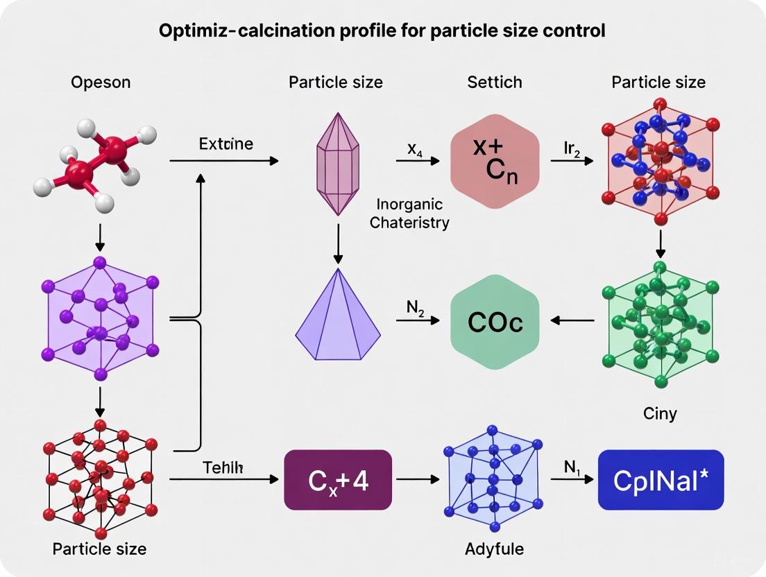
Experimental Workflow and Thermal Pathways
The following diagram illustrates the key decision points and thermal pathways in optimizing a calcination process for particle size and morphology control.
Diagram 1: Thermal pathway for calcination optimization.
Polymorph Selection Based on Thermal History
This diagram outlines the thermal pathway for controlling polymorphic outcomes in enantiotropic systems, using Polyoxymethylene (POM) as an example.
Diagram 2: Thermal control of polymorph selection.
Troubleshooting Guides and FAQs
Frequently Asked Questions
Q1: What are the most common signs of undesirable phase decomposition in my ceramic material?
- Visible changes in microstructure, such as the coarsening of a secondary phase at grain boundaries, is a key indicator [7].
- A significant drop in material hardness can signal phase decomposition, often resulting from the coarsening of a softer phase and changes in the composition of the matrix [7].
- Altered oxidation behavior, for instance, a shift from a continuous mass gain to a stabilized mass, can indicate that decomposition has affected the material's ability to form a protective layer [7].
Q2: How does the calcination temperature specifically influence the final properties of a metal oxide photocatalyst? Calcination temperature is a critical processing parameter that directly controls several key physicochemical properties [8]:
- Phase Composition: It can induce phase transformations (e.g., from the anatase to the rutile phase in TiO₂), which directly impact catalytic activity [8].
- Surface Area and Particle Size: Higher temperatures cause sintering, leading to a drastic reduction in surface area and an increase in particle size, which reduces the active sites available for reaction [8].
- Photocatalytic Performance: Optimal calcination temperature yields the highest photodegradation rate by balancing a high surface area with efficient charge separation and low charge carrier recombination [8].
Q3: My supported nanoparticle catalyst is losing activity. How can I determine if sintering is occurring and by which mechanism? Sintering, a primary deactivation mechanism, can occur via two main pathways [9]:
- Ostwald Ripening (OR): This involves the migration of single atoms or molecular species from smaller particles to larger ones, driven by differences in surface energy. It is often the predominant mechanism at low to moderate temperatures.
- Particle Migration and Coalescence (PMC): This involves the physical movement and merging of whole nanoparticles.
- Advanced in-situ electron microscopy techniques, such as environmental STEM, can track these dynamic processes at the single-atom level in real-time, allowing you to visualize which mechanism is active under your specific reaction conditions [9].
Q4: I am introducing a new phase into my material for toughening. How can I ensure it remains stable during high-temperature service?
- Dopant Selection: Choosing appropriate dopants is crucial. For example, in ZrO₂-based systems, Yb³⁺ is an excellent stabilizer that can suppress the formation of undesirable monoclinic phase content during long-term annealing [10].
- Phase Content Monitoring: The key is not necessarily to eliminate a toughening phase but to select a composition where its content remains stable under service conditions. An appropriate amount of a stable monoclinic phase can provide a toughening effect without leading to destructive phase transformations [10].
Troubleshooting Common Experimental Issues
Problem: Unexpected Softening or Reduction in Hardness After Heat Treatment
- Potential Cause: Phase decomposition leading to the formation and coarsening of a softer secondary phase (e.g., a Cr-rich phase in a W-based alloy) and a reduction of solid-solution strengthening elements in the matrix [7].
- Solution:
- Investigate Microstructure: Use SEM/EDS to characterize the grain boundaries and phases present. Look for evidence of coarsened precipitates [7].
- Optimize Annealing Parameters: Adjust the annealing temperature and time to avoid the regime where significant coarsening occurs. The chemical composition of phases often stabilizes after a certain duration (e.g., ~75 hours in the WCrY system) [7].
Problem: Rapid Deactivation of a Supported Nanoparticle Catalyst
- Potential Cause: Sintering of the metal nanoparticles, leading to a loss of active surface area [9].
- Solution:
- Confirm via Characterization: Use TEM to analyze the particle size distribution before and after reaction. An increase in mean particle size confirms sintering.
- Understand the Mechanism: Perform in-situ studies to determine if OR or PMC is the dominant mechanism. This informs the mitigation strategy [9].
- Mitigation Strategies: Consider modifying the support to create stronger metal-support interactions (to suppress single-atom migration for OR) or introducing spatial constraints to physically prevent particle migration (for PMC).
Problem: Poor Photocatalytic Degradation Performance After Calcination
- Potential Cause: The calcination temperature is not optimized, leading to excessive sintering, phase transformation, or a combination of both [8].
- Solution:
- Systematic Temperature Study: Synthesize and calcine the photocatalyst at a range of temperatures (e.g., 500°C, 600°C, 700°C, 800°C) [8].
- Comprehensive Characterization: Correlate the photocatalytic performance with structural and electronic properties as shown in the table below.
Data Presentation: Calcination Temperature Effects
The following table summarizes the quantitative impact of calcination temperature on a ternary Cu₂O/WO₃/TiO₂ (CWT) photocatalyst, demonstrating the critical need for temperature optimization [8].
Table 1: Influence of Calcination Temperature on the Properties of CWT Photocatalyst
| Calcination Temperature (°C) | Primary Crystal Phase | Surface Area (m²·g⁻¹) | Average Particle Size (nm) | Photodegradation Rate Constant for RB5 (×10⁻² min⁻¹) |
|---|---|---|---|---|
| 500 | Anatase | 35.77 | 39.11 | 0.70 |
| 600 | Anatase | Data Not Provided | Data Not Provided | Data Not Provided |
| 700 | Rutile | Data Not Provided | Data Not Provided | Data Not Provided |
| 800 | Rutile | 8.09 | 180.25 | Data Not Provided |
Experimental Protocols
Protocol 1: Assessing Phase Decomposition and Its Mechanical Impact
This methodology is adapted from studies on WCrY SMART material [7].
1. Sample Preparation:
- Fabricate the initial material via ball milling and field-assisted sintering to achieve a homogeneous microstructure.
- Subject the as-sintered material to isothermal annealing at a target temperature (e.g., 1000°C) for varying durations (e.g., 0-100 hours) to induce decomposition.
2. Microstructural and Chemical Characterization:
- Use Scanning Electron Microscopy (SEM) and Energy Dispersive X-ray Spectroscopy (EDS) to:
- Observe the formation and coarsening of secondary phases (e.g., Cr-rich phases) at grain boundaries.
- Quantify the chemical composition of the different phases (e.g., W-rich and Cr-rich phases) over annealing time.
- Use X-ray Diffraction (XRD) to identify the crystallographic phases present.
3. Mechanical Property Evaluation:
- Perform Vickers microhardness tests at room temperature to track softening.
- Conduct flexural strength (e.g., 3-point bend) and fracture toughness tests at both room and elevated temperatures.
4. Functional Property Testing:
- Evaluate oxidation resistance in a controlled atmosphere (e.g., humid air at 1000°C) using Thermogravimetric Analysis (TGA) to monitor mass gain over time.
Protocol 2: Evaluating Calcination Temperature on Photocatalyst Properties
This methodology is adapted from the synthesis of Cu₂O/WO₃/TiO₂ composites [8].
1. Synthesis:
- Prepare the ternary composite via an ultrasonic-assisted hydrothermal technique.
- Divide the final product and calcine separate portions at different temperatures (e.g., 500°C, 600°C, 700°C, 800°C) for a fixed duration.
2. Physicochemical Characterization:
- Thermogravimetric Analysis (TGA): Determine the thermal stability and appropriate calcination temperature range.
- X-ray Diffraction (XRD): Identify crystal phases and use the Scherrer equation to calculate crystallite size.
- Surface Area and Porosity (BET/BJH): Measure the specific surface area, pore volume, and pore size distribution.
- Electron Microscopy (SEM/TEM): Analyze surface morphology, particle size, and distribution of different phases.
- UV-Vis Spectroscopy: Determine the band gap energy using Tauc's plot.
3. Photoelectrochemical (PEC) Study:
- Fabricate a working electrode by coating the photocatalyst on FTO glass.
- Perform Electrochemical Impedance Spectroscopy (EIS) to determine charge-transfer resistance.
- Perform Linear Sweep Voltammetry (LSV) to measure current density.
- Perform Mott-Schottky analysis to study the semiconductor type and flat-band potential.
4. Performance Testing:
- Use a target pollutant (e.g., Reactive Black 5 dye) in an aqueous solution under simulated sunlight.
- Monitor the degradation rate via UV-Vis spectroscopy and calculate the pseudo-first-order rate constant.
Material Transformation Workflows
Diagram: Troubleshooting Phase Stability & Sintering
The Scientist's Toolkit: Research Reagent Solutions
Table 2: Essential Materials for Synthesis and Characterization
| Item | Function/Application | Example from Research |
|---|---|---|
| Yttria-Stabilized Zirconia (YSZ) | Base material for thermal barrier coatings (TBCs); studied for its phase stability under high temperatures [10]. | Used as a benchmark in developing co-doped Gd₂O₃/Yb₂O³ ZrO₂ for improved stability [10]. |
| Rare-Earth Oxides (Gd₂O₃, Yb₂O₃, Y₂O₃) | Used as dopants/stabilizers to control phase composition (cubic/monoclinic), enhance phase stability, and reduce thermal conductivity in ZrO₂ ceramics [10]. | Co-doping was shown to suppress m-phase formation and improve sintering resistance [10]. |
| Titanium Isopropoxide (TTIP) | A common metal-alkoxide precursor for the sol-gel or hydrothermal synthesis of TiO₂-based photocatalysts [8]. | Used as the Ti source in the synthesis of the Cu₂O/WO₃/TiO₂ ternary composite [8]. |
| Sodium Tungstate Dihydrate | A source of tungsten for the synthesis of WO₃, which is used to form heterojunctions with TiO₂ to enhance visible-light absorption [8]. | Used in the preparation of the WO₃ component of the CWT photocatalyst [8]. |
| Copper Nitrate Trihydrate | A common copper salt precursor used for incorporating Cu₂O into composite materials to create p-n heterojunctions [8]. | Used as the Cu source in the CWT photocatalyst [8]. |
| Platinum Precursors (e.g., H₂PtCl₆) | Used for the synthesis of supported Pt nanoparticle model catalysts for sintering and single-atom dynamics studies [9]. | Aqueous H₂PtCl₆ solution was deposited onto carbon supports to create model systems [9]. |
This guide provides technical support for researchers optimizing calcination profiles to control the crystallite size of inorganic nanomaterials. Calcination, a critical thermal treatment process, directly influences key material properties by controlling crystallinity and particle size. The following sections offer quantitative data, detailed methodologies, and troubleshooting advice to help you achieve precise control over your material's characteristics for applications in pharmaceuticals, catalysis, and advanced materials.
The following tables consolidate experimental data from published research, demonstrating the consistent trend of increasing crystallite size with higher calcination temperatures across various metal oxide systems.
Table 1: Crystallite Size vs. Calcination Temperature for Various Metal Oxides
| Material | Synthesis Method | Calcination Temperature (°C) | Crystallite Size (nm) | Characterization Technique | Source |
|---|---|---|---|---|---|
| MgO Nanoflakes | Co-precipitation | 400 | 8.80 | XRD (Scherrer's Formula) | [11] |
| 500 | 8.88 | ||||
| 600 | 10.97 | ||||
| CoFe₂O₄ in SiO₂ | Sol-gel | 400 | 5.9 | XRD (Scherrer's Formula) | [12] |
| 500 | 8.6 | ||||
| 600 | 9.3 | ||||
| 900 | 29.6 | ||||
| 1000 | 34.3 | ||||
| ZnO Nanoparticles | Chemical Precipitation | 400 | ~40-90 (Avg. ~62) | XRD, Particle Size Analysis | [13] |
| 600 |
Table 2: Associated Property Changes with Calcination Temperature
| Material | Calcination Temperature (°C) | Other Property Changes Observed | Source |
|---|---|---|---|
| MgO Nanoflakes | 400 → 600 | • Surface Area: Decreases• Thermal Stability: Increases• Antimicrobial Activity: Superior at 400°C & 500°C vs. 600°C• Cytotoxicity: MgO-400°C shows slight cytotoxicity; MgO-500/600°C are biocompatible. | [11] |
| CoFe₂O₄ in SiO₂ | 400 → 1000 | • Saturation Magnetization (Ms): Increases from 0.21 to 12.01 emu/g• Coercivity (Hc): Non-linear increase from 27 to 220 Oe. | [12] |
| ZnO Nanoparticles | 400 → 600 | • Optical Band Gap: Decreases from 3.15 eV to 3.05 eV. | [13] |
| Low-Grade Kaolinite Clay | 800 (for 180 mins) | • Pozzolanic Reactivity: Highest achieved.• Workability in Cement: Ideal, enhancing slump by up to 40%. | [14] |
Experimental Protocols & Workflows
Detailed Synthesis and Characterization Methodology
The following workflow outlines the general process for synthesizing materials and investigating the effect of calcination temperature.
Protocol A: Synthesis of MgO Nanoflakes via Co-precipitation [11]
- Objective: To synthesize MgO nanoflakes and study the effect of calcination temperature (400°C, 500°C, 600°C) on their properties.
- Materials: Magnesium precursor (e.g., nitrate or chloride), precipitating agent (e.g., sodium hydroxide or carbonate), deionized water.
- Procedure:
- Dissolve the magnesium precursor in deionized water under constant stirring.
- Slowly add the precipitating agent solution to form a precipitate of magnesium hydroxide (Mg(OH)₂).
- Maintain the reaction with controlled stirring (e.g., 90 minutes).
- Filter and wash the precipitate thoroughly with deionized water to remove impurities.
- Dry the filtered cake in an oven to obtain the precursor.
- Divide the dried precursor into three portions.
- Calcine each portion in a muffle furnace at a pre-determined temperature (400°C, 500°C, and 600°C) for a fixed duration (e.g., 2 hours) using a standard heating rate (e.g., 10°C/min).
Protocol B: Synthesis of CoFe₂O₄-SiO₂ Nanocomposites via Sol-Gel [12]
- Objective: To prepare magnetic nanocomposites and investigate calcination effects from 400°C to 1000°C.
- Materials: Tetrakis(2-hydroxyethyl) orthosilicate (THEOS) as silica precursor, Fe(NO₃)₃·9H₂O, Co(NO₃)₂·6H₂O, deionized water.
- Procedure:
- Dissolve the metal nitrates in deionized water.
- Add an aqueous solution of THEOS to the nitrate solution under vigorous stirring.
- Stir the mixture for 1 hour to form a homogeneous sol.
- Allow the sol to gel at room temperature for several days in a partially closed vessel.
- Dry the obtained alcogel in an oven at 110°C for 24 hours to form a xerogel.
- Crush the xerogel and calcine the powder at different temperatures (e.g., 400, 500, 600, 900, 1000°C) for 2 hours.
Protocol for Crystallite Size Determination by XRD
The crystallite size is most commonly determined from X-ray Diffraction (XRD) data using the Scherrer Equation [15] [16].
Scherrer Equation:
D = (K * λ) / (β * Cosθ)
- D: Volume-weighted mean crystallite size (nm)
- K: Dimensionless shape factor (often ~0.94)
- λ: X-ray wavelength (nm) (e.g., Cu Kα = 0.15418 nm)
- β: Line broadening at half the maximum intensity (FWHM) in radians
- θ: Bragg angle (half of the 2θ peak position) [15]
Step-by-Step Calculation Tutorial [15]:
- Obtain XRD Pattern: Run the XRD for your calcined powder samples.
- Identify Peak: Zoom in on the diffraction peak you wish to analyze.
- Measure FWHM: Determine the Full Width at Half Maximum (FWHM, β) of the peak in degrees.
- Note Peak Position: Record the Bragg angle (2θ) at the peak's maximum.
- Use Calculator: Input the peak position (2θ), FWHM (β), and X-ray wavelength (λ) into an online calculator or perform the calculation manually, ensuring FWHM is converted to radians.
Note on Limitations: The Scherrer equation is most reliable for crystallite sizes between a few nanometers and about 100 nm. For more accurate results, the Modified Scherrer method is recommended, which involves calculating the size using multiple diffraction peaks and specific rules to minimize error [17].
Troubleshooting FAQs
FAQ 1: My crystallite size is larger than expected at my target temperature. What could be wrong?
- Potential Cause: The actual temperature in the furnace might be higher than the set point, or the holding time might be too long.
- Solution: Calibrate your furnace regularly using a thermocouple. Optimize the holding time; longer times can lead to excessive crystal growth and coarsening [18].
- Preventive Measure: Establish a precise temperature profile and stick to it for all experiments to ensure reproducibility.
FAQ 2: My powder particles are aggregating heavily during calcination, making characterization difficult. How can I prevent this?
- Potential Cause: The precursor powder was not finely ground or has high moisture content, leading to sintering.
- Solution:
- Grind Precursor: Ensure the pre-calcined powder is finely ground to a uniform particle size [18].
- Control Atmosphere: In some cases, using a controlled atmosphere during calcination can reduce aggregation.
- Use a Matrix: As demonstrated in the sol-gel synthesis, a silica matrix can effectively confine nanoparticle growth and prevent aggregation [12].
FAQ 3: I am getting inconsistent crystallite size results from XRD. What should I check?
- Potential Cause: Inaccurate measurement of the FWHM (β) from the XRD peak.
- Solution:
- Ensure the XRD instrument is properly aligned and calibrated.
- Use appropriate software to accurately determine the FWHM, typically after subtracting the background and accounting for instrumental broadening.
- Apply the Scherrer equation consistently, using the same peak (often the most intense one) for all samples [11] [12]. Consider using the Modified Scherrer method for higher accuracy [17].
FAQ 4: Is it possible to remove free moisture from my sample in the calciner?
- Answer: While physically possible, it is highly inefficient and costly. The lower rate of heat transfer in a calciner makes moisture removal slow, driving up fuel and energy costs.
- Best Practice: Always include a dedicated drying step (e.g., in a rotary dryer or oven) to remove free moisture before the calcination process begins [18].
The Scientist's Toolkit: Research Reagent Solutions
Table 3: Essential Materials and Equipment for Calcination Studies
| Item | Function/Benefit | Example from Context |
|---|---|---|
| THEOS (Tetrakis(2-hydroxyethyl) orthosilicate) | A water-soluble silica precursor for sol-gel synthesis. Eliminates the need for organic solvents, simplifying the process and enabling a better-dispersed nanocomposite. [12] | Used to create a silica matrix for confining CoFe₂O₄ nanoparticles. |
| Diamond ATR Crystal | The most common and durable crystal for FT-IR analysis, resistant to most chemicals and suitable for pressing solid samples. Standard range is 7800-400 cm⁻¹. [19] | Used for characterizing chemical bonds and functional groups in synthesized powders. |
| Rotary Kiln/Calciner | A standard piece of equipment for industrial-scale calcination. Can be direct-fired (material contacts combustion gases) or indirect-fired (material heated through the drum wall). [18] | Used for thermal processing of various materials under controlled atmospheres and temperatures. |
| Scherrer Equation Calculator | Online tool to quickly determine crystallite size from XRD data (peak position and FWHM). Simplifies the calculation process. [15] | InstaNANO and other online resources provide accessible calculators for researchers. |
Troubleshooting Guides
Guide 1: Addressing Low Saturation Magnetization
Problem: The synthesized ferrite nanoparticles exhibit lower-than-expected saturation magnetization (Ms), reducing their effectiveness for applications like magnetic hyperthermia.
Solutions:
- Increase Calcination Temperature: Higher calcination temperatures often improve crystallinity and cation ordering, leading to higher Ms. For instance, Co-ZnFe₂O₄ nanoparticles calcined at 1000°C achieved an Ms of 22.12 emu/g, significantly higher than those calcined at 600°C or 800°C [20].
- Optimize Calcination Duration: Excessively long calcination can lead to impurity phases that degrade magnetic properties. Hold times of 3-6 hours are often optimal. For MnZn ferrites, a 3-hour calcination at 1060°C produced the highest Ms (53.4 emu/g), while longer times induced α-Fe₂O₃ impurities [21].
- Verify Cation Distribution: Use techniques like X-ray diffraction (XRD) and Mössbauer spectroscopy to confirm correct cation distribution between tetrahedral and octahedral sites in the spinel structure, as this directly governs magnetic moment [20] [22].
Guide 2: Controlling Particle Size and Agglomeration
Problem: Particles are too large, overly agglomerated, or have a broad size distribution, which negatively impacts suspension stability and hyperthermia performance.
Solutions:
- Modify Thermal Profile: A lower initial calcination temperature can suppress excessive crystal growth. A modified co-precipitation method conducted at a maximum of 60°C successfully produced nanoparticles without the need for coating agents [23].
- Control Calcination Time: The duration of thermal treatment directly influences particle size and agglomeration. An intermediate calcination time (e.g., 3 hours) can yield a unimodal particle size distribution, whereas shorter or longer times may result in broader, non-uniform distributions [21].
- Introduce a Capping Agent: During synthesis, use polymers like poly(vinyl pyrrolidone) (PVP) or polyethylene glycol (PEG) to coat particles and create a physical barrier that minimizes agglomeration during subsequent thermal treatment [23] [24].
Guide 3: Managing Phase Instability and Impurity Formation
Problem: Impurity phases, such as α-Fe₂O₃ (hematite), appear in the XRD pattern after calcination, indicating degradation of the spinel structure.
Solutions:
- Avoid Over-oxidation: Perform calcination in a controlled atmosphere (e.g., nitrogen) for compositions prone to oxidation, especially those containing Fe²⁺ ions. Studies on high-entropy ferrites show a nitrogen atmosphere is crucial for maintaining phase stability at high temperatures [25].
- Dope with Stable Cations: Incorporate dopant ions like Ni²⁺ to enhance thermal stability against oxidation. Research shows Ni-doped ferrite nanoparticles more effectively suppress the temperature-induced oxidation process compared to Co or Mn doping [26].
- Optimize Calcination Time: An excessively long calcination time can destabilize the system. For MnZn ferrites, calcination beyond 3 hours at 1060°C led to the formation of α-Fe₂O₃ and γ-Fe₂O₃ impurities [21].
Frequently Asked Questions (FAQs)
FAQ 1: How does calcination temperature specifically affect the magnetic properties of cobalt-zinc ferrite?
Calcination temperature directly influences crystallinity, cation distribution, and ultimately, magnetic properties. For Co-ZnFe₂O₄, saturation magnetization (Ms) increases with rising calcination temperature. One study showed Ms values rose to a peak of 22.12 emu/g at 1000°C, attributed to enhanced crystallinity and redistribution of Fe, Co, and Zn cations within the spinel lattice. Higher temperatures also typically increase particle size and can change morphology (e.g., from nanorods to more spherical particles) [20].
FAQ 2: What is the optimal calcination time for nickel-zinc ferrite to achieve high saturation magnetization?
For Ni₀.₅Zn₀.₅Fe₂O₄ synthesized via solid-state reaction, a calcination time of 6 hours at 1200°C yielded an excellent saturation magnetization of 80.07 emu/g. This duration, at a sufficiently high temperature, allows for complete diffusion and formation of a pure spinel phase with optimal magnetic characteristics [27].
FAQ 3: Can I achieve a pure ferrite phase with low-temperature calcination to save energy?
Yes, it is possible with method optimization. A modified co-precipitation process has been used to synthesize CoFe₂O₄ and ZnCoFe₂O₄ nanoparticles at a maximum temperature of 60°C in air. These nanoparticles demonstrated good crystallinity and magnetic properties suitable for hyperthermia, proving that high-temperature calcination is not always mandatory [23].
FAQ 4: How does zinc substitution influence the properties of cobalt ferrite?
Zinc substitution in cobalt ferrite (forming Co₁₋ₓZnₓFe₂O₄) significantly alters magnetic properties. Zn²⁺ ions preferentially occupy tetrahedral (A) sites, displacing Fe³⁺ ions to octahedral (B) sites. This strengthens the A-B super-exchange interaction, typically leading to an increase in saturation magnetization up to an optimal zinc concentration. Beyond this point, further zinc addition can weaken the interaction and reduce magnetization [20] [22].
The following tables consolidate key experimental data from research on thermal processing of ferrites.
Table 1: Effect of Calcination Temperature on Ferrite Properties
| Ferrite Type | Calcination Temperature (°C) | Crystallite Size (nm) | Saturation Magnetization (Ms) | Key Findings | Source |
|---|---|---|---|---|---|
| Co-ZnFe₂O₄ | 600 | Not Specified | < 22.12 emu/g | Elongated nanorod morphology. | [20] |
| Co-ZnFe₂O₄ | 800 | Not Specified | < 22.12 emu/g | Transition in morphology. | [20] |
| Co-ZnFe₂O₄ | 1000 | Not Specified | 22.12 emu/g | Peak Ms, spherical particles. | [20] |
| Ni₀.₅Zn₀.₅Fe₂O₄ | 900 | Not Specified | < 80.07 emu/g | Lower Ms, incomplete phase formation. | [27] |
| Ni₀.₅Zn₀.₅Fe₂O₄ | 1200 | Not Specified | 80.07 emu/g | Excellent Ms, pure spinel phase. | [27] |
| CuFe₂O₄ | 773 (500°C) | 24 | Not Specified | - | [24] |
| CuFe₂O₄ | 1173 (900°C) | 34 | Not Specified | - | [24] |
Table 2: Effect of Calcination Time on Ferrite Properties
| Ferrite Type | Calcination Time (Hours) | Particle Size | Saturation Magnetization (Ms) | Key Findings | Source |
|---|---|---|---|---|---|
| MnZn Ferrite | 1 | Non-unimodal distribution | < 53.4 emu/g | Non-optimal particle distribution. | [21] |
| MnZn Ferrite | 3 | Unimodal distribution | 53.4 emu/g | Optimal Ms and particle size. | [21] |
| MnZn Ferrite | 5-7 | Increased agglomeration | Decreased | Appearance of α-Fe₂O₃ impurities. | [21] |
| MnZn Ferrite | 9 | Largest particle size | Decreased | Appearance of γ-Fe₂O₃ impurities. | [21] |
| Ni₀.₅Zn₀.₅Fe₂O₄ | 6 | Not Specified | 80.07 emu/g | Optimal for this system. | [27] |
| Ni₀.₅Zn₀.₅Fe₂O₄ | 12 | Not Specified | Not Specified | Prolonged time may not offer significant benefit. | [27] |
Experimental Protocols
Objective: To synthesize cytocompatible cobalt-zinc ferrite nanoparticles with high specific loss power (SLP) for hyperthermia applications without high-temperature calcination.
Materials: Iron(III) chloride hexahydrate, Iron(II) chloride tetrahydrate, Cobalt(II) chloride hexahydrate, Zinc chloride, Ammonium hydroxide (30%), Sodium citrate, Deionized water.
Step-by-Step Workflow:
- Precursor Solution: Dissolve the metal salts in 50 mL deionized water with a molar ratio of Fe³⁺:Fe²⁺:Co²⁺/Zn²⁺ = 3:2:1. Stir for 15 minutes at room temperature.
- Heating: Heat the solution to 60°C under vigorous stirring and maintain for 5 minutes.
- Precipitation: Add 20 mL of ammonium hydroxide (30%) dropwise to the heated solution to initiate particle growth.
- Aging and Coating: Continue stirring for 30 minutes at 60°C. For coating, add 0.5 g of sodium citrate and stir further.
- Washing and Drying: Collect the black precipitate by centrifugation or magnetic separation. Wash several times with distilled water to remove ammonium salts. Dry the resulting powder for 24 hours.
Objective: To prepare high-purity, high-magnetization Ni₀.₅Zn₀.₅Fe₂O₄ microparticles via an optimized solid-state reaction for composite filler applications.
Materials: Nickel oxide (NiO), Zinc oxide (ZnO), Iron oxide (Fe₂O₃), high-purity powders.
Step-by-Step Workflow:
- Milling: Mix the raw oxide powders according to the stoichiometric formula. Use a high-energy planetary mill (e.g., Pulverisette 4) with a ball-to-powder mass ratio of 20:1 and a rotation speed of 500 rpm for 30-60 minutes.
- Pelletizing: Press the milled powder into pellets.
- Calcination: Calcine the pellets in air at a temperature of 1200°C for 6 hours (using a ramp rate of, for example, 5°C/min).
- Final Milling (Optional): Gently mill the calcined product to obtain a fine powder for further use or characterization.
Experimental Workflow Visualization
Diagram Title: Ferrite Thermal Optimization Workflow
The Scientist's Toolkit: Research Reagent Solutions
Table 3: Essential Materials for Ferrite Synthesis and Analysis
| Reagent / Equipment | Function / Role | Example Use Case |
|---|---|---|
| Metal Chlorides/Nitrates | Provide metal cation precursors (Fe³⁺, Fe²⁺, Co²⁺, Zn²⁺, Ni²⁺) in solution-based synthesis. | Co-precipitation of CoFe₂O₄ using FeCl₃, FeCl₂, and CoCl₂ [23]. |
| Ammonium Hydroxide (NH₄OH) | Precipitating agent in aqueous synthesis to form metal hydroxides. | Initiating particle growth in the modified co-precipitation method [23]. |
| Sodium Citrate | Coating agent to functionalize nanoparticle surface, improve dispersion, and cytocompatibility. | Coating CoFe₂O₄ nanoparticles for enhanced biocompatibility [23]. |
| Poly(vinyl pyrrolidone) - PVP | Capping agent to control particle growth and prevent agglomeration during synthesis. | Synthesizing closely packed CuFe₂O₄ nanocrystals [24]. |
| Planetary Ball Mill | High-energy milling to homogenize and reduce particle size of solid precursors. | Mixing oxide powders for solid-state reaction of Ni₀.₅Zn₀.₅Fe₂O₄ [27]. |
| Vibrating Sample Magnetometer (VSM) | Measures magnetic properties (saturation magnetization, coercivity) of the synthesized powder. | Determining Ms of samples calcined at different temperatures [27] [20]. |
| X-ray Diffractometer (XRD) | Confirms crystal structure, phase purity, and estimates crystallite size. | Identifying spinel phase and detecting α-Fe₂O₃ impurities [27] [21]. |
Troubleshooting Guides
FAQ 1: How does calcination temperature specifically affect the saturation magnetization of my ferrite nanoparticles?
The Problem: You have synthesized ferrite nanoparticles, but the magnetic strength (saturation magnetization) is lower than required for your application, such as magnetic hyperthermia or data storage.
The Solution: Increasing the calcination temperature enhances crystallinity and causes a redistribution of cations within the spinel lattice, which can significantly boost saturation magnetization.
Detailed Explanation: The saturation magnetization (Ms) of ferrite nanoparticles is highly dependent on their crystallinity and the specific arrangement (distribution) of metal cations between the tetrahedral and octahedral sites of the crystal lattice. Higher calcination temperatures promote better crystallinity and can drive this cation redistribution, optimizing the magnetic moments within the material.
Supporting Experimental Data: The table below summarizes quantitative findings from recent studies on how calcination temperature influences saturation magnetization.
Table 1: Effect of Calcination Temperature on Saturation Magnetization
| Material | Calcination Temperature | Saturation Magnetization (Ms) | Citation |
|---|---|---|---|
| Co–ZnFe₂O₄ | 1000 °C | 22.12 emu/g | [20] |
| Cobalt Ferrite (CoFe₂O₄) | 1000 °C | 85 emu/g | [28] |
| Nickel Ferrite (NiFe₂O₄) | 500-900 °C | High coercivity, indispensible for storage devices | [29] |
Protocol:
- Synthesis: Co–ZnFe₂O₄ nanoparticles were synthesized via a wet chemical co-precipitation method using chlorides of cobalt, zinc, and iron as precursors [20].
- Calcination: The resulting gel was finely ground and divided into portions, which were then calcined at systematically increased temperatures (e.g., 600 °C, 800 °C, and 1000 °C) for a set duration [20] [29].
- Characterization: The magnetic properties were measured at room temperature using a Vibrating Sample Magnetometer (VSM) [29] [28].
FAQ 2: Why does the band gap of my semiconductor nanopowder decrease after high-temperature calcination?
The Problem: You observe a reduction in the band gap of your photocatalyst after high-temperature sintering, which seems counterintuitive to achieving high photocatalytic activity.
The Solution: The band gap narrowing is a direct consequence of particle size increase and improved crystallinity at higher calcination temperatures. While a smaller band gap can be beneficial for absorbing a broader spectrum of light, its overall effect on photocatalytic efficiency must be evaluated against other factors like charge recombination.
Detailed Explanation: Calcination at elevated temperatures causes nanoparticles to grow and crystallize further. This increase in particle size reduces the quantum confinement effect, which is prominent at very small particle sizes and leads to band gap widening. Therefore, as particles grow, the band gap typically decreases.
Supporting Experimental Data: The following table illustrates the inverse relationship between sintering temperature, particle size, and band gap.
Table 2: Effect of Sintering Temperature on Band Gap and Particle Size
| Material | Sintering Temperature | Average Particle Size | Band Gap | Citation |
|---|---|---|---|---|
| NiSnO₃ | 250 °C | ~5.05 nm | ~3.38 eV | [30] |
| NiSnO₃ | 400 °C | ~8.05 nm | ~2.90 eV | [30] |
| CoFe₂O₄ | 500-1000 °C | 33 - 169 nm | 3.00 - 3.52 eV | [28] |
Protocol:
- Synthesis: NiSnO₃ nanopowder was synthesized using a co-precipitation method [30].
- Sintering: The precipitated powder was sintered at different temperatures (250 °C to 400 °C) [30].
- Characterization: The band gap was determined from data obtained using Ultraviolet-Visible (UV-Vis) Spectroscopy and the Tauc plot method. Particle size was confirmed via Transmission Electron Microscopy (TEM) [30].
FAQ 3: My catalyst's performance has dropped after calcination. Did I lose surface area?
The Problem: After a high-temperature calcination step intended to improve crystallinity, your catalyst shows reduced activity, likely due to a loss of specific surface area.
The Solution: Yes, high-temperature calcination often leads to particle coarsening and sintering, which drastically reduces the specific surface area, a critical parameter for catalytic activity. Exploring alternative synthesis routes or optimizing the calcination profile is necessary.
Detailed Explanation: Specific surface area has an inverse relationship with particle size. As calcination temperature increases, particles sinter and grow, leading to a direct reduction in the total surface area available for reactions [31]. This is a primary reason for performance degradation in catalysts and adsorbents after high-temperature treatment. Powders with a broad particle size distribution can show a significant surface area reduction early in the sintering process [32].
Protocol:
- Measurement: The specific surface area is typically measured using gas adsorption methods (e.g., BET theory) [31].
- Mitigation: To lower the required calcination temperature and preserve a higher surface area, consider innovative synthesis methods. For example, the glucose-urea method for synthesizing La₀.₆Sr₀.₄Co₀.₂Fe₀.₈O₃−δ (LSCF) air electrodes significantly reduces sintering temperatures while producing smaller, more uniform particles compared to conventional sol-gel methods [33].
Essential Visualizations
Cascading Effects of Calcination Temperature
(Diagram: The cascading effects of increasing calcination temperature on key material properties.)
Experimental Workflow for Optimization
(Diagram: A standard workflow for optimizing calcination profiles, from synthesis to multi-faceted characterization.)
The Scientist's Toolkit: Research Reagent Solutions
Table 3: Essential Reagents for Ferrite Nanoparticle Synthesis via Wet-Chemical Routes
| Reagent / Material | Function in Synthesis | Example |
|---|---|---|
| Metal Precursors | Source of metal cations for the ferrite crystal lattice. | Cobalt chloride (CoCl₂·6H₂O), Iron chloride (FeCl₃·6H₂O), Zinc chloride (ZnCl₂·6H₂O) [20]. |
| Precipitating Agent | Controls pH to facilitate the co-precipitation of metal hydroxides. | Sodium hydroxide (NaOH) or Ammonium hydroxide (NH₄OH) [20] [28]. |
| Stabilizing / Chelating Agent | Controls particle growth, limits agglomeration, and improves dispersion. | Citric Acid, Polyethylene Glycol (PEG), Polypropylene Glycol [20] [29] [28]. |
| Solvent | Medium for the chemical reaction and dissolution of precursors. | Deionized Water, Ethanol [29] [28]. |
From Lab to Production: Designing Calcination Protocols for Targeted Particle Sizes in Specific Applications
This technical support center provides troubleshooting guides and FAQs for researchers navigating common challenges in materials synthesis. The content is framed within the broader context of optimizing calcination profiles for particle size control.
Comparison of Synthesis Methods
The table below summarizes the core characteristics, outcomes, and optimal use cases for the three primary chemical synthesis routes.
| Synthesis Method | Typical Precursors | Key Process Parameters | Reported Particle Size/Characteristics | Ideal Applications |
|---|---|---|---|---|
| Co-precipitation [34] [35] | Metal salts (e.g., chlorides, nitrates), precipitating agent (e.g., NaOH, oxalate) [36] | pH control, precipitation temperature, stirring rate, calcination profile [35] | High purity, better crystallinity, and more homogeneous cation mixing compared to sol-gel [34] [35] | Producing high-purity, crystalline powders for battery materials (e.g., LMNO, ZnFe₂O₄) where atomic-level mixing is critical [34] [35] [36] |
| Sol-Gel [34] [28] [37] | Metal alkoxides or metal salts, solvent (e.g., ethanol), complexing agent (e.g., citric acid) [34] [28] | Type of precursor, pH, solvent polarity, temperature, calcination profile [28] [37] | Crystallite size can be tuned from ~33 nm to ~169 nm by varying calcination temperature (500°C–1000°C) [28]. Can yield various morphologies (nanoparticles, sheets, ribbons) [37] | Synthesizing size- and shape-controlled nanostructures (e.g., Mn₃O₄, CuO) and highly mesoporous materials for catalysis and sensors [34] [37] |
| Solid-State [36] | Solid metal oxides and carbonates [36] | Milling time/energy, calcination temperature/duration, heating rate [36] | Larger particle size, often requires high temperatures and prolonged heating, can lead to impurities [36] | Simpler compositions where high temperatures are not detrimental, and precise nano-scale control is not the primary goal [36] |
Troubleshooting Guides & FAQs
Co-precipitation
Problem: Inhomogeneous Cation Mixing in Final Product
- Cause: Inadequate control during the precipitation step, leading to uneven distribution of metal ions in the precursor [35].
- Solution: Use a chelating agent (e.g., oxalate) that forms stable complexes with all metal ions across the stoichiometric ratio. Ensure vigorous and uniform stirring during precursor addition [35].
Problem: Formation of ZrO₂ Impurity in NASICON Phases
- Cause: Calcination temperature is either too low, preventing complete reaction, or too high, causing decomposition of the NASICON phase [34].
- Solution: Systematically optimize the calcination temperature. For example, one study found that a temperature of 1000°C for 10 hours yielded better electrochemical properties, while other temperatures promoted ZrO₂ formation [34].
Sol-Gel
Problem: Inability to Control Crystallite Size
- Cause: The calcination profile is not properly tuned. Higher temperatures lead to increased crystallite size [28].
- Solution: Precisely control the final calcination temperature. For instance, cobalt ferrite (CoFe₂O₄) nanoparticles synthesized via sol-gel showed crystallite sizes from 33 nm (500°C) to 169 nm (1000°C) [28].
Problem: Low Morphology Yield or Irregular Shapes
- Cause: The solvent system and precursor-to-base concentration significantly influence nanocrystal self-assembly and final morphology [37].
- Solution: Experiment with different solvent systems (e.g., water, 70% ethanol, DMF, toluene) and molar ratios of metal precursor to base (e.g., 1:5, 1:10, 1:15) to control the growth of specific shapes like hexagonal plates or ribbons [37].
General Synthesis & Calcination
Problem: Low Ionic Conductivity in Solid Electrolyte
- Cause: Low final density of the sintered pellet and the presence of impurity phases that act as barriers for ion movement [34].
- Solution: Employ advanced sintering techniques like Spark Plasma Sintering (SPS). SPS allows for lower sintering temperatures and shorter times, resulting in higher density, nano-sized grains, and significantly higher conductivity (e.g., 1.7 × 10⁻³ S cm⁻¹ for NASICON) compared to conventional sintering [34].
Problem: Formation of Amorphous Phases or Loss of Volatile Elements
- Cause: Excessively high sintering temperatures required in solid-state reactions can reduce powder reactivity and lead to the loss of volatile elements like phosphorus and sodium, forming detrimental amorphous phases [34].
- Solution: Utilize wet-chemical methods (co-precipitation or sol-gel) that offer higher powder reactivity, enabling lower processing temperatures and preserving stoichiometry [34].
Experimental Workflows
The following diagrams illustrate the general workflows for the featured synthesis methods, highlighting the critical steps for controlling the calcination profile and final particle size.
Research Reagent Solutions
This table details key reagents and their functions in the synthesis processes discussed.
| Reagent | Function/Application | Key Consideration |
|---|---|---|
| Metal Alkoxides (e.g., Zirconium tetrapropoxide, TEOS) [34] | High-purity precursors in sol-gel synthesis for oxides like NASICON [34]. | Can be expensive, sensitive to moisture, and require careful handling [37]. |
| Metal Salts (e.g., Chlorides, Nitrates) [28] [36] | Common, often cheaper precursors for both co-precipitation and sol-gel methods [28] [36]. | Anion type (e.g., chloride, nitrate) can influence the synthesis and may leave residues. |
| Precipitating Agents (e.g., NaOH, Oxalic acid) [35] [36] | Initiate the formation of solid precursors from solution in co-precipitation [35]. | Choice of agent (e.g., oxalate over carbonate) can prevent impurity phases and improve cation mixing [35]. |
| Chelating Agents / Solvents (e.g., Citric acid, Glycerol, Ethanol) [28] [37] | Control hydrolysis/condensation rates in sol-gel; can act as fuel in combustion synthesis; influence morphology [28] [37]. | Solvent polarity (e.g., water vs. toluene) is a key factor in directing shape-controlled crystal growth [37]. |
Frequently Asked Questions
What is a calcination profile and why is it critical for my research? A calcination profile is the carefully controlled sequence of temperature changes—including the ramp rate, target temperature (dwell temperature), and dwell time—applied to a material [38]. This profile directly determines critical material properties such as phase composition, crystallite size, specific surface area, and porosity [39] [40]. Precise control is essential for reproducibility and for tailoring materials for specific applications, such as creating catalysts with high activity or pigments with desired color properties [41] [40].
How does dwell temperature influence the final product's characteristics? The dwell temperature is one of the most influential parameters. It controls chemical decomposition, phase transitions, and the removal of volatile components [42] [38].
- Too low: Reactions may not complete, leaving unreacted precursors or failing to achieve the desired crystalline phase [39].
- Optimal range: Maximizes desired properties, such as amorphous content for reactivity or a specific crystal phase for catalytic activity [41] [1].
- Too high: Can lead to the formation of inert crystalline phases, significant particle coarsening (sintering), and a dramatic reduction in surface area, which often diminishes reactivity [39] [1] [40].
The table below summarizes the effects of temperature on different material systems, as observed in research:
| Material System | Observed Effect of Calcination Temperature | Optimum / Key Range | Citation |
|---|---|---|---|
| Metakaolin (from Kaolinite Clay) | Amorphous content peaks at 750-800°C; higher temperatures form inert phases (cristobalite, mullite), reducing reactivity. | 700°C - 800°C | [1] |
| TiO₂ | Crystallinity and crystallite size increase with temperature; anatase to rutile phase transition occurs at higher temperatures. | Varies by application; anatase phase often preferred for photocatalysis. | [41] [39] [40] |
| SnO₂ & Composites | Specific surface area decreases dramatically; isoelectric point (IEP) shifts due to surface dehydration and phase changes. | Lower temperatures (e.g., 300°C) preserve high surface area. | [39] [40] |
| Bismuth-based Photocatalysts | Directly determines the type of photocatalyst formed (e.g., BiOIO₃, Bi₅O₇I, Bi₂O₃) through phase transformations. | Specific to target phase; e.g., 500–600°C for Bi₅O₇I. | [41] |
| Cadmium Pigments | Lower temperatures yield lighter, brighter shades; higher temperatures produce deeper, stronger shades. | Chosen based on target color. | [40] |
What is the purpose of dwell time, and how is it determined? Dwell time, or residence time, is the period the material is held at the target temperature [43]. Its primary purpose is to allow the entire sample to reach thermal equilibrium, ensuring the desired chemical or physical transformation goes to completion uniformly [43]. Insufficient dwell time can result in a partially reacted product with a gradient of properties. The required dwell time depends on the sample's mass, geometry, and the nature of the reaction, and is often controlled in rotary kilns by adjusting drum speed and slope [38].
Why is the ramp rate important, and what are the trade-offs? The ramp rate, or heating rate, controls how quickly the material reaches the dwell temperature.
- Slow Ramp Rate: Allows for more uniform heating throughout the sample, minimizes thermal stress that can cause cracking, and can promote better crystallinity by allowing a gradual reorganization of the structure [39].
- Fast Ramp Rate: Can lead to a rapid release of volatile gases (e.g., from polymer decomposition), which may help create a more porous structure. However, it also risks fracturing particles or creating an uneven temperature profile within the sample [39].
A common problem in my lab is that calcined powders become heavily aggregated and lose surface area. How can this be prevented? This is a classic symptom of sintering, where elevated temperatures cause small particles to fuse. To mitigate this:
- Optimize Temperature: Use the lowest effective temperature that achieves your desired transformation [1] [40].
- Control Dwell Time: Avoid unnecessarily long dwell times at peak temperature [41].
- Consider Atmosphere: In some cases, using a controlled atmosphere can influence sintering kinetics.
- Use Templates or Precursors: Some synthesis methods use organic templates that burn out during calcination, preserving porosity and preventing dense aggregation [39].
Troubleshooting Common Calcination Problems
| Problem | Possible Causes | Solutions |
|---|---|---|
| Unexpected Crystalline Phase | Dwell temperature too high or too low; incorrect for the target phase. | Consult phase diagrams; run a temperature series to identify the correct window for your material [41] [39]. |
| Low Specific Surface Area | Over-sintering due to excessive dwell temperature or time [39]. | Lower the dwell temperature and/or shorten the dwell time [41] [1]. |
| Powder Agglomeration into Hard Lumps | Sintering; may also be due to incomplete removal of volatile components creating liquid phases. | Ensure sufficient atmosphere flow to remove volatiles; use a crucible with a large surface area; consider adding a brief grinding step between ramps. |
| Incomplete Reaction | Insufficient dwell temperature or time; sample too large, preventing core from reaching temperature. | Increase temperature/time; ensure a slow enough ramp rate for thermal equilibrium; use a thinner sample bed [43]. |
| Cracked or Fractured Monoliths | Excessive thermal stress from a ramp rate that is too fast. | Implement a slower ramp rate, especially through known phase transition temperatures [39]. |
Experimental Protocol: Determining an Optimal Calcination Profile
This protocol outlines a systematic approach to defining the calcination profile for a new material, using the example of maximizing the amorphous content and reactivity of metakaolin from kaolinite clay [1].
1. Hypothesis We hypothesize that calcining a kaolinite clay sample at 800°C for 2 hours will produce metakaolin with a higher amorphous content and greater pozzolanic reactivity than samples calcined at lower or higher temperatures.
2. Materials and Equipment
- Source Material: Kaolinite clay (e.g., ADU or CCC types from [1]).
- Equipment: High-temperature muffle furnace with programmable temperature controller, alumina crucibles, mortar and pestle, desiccator.
- Characterization: X-ray Diffraction (XRD) to quantify amorphous/inert phases, Scanning Electron Microscopy (SEM) for morphology.
3. Step-by-Step Procedure
- Sample Preparation: Dry the raw kaolinite clay. Grind it gently and sieve to obtain a consistent particle size fraction.
- Furnace Programming: Program the furnace with the following profiles for different batches. Use a moderate ramp rate of 5-10°C/min to the target dwell temperature.
- Calcination: Place identical masses of the prepared clay into separate, labeled alumina crucibles. Load them into the furnace at room temperature and run the programmed cycles. Allow the furnace to cool naturally after the dwell time.
- Post-processing: Carefully remove the crucibles and transfer the calcined product (now metakaolin) to a desiccator to cool completely and avoid moisture absorption.
- Characterization: Perform XRD analysis on each sample to determine the amorphous content and identify any inert crystalline phases that have formed.
4. Expected Outcomes As reported in [1], you should observe a peak in amorphous content at a specific temperature (e.g., 94% for ADU clay at 800°C). At temperatures above this optimum, the XRD will show the emergence of inert phases like cristobalite, and SEM will show a denser, less porous microstructure.
The Scientist's Toolkit: Essential Research Reagents & Materials
| Item | Function / Relevance to Calcination |
|---|---|
| Alumina (Al₂O₃) Crucibles | High-temperature containers that are inert and suitable for most materials up to ~1700°C. |
| Programmable Muffle Furnace | Provides precise control over the entire calcination profile (ramp, dwell, cool) in an air atmosphere. |
| Tube Furnace | Essential for calcinations requiring a controlled or inert atmosphere (e.g., N₂, Ar). |
| Mortar and Pestle | For gentle grinding of precursor materials to ensure uniformity and of calcined products to break up soft aggregates. |
| Kaolinite Clay (Al₂Si₂O₅(OH)₄) | A common model precursor for studying dehydroxylation and the formation of reactive metakaolin [1]. |
| Metal Salt Precursors (e.g., Carbonates, Nitrates) | Common starting materials for the synthesis of metal oxide nanoparticles via thermal decomposition [39]. |
Visual Guide: The Calcination Parameter Interplay
The following diagram illustrates the logical relationship between the three key calcination parameters and their primary effects on the final material's properties.
Troubleshooting Guides & FAQs
This section addresses common experimental challenges in calcination processes for ferrites, hydroxyapatite, and magnesium oxide-based materials, providing targeted solutions for researchers.
Ferrites Calcination Optimization
Q: How do calcination temperature and time influence the phase purity and magnetic properties of soft ferrites like Ni-Zn and Mn-Zn ferrites?
A: The formation of a pure spinel ferrite phase with optimal magnetic properties is highly sensitive to both calcination temperature and time. Inadequate or excessive heating can lead to impurity phases or degraded magnetic performance.
- Issue: Appearance of impurity phases (e.g., α-Fe₂O₃) in Mn-Zn ferrite.
- Cause: Excessively long calcination times can alter the equilibrium in the system, leading to the formation of secondary phases like α-Fe₂O₃ or γ-Fe₂O₃ [21].
- Solution: Optimize the calcination duration. For Mn-Zn ferrite powders prepared via a sol-spray drying method, a calcination time of 3 hours at 1060°C was found to yield a pure spinel phase without impurities [21].
- Issue: Low saturation magnetization (Ms) in Ni-Zn ferrite.
- Cause: Insufficient calcination temperature or time, which prevents the complete formation of the spinel structure and optimal cation distribution between tetrahedral and octahedral sites [27].
- Solution: Increase the calcination temperature within an optimal range. For Ni₀.₅Zn₀.₅Fe₂O₄, calcination at 1200°C for 6 hours achieved a high saturation magnetization of 80.07 emu/g [27]. Note that for Co-Zn ferrites, a temperature of 1000°C was optimal for maximizing Ms [20].
- Issue: Inhomogeneous particle size and poor magnetic performance.
- Cause: Inadequate mixing of precursor oxides before calcination.
- Solution: Ensure a fine and homogeneous mixture of raw materials using optimized ball milling. A milling speed of 500 rpm for 30-60 minutes has been used effectively for Ni-Zn ferrite synthesis [27].
Hydroxyapatite (HAp) Calcination Optimization
Q: What are the key calcination-related factors for synthesizing hydroxyapatite with the desired stoichiometry, crystallinity, and sorption or mechanical properties?
A: The optimal calcination profile for HAp depends heavily on the source material (chemical or biogenic) and the target application (e.g., sorbent or biomaterial).
- Issue: Non-stoichiometric Ca/P ratio in HAp derived from biowaste.
- Cause: The use of non-optimal calcination temperatures fails to fully remove organic compounds and achieve the proper crystalline phase.
- Solution: Calcine bovine bone powder at the optimal temperature for stoichiometric HAp. A Ca/P ratio of 1.7058, closest to the stoichiometric value of 1.67, was achieved at 950°C for 2 hours [44].
- Issue: HAp with low sorption capacity for heavy metals like Cadmium (Cd²⁺).
- Cause: High synthesis temperature and long aging times lead to high crystallinity and large crystallites, which reduce specific surface area and sorption efficiency [45].
- Solution: For HAp intended as a sorbent, synthesize at room temperature via neutralization and avoid aging the product. This yields smaller crystallites, higher surface area, and superior sorption properties [45].
- Issue: Poor mechanical strength of HAp scaffolds.
- Cause: Inadequate sintering temperature and compaction load during post-calcination processing.
- Solution: For HAp scaffolds derived from a mixture of catfish and bovine bones, optimize multiple parameters simultaneously. A sintering temperature of 900°C, a holding time of 1 hour 18 minutes, and a compaction load of 311.73 Pa were identified as optimal for enhancing compressive strength and elastic modulus [46].
Magnesium Oxide (MgO) Preparation and Integration
Q: How does the particle size of MgO-bearing fluxes affect the strength of composite materials like sinters?
A: The particle size of MgO additives directly influences the reactivity and microstructural homogeneity of the final product.
- Issue: Decreased compressive strength in Fe₂O₃-MgO sinters after reduction.
- Cause: The use of coarse MgO-bearing flux (e.g., Light Calcined Magnesite, LCM) leads to less complete mineralization reactions and non-uniform microstructures [47].
- Solution: Use a finer MgO-bearing flux. Replacing coarse LCM (median size 143 μm) with fine LCM (median size 46.8 μm) significantly increased the compressive strength of Fe₂O₃-MgO-CaO series sinters after reduction, from 4.00 MPa to 6.23 MPa [47].
- Issue: Poor diffusion and mineralization during sintering with MgO.
- Cause: Large particle size of the MgO source slows down the solid-state diffusion and reaction kinetics.
- Solution: Fine-grind the MgO-bearing flux. Experiments showed that fine LCM had a diffusion layer thickness of 397.1 μm and a diffusion rate of 19.86 μm/min, compared to 250.8 μm and 12.54 μm/min for coarse LCM, explaining the enhanced strength with finer particles [47].
The following tables consolidate key experimental data from the literature to guide the optimization of calcination parameters.
Table 1: Optimal Calcination Parameters for Magnetic Ferrites
| Material | Synthesis Method | Optimal Calcination Temperature | Optimal Calcination Time | Key Outcome | Citation |
|---|---|---|---|---|---|
| Ni₀.₅Zn₀.₅Fe₂O₄ | Solid-state reaction | 1200 °C | 6 hours | Saturation Magnetization (Ms) = 80.07 emu/g | [27] |
| Mn₀.₅Zn₀.₅Fe₂O₄ | Sol-spray drying | 1060 °C | 3 hours | Pure spinel phase; Ms = 53.4 emu/g; Minimal particle size | [21] |
| Co–ZnFe₂O₄ | Wet chemical | 1000 °C | Not Specified | Saturation Magnetization (Ms) = 22.12 emu/g | [20] |
Table 2: Optimal Calcination & Sintering Parameters for Hydroxyapatite (HAp)
| HAp Source | Primary Goal | Optimal Temperature | Optimal Time | Key Outcome | Citation |
|---|---|---|---|---|---|
| Bovine Femur Bone | Stoichiometry | 950 °C | 2 hours | Ca/P Ratio = 1.71 (closest to 1.67) | [44] |
| Chemical Precursors | Cd²⁺ Sorption | Room Temp. (Synthesis) | No Aging | Highest sorption capacity; small crystallites; high surface area | [45] |
| Catfish/Bovine Bone Mix | Scaffold Strength | 900 °C (Sintering) | 1 h 18 min | Compressive Strength = 13 MPa; Porosity = 49.45% | [46] |
Table 3: Effect of MgO Particle Size on Sinter Properties
| MgO-Bearing Flux | Median Particle Size | Sample Series | Compressive Strength (Before Reduction) | Compressive Strength (After Reduction) | Citation |
|---|---|---|---|---|---|
| Coarse LCM | 143 μm | Fe₂O₃–MgO | 5.66 MPa | 2.49 MPa | [47] |
| Fine LCM | 46.8 μm | Fe₂O₃–MgO | 7.42 MPa | 6.03 MPa | [47] |
| Coarse LCM | 143 μm | Fe₂O₃–MgO-CaO | 4.62 MPa | 4.00 MPa | [47] |
| Fine LCM | 46.8 μm | Fe₂O₃–MgO-CaO | 7.01 MPa | 6.23 MPa | [47] |
Experimental Protocols
Protocol: Solid-State Synthesis of Ni-Zn Ferrite
- Objective: To synthesize Ni₀.₅Zn₀.₅Fe₂O₄ ferrite powder with high saturation magnetization [27].
- Materials: Nickel oxide (NiO), zinc oxide (ZnO), and iron oxide (Fe₂O₃) powders.
- Equipment: High-energy planetary mill (e.g., Pulverisette 4), furnace.
- Procedure:
- Mixing: Weigh the precursor oxides according to the stoichiometric ratio.
- Ball Milling: Load the powder mixture into Agate vials with balls. Use a ball-to-powder mass ratio of 20:1. Mill at 500 rpm for 30 minutes.
- Calcination: Place the milled powder in a suitable crucible and calcine in air at 1200 °C for 6 hours.
- Characterization: Use XRD to confirm the formation of a pure spinel phase and a vibrating sample magnetometer (VSM) to measure saturation magnetization.
Protocol: Optimizing HAp from Biowaste for Stoichiometry
- Objective: To produce stoichiometric HAp nano powder from bovine femur bone [44].
- Materials: Cleaned and dried bovine femur bone.
- Equipment: Furnace, mortar and pestle or mill.
- Procedure:
- Preparation: Clean the bovine bone, dry it, and crush it into a fine powder.
- Calcination: Transfer the powder to a furnace and calcine at 950 °C for 2 hours, using a heating rate of 10 °C/min. Allow the powder to cool slowly inside the furnace.
- Characterization:
- XRD: To determine crystallinity and phase purity. Calculate the lattice parameters.
- FTIR: To confirm the presence of phosphate (PO₄)³⁻ and hydroxide (OH⁻) groups.
- EDX: To measure the Ca/P ratio and confirm it is close to 1.67.
Optimization Workflows
The following diagram illustrates the decision-making workflow for optimizing calcination profiles across different materials, based on the cited research.
Diagram 1: A generalized workflow for optimizing calcination parameters for ferrites, hydroxyapatite, and MgO-composites, based on research objectives.
The Scientist's Toolkit: Research Reagent Solutions
This table lists key reagents and materials used in the featured experiments, along with their specific functions in the calcination and synthesis processes.
Table 4: Essential Materials for Calcination Optimization Experiments
| Material / Reagent | Function in Research Context | Citation |
|---|---|---|
| Oxide Precursors (NiO, ZnO, Fe₂O₃) | Raw materials for the solid-state synthesis of Ni-Zn ferrite powders. | [27] |
| Light Calcined Magnesite (LCM) | A source of MgO flux used to study its effect on the strength and microstructure of iron ore sinters. | [47] |
| Bovine Femur Bone | A natural biogenic source of calcium phosphate for the synthesis of biomimetic hydroxyapatite. | [44] |
| Catfish & Bovine Bone Mix | A novel combination of biowastes used as a precursor for creating HAp scaffolds with enhanced mechanical properties. | [46] |
| Cetyltrimethyl Ammonium Bromide (CTAB) | A surfactant used in the sol-spray drying synthesis of Mn-Zn ferrite to control particle morphology and prevent agglomeration. | [21] |
| Ammonium Phosphate ((NH₄)₂HPO₄) | A phosphorus source used in the solid-state synthesis of HAp from eggshell (Ca-source). | [48] |
Troubleshooting Guides and FAQs
Frequently Asked Questions
Q1: Why does my final calcined powder have a much larger particle size than the precursor, even though the calcination temperature was controlled? This is typically caused by particle agglomeration and sintering during calcination. As the temperature increases, crystallites grow and inter-particle porosity collapses, leading to a significant reduction in specific surface area and an increase in average particle size [40]. Using an oxalate precursor, which can lead to a better-crystallized powder with a smooth surface and small specific area, has been shown to result in lower apparent leachability, indirectly suggesting better control over final particle morphology [49].
Q2: How does the choice of precursor chemically influence the final particle size after calcination? Different precursors decompose and react along distinct pathways, directly impacting the nucleation and growth of final particles. For instance, in the synthesis of thorium dioxide, an oxalate precursor was found to yield a better-crystallized powder with a smooth surface and small specific area, leading to the lowest apparent leachability. In contrast, a hydroxide precursor produced rough surfaces and significant aggregation [49]. The chemical composition of the precursor (e.g., hydroxide, oxalate, carbonate) dictates the gaseous by-products released during calcination (e.g., H₂O, CO₂), which in turn influences the porosity and density of the final particles [40].
Q3: I need a calcined material with high crystallinity but a small particle size. Is this possible, and what precursor characteristic is most critical? This is a key challenge, as higher calcination temperatures generally improve crystallinity but also promote particle coarsening [50] [40]. The most critical precursor characteristic is a uniform and narrow primary particle size distribution before calcination. As demonstrated in the synthesis of LiMn₀.₅Fe₀.₅PO₄ cathodes, refining the precursor particle size (e.g., the oxalate precursor Mn₀.₅Fe₀.₅C₂O₄·2H₂O) is a foundational strategy for obtaining a uniform final product with shortened ion diffusion paths, which is crucial for performance [51]. Precise control over the precursor's morphology is essential to allow for high crystallinity at the lowest possible calcination temperature.
Q4: My precursor powder is highly cohesive and does not mix or feed uniformly. How can this impact the calcination outcome? Poor powder flowability, often caused by very fine, cohesive particles (e.g., SrCO₃ and BaCO₃ powders with a mean size of ~5.5 μm), leads to the formation of hard agglomerates and inhomogeneous mixing [52]. During calcination, this results in incomplete chemical reactions, localized sintering, and ultimately, a final product with non-uniform phase composition and a broad, unpredictable particle size distribution. Granulation of the mixture before calcination can significantly improve handling and reaction homogeneity [52].
Troubleshooting Common Experimental Issues
Problem: Inconsistent Final Particle Size Between Batches
- Potential Cause: Uncontrolled precursor precipitation conditions leading to variations in primary particle size and morphology.
- Solution: Implement strict control over synthesis parameters during precursor preparation. For hydroxide co-precipitation, key parameters include pH, ammonia-to-salt ratio, stirring speed, and feed rate [53]. For example, maintaining a pH of 11.1, an ammonia-to-salt ratio of 1.0, and a stirring speed of 1200 rpm can promote uniform morphology [53].
Problem: Appearance of Undesired Impurity Phases After Calcination
- Potential Cause: The use of a precursor that decomposes or reacts incompletely at the selected calcination temperature, or an inhomogeneous precursor mixture.
- Solution: Ensure the calcination temperature and duration are sufficient for complete decomposition and reaction of the precursor. For example, in hexaferrite synthesis, a calcination temperature of 1300°C was needed to form the desired phase and achieve optimal magnetic properties, while lower temperatures resulted in significant hematite impurities [52]. Repeated calcination may sometimes be necessary to obtain a single-phase material [40].
Problem: Excessive Agglomeration in Final Powder, Making Further Processing Difficult
- Potential Cause: Precursor particles with high surface energy or the use of a calcination temperature that is too high, inducing severe sintering.
- Solution: Consider using a precursor that sinters less readily. Research on ThO₂ showed that the oxalate precursor led to powders with lower apparent leachability, which is linked to favorable surface properties and less aggregation [49]. Alternatively, introduce a calcination aid or modify the thermal profile to reduce particle coalescence.
Experimental Data & Protocols
Quantitative Influence of Calcination on Particle Size
Table 1: Effect of Calcination Temperature on Crystallite and Particle Size in Metal Oxide Synthesis
| Material Synthesized | Precursor Used | Calcination Temperature | Crystallite Size (nm) | Specific Surface Area | Key Finding |
|---|---|---|---|---|---|
| MgO Nanoflakes [50] | Co-precipitated Hydroxide | 400 °C | ~8.5* | Highest | Lower temperature → smaller size, higher surface area. |
| 500 °C | ~12.5* | Medium | |||
| 600 °C | ~19.5* | Lowest | Higher temperature → larger size, lower surface area. | ||
| Fe₂O₃–ZrO₂ NC [54] | Mechanochemical Mix | 300 °C | -- | 31.4 m²/g | Higher temperature → increased crystallinity & reduced surface area. |
| 600 °C | -- | 16.8 m²/g | |||
| 900 °C | -- | 6.5 m²/g | |||
| SrFe₁₂O₁₉ [52] | Carbonate Mix (SrCO₃+Fe₂O₃) | 1100 °C | -- | ~80-90 µm (particle size) | Calcination temperature critically influences final magnetic properties. |
| 1300 °C | -- | ~80-90 µm (particle size) |
Note: Crystallite sizes for MgO were estimated from graphical data in [50].
This protocol outlines the integrated strategy of precursor particle refinement followed by calcination.
Objective: To synthesize a high-performance LMFP/C cathode material with uniform particle size and a stabilized interface.
Part A: Synthesis of Mn₀.₅Fe₀.₅C₂O₄·2H₂O Precursor
- Solution Preparation: Dissolve FeSO₄·7H₂O and MnSO₄·H₂O in deionized water in a 1:1 molar ratio to form a clear metal salt solution.
- Precipitation: Use a peristaltic pump to simultaneously feed the metal salt solution and an ammonium oxalate ((NH₄)₂C₂O₄) solution into a three-necked flask under continuous stirring.
- pH Control: Maintain the reaction pH at 3 by adding dilute H₂SO₄ or NaOH solutions as needed. Precise pH control is critical for obtaining the desired precursor particle size and morphology.
- Aging & Filtration: Allow the precipitate to age for 1 hour. Then, filter the precipitate and wash it thoroughly with deionized water and ethanol.
- Drying: Dry the filtered precipitate in a vacuum oven at 80°C for 12 hours to obtain the Mn₀.₅Fe₀.₅C₂O₄·2H₂O precursor.
Part B: Calcination and Carbon Coating to Form LMFP/C
- Mixing: Mix the dry oxalate precursor with lithium carbonate (Li₂CO₃) and a carbon source (e.g., glucose) in a stoichiometric ratio. For N,S co-doping, a nitrogen and sulfur-containing compound (e.g., thiourea) is added.
- Ball Milling: Subject the mixture to ball milling to ensure homogeneity.
- Calcination: Transfer the mixture to a tube furnace. Calcinate under an inert atmosphere (e.g., argon gas) with a controlled thermal profile:
- Ramp: Heat from room temperature to 350°C at a rate of 2°C/min and hold for 2 hours.
- Final Sintering: Further increase the temperature to 600-700°C at 5°C/min and hold for 8-10 hours.
- Cooling: Allow the sample to cool naturally to room temperature under the inert atmosphere. The final product is LiMn₀.₅Fe₀.₅PO₄/C (LMFP/C).
The Scientist's Toolkit: Essential Research Reagents & Materials
Table 2: Key Reagents for Precursor Synthesis and Calcination
| Reagent/Material | Function in Experiment | Key Consideration |
|---|---|---|
| Ammonium Oxalate ((NH₄)₂C₂O₄) | A common precipitating agent for forming oxalate precursors [51]. | Allows effective co-precipitation of multiple metal ions and facilitates a relatively slow reaction rate, promoting uniform nucleation [53]. |
| Metal Carbonates (SrCO₃, BaCO₃) | Feedstock for strontium or barium in hexaferrite synthesis [52]. | Often very cohesive powders with poor flowability; require granulation for homogeneous calcination [52]. |
| Metal Nitrates (Th(NO₃)₄·5H₂O) | A common precursor for synthesizing metal oxides like ThO₂ [49]. | The anion and hydration state influence the decomposition pathway and final particle surface texture [49]. |
| Ammonia Solution (NH₄OH) | Chelating and pH-control agent in hydroxide co-precipitation [53]. | Concentration and ratio to metal salts must be precisely controlled to balance nucleation and growth rates [53]. |
| Inert Gas (Argon/Nitrogen) | Atmosphere control during calcination for non-oxide materials or to prevent oxidation [51]. | Essential for preventing the formation of unwanted oxides during the calcination of oxygen-sensitive materials. |
Experimental Workflow and Precursor Selection
The following diagram illustrates the logical workflow for selecting a precursor and optimizing the calcination process to achieve target final particle properties.
Precursor to Product Workflow
Mechanism of Particle Growth and Fusion During Calcination
The transformation from precursor to final product during calcination involves several key stages, which are influenced by the initial precursor properties.
Particle Transformation Mechanism
Controlling particle size and crystallinity through calcination is a cornerstone of materials science, directly determining the efficacy of nanomaterials in advanced biomedical applications. The calcination profile—particularly the peak temperature and duration—dictates critical properties such as crystallite size, surface area, phase purity, and magnetic response. This technical support center provides targeted guidance for researchers optimizing calcination parameters to tailor nanoparticles for magnetic hyperthermia, drug delivery, and antimicrobial uses, framed within a broader thesis on particle size control.
Troubleshooting Guides and FAQs
Frequently Asked Questions (FAQs)
Q1: Why does my nanoparticle aggregate after calcination at higher temperatures? A: Aggregation is a direct consequence of excessive surface energy reduction and sintering. As calcination temperature increases, atomic diffusion accelerates, leading to crystallite growth and fusion of adjacent particles. For instance, MgO nanoflakes exhibited a particle size increase from 102 nm to 150 nm when the calcination temperature was raised from 400°C to 600°C [55]. Similarly, CoFe₂O₄ nanoparticle crystallites grew from 33 nm to 169 nm as temperature increased from 500°C to 1000°C [28].
- Solution: Optimize the calcination temperature to the minimum required for achieving the desired crystalline phase. Introduce a sacrificial template or surfactant during synthesis that decomposes cleanly upon calcination, creating physical barriers against particle coalescence.
Q2: My antimicrobial nanoparticles show reduced efficacy after high-temperature calcination. What is the cause? A: This is frequently due to a reduction in specific surface area and the loss of surface-active functional groups. Research on CuO nanoparticles green-synthesized with Camellia sinensis extract demonstrated that samples calcined at 300°C possessed superior antibacterial activity compared to those calcined at higher temperatures. This was attributed to larger pore sizes and the preservation of key functional groups, such as CC bonds, which are lost at elevated temperatures [56]. Similarly, MgO nanoflakes calcined at 400°C and 500°C showed superior antimicrobial activity over those calcined at 600°C, correlating with higher surface area [55].
- Solution: Identify the optimal calcination window that ensures crystallinity without degrading functional surface chemistry. For antimicrobial applications, lower calcination temperatures (e.g., 300-500°C) are often preferable.
Q3: How does calcination temperature specifically impact heating efficiency in magnetic hyperthermia? A: Calcination temperature profoundly affects magnetic properties—saturation magnetization (Ms) and coercivity (Hc)—which govern Specific Loss Power (SLP). An optimal temperature produces a high Ms and appropriate anisotropy. For example, Zn₀.₆Co₀.₄Fe₂O₄ nanoparticles synthesized hydrothermally and optimized through calcination demonstrated high SLP and Intrinsic Loss Power (ILP), making them efficient for hyperthermia [57]. Conversely, under-calcined particles may have poor crystallinity and low Ms, while over-calcined particles can grow too large, exiting the superparamagnetic single-domain regime and reducing relaxation losses [28] [58].
- Solution: Systematically study the magnetic properties and SLP across a range of calcination temperatures to find the peak performance point, typically where crystallinity is high but excessive grain growth has not occurred.
Q4: Why is the amorphous content important in metakaolin, and how is it controlled by calcination? A: The reactivity of metakaolin in concrete is directly linked to its amorphous, disordered structure. Controlled calcination transforms crystalline kaolinite into reactive amorphous aluminosilicate. The amorphous content peaks at a specific temperature—750°C for one clay (CCC) and 800°C for another (ADU)—achieving up to 94% amorphous phase. Beyond these optimal temperatures, inert crystalline phases (cristobalite, mullite) form, diminishing reactivity [1].
- Solution: Use thermal analysis (TGA/DTA) to identify the dehydroxylation temperature of your specific kaolinite source and calibrate calcination slightly above this point to maximize amorphous content.
The table below summarizes optimized calcination conditions for various nanomaterials and their resulting properties in target applications.
Table 1: Application-Driven Optimization of Calcination Temperature
| Material | Target Application | Optimal Calcination Temperature | Key Outcome | Citation |
|---|---|---|---|---|
| Metakaolin (ADU Clay) | Concrete Reinforcement | 800 °C | 94% amorphous content; 59% increase in 28-day compressive strength | [1] |
| MgO Nanoflakes | Antimicrobial Packaging | 400-500 °C | Superior antimicrobial activity against E. coli and S. aureus; higher surface area | [55] |
| Copper Oxide (CuO) NPs | Antimicrobial Agents | 300 °C | Largest pore size and preserved functional groups; highest inhibition zone (29 mm vs. E. coli) | [56] |
| Cobalt Ferrite (CoFe₂O₄) | Magnetic Materials | 500-800 °C | Crystallite size control (33-169 nm); tuning of magnetic properties for hyperthermia | [28] |
| Zn₀.₆Co₀.₄Fe₂O₄ | Magnetic Hyperthermia | Hydrothermal + Calibration | Highest Specific Loss Power (SLP) and Intrinsic Loss Power (ILP) | [57] |
| Ni₀.₉Mn₀.₁Fe₂O₄ | Dye Adsorption | 400 °C | High surface area (136.5 m²/g), superparamagnetism, excellent regeneration | [59] |
| Cu₂O/WO₃/TiO₂ (CWT) | Photocatalysis | 500 °C | Highest surface area (35.77 m²/g) and photodegradation rate for Reactive Black 5 | [8] |
Experimental Protocols
Protocol: Sol-Gel Synthesis and Calcination of Cobalt Ferrite (CoFe₂O₄) Nanoparticles
This protocol is adapted from the study on the effect of calcination temperature on nano-cobalt ferrite [28].
1. Reagents:
- Cobalt nitrate [Co(NO₃)₂·6H₂O]
- Ferric nitrate [Fe(NO₃)₃·9H₂O]
- Citric acid (C₆H₈O₇·H₂O)
- Glycerol (C₃H₈O₃)
- Ammonium hydroxide (NH₄OH)
- Deionized distilled water
2. Procedure:
- Solution Preparation: Dissolve cobalt nitrate and ferric nitrate in deionized water in a 1:2 molar ratio (Co:Fe). Use a magnetic stirrer to achieve a homogeneous solution.
- Chelation: Add citric acid to the metal nitrate solution as a chelating agent. The molar ratio of citric acid to total metal ions is typically 1:1.
- pH Adjustment: Slowly add ammonium hydroxide dropwise to the mixture under constant stirring until the solution reaches a pH of ~7.
- Gel Formation: Add glycerol as a fuel and continue heating the mixture at 80-90°C with continuous stirring until a viscous gel forms.
- Pre-calcination: Increase the temperature to ~200°C. The gel will auto-ignite, leading to a self-propagating combustion reaction, resulting in a fluffy solid precursor.
- Calcination: Grind the obtained precursor powder into a fine consistency using an agate mortar and pestle. Divide the powder into several aliquots. Calcine each aliquot in a muffle furnace at different target temperatures (e.g., 500°C, 600°C, 700°C, 800°C, 900°C, 1000°C) for a fixed duration (e.g., 2-4 hours) with a controlled heating rate (e.g., 5°C/min).
3. Characterization:
- XRD: Confirm the formation of the single spinel phase and calculate crystallite size using the Scherrer equation.
- SEM/TEM: Analyze particle size, morphology, and distribution.
- VSM: Measure saturation magnetization (Ms) and coercivity (Hc).
Protocol: Hydrothermal Synthesis of Zn-Substituted Cobalt Ferrite for Hyperthermia
This protocol is based on the synthesis of optimized Zn-substituted CoFe₂O₄ nanoparticles [57].
1. Reagents:
- Zinc nitrate (Zn(NO₃)₂·6H₂O)
- Cobalt nitrate (Co(NO₃)₂·6H₂O)
- Iron nitrate (Fe(NO₃)₃·9H₂O)
- Sodium hydroxide (NaOH)
- Double-distilled water
2. Procedure:
- Precursor Solution: Dissolve stoichiometric amounts of zinc, cobalt, and iron nitrates in 20 mL of double-distilled water to create a 1 M total metal ion solution. Stir for 1 hour.
- Precipitation: Slowly add 10 mL of a 3 M NaOH solution dropwise to the stirred precursor solution to initiate coprecipitation.
- Hydrothermal Reaction: Transfer the resultant mixture into a Teflon-lined stainless-steel autoclave, filling it to 60% capacity. Seal the autoclave and maintain it at 200°C for 18 hours in an oven.
- Washing and Drying: After natural cooling, collect the precipitate by centrifugation. Wash several times with double-distilled water and ethanol to remove residual ions and by-products. Dry the final product in an oven at 60-80°C.
- Calcination (Optional): The as-synthesized powder may be calcined at a determined optimal temperature (e.g., 400-600°C) to fine-tune crystallinity and magnetic properties.
3. Characterization:
- XRD with Rietveld Refinement: Confirm phase purity and analyze cation redistribution.
- VSM: Characterize the transition from ferrimagnetic to superparamagnetic behavior with increasing Zn substitution.
- Hyperthermia Setup: Measure SLP under an alternating magnetic field (e.g., 65–125 Oe amplitude, 250–350 kHz frequency).
The Scientist's Toolkit: Research Reagent Solutions
Table 2: Essential Materials for Nanomaterial Synthesis and Calcination
| Reagent / Material | Function in Synthesis | Example Use Case |
|---|---|---|
| Metal Nitrates (e.g., Fe(NO₃)₃·9H₂O, Co(NO₃)₂·6H₂O) | Precursor sources of metal cations. | Sol-gel synthesis of CoFe₂O₄ [28]; Hydrothermal synthesis of ZnₓCo₁₋ₓFe₂O₄ [57]. |
| Citric Acid | Chelating agent in sol-gel; complexes metal ions to form a uniform gel. | Prevents premature precipitation and ensures cation homogeneity in CoFe₂O₄ synthesis [28]. |
| Sodium Hydroxide (NaOH) | Precipitating agent; adjusts pH to initiate nanoparticle formation. | Hydrothermal synthesis of ferrites [57]; Co-precipitation of MgO nanoflakes [55]. |
| Triethoxyvinylsilane (TEVS) | Silica precursor for controlled nanoparticle synthesis. | Forming size-tunable silica nanoparticles (15-1800 nm) for drug delivery [60]. |
| Plant Extracts (e.g., Camellia sinensis) | Green reducing and capping agents in biosynthesis. | Synthesis of CuO NPs, where extract biomolecules control growth and morphology [56]. |
| Tween 80 | Surfactant; controls particle size and prevents agglomeration. | Used as an anionic surfactant in the micelle entrapment method for silica NPs [60]. |
Workflow and Pathway Visualizations
Calcination Optimization Decision Pathway
The following diagram outlines the logical decision process for optimizing calcination profiles based on desired application outcomes.
Sol-Gel Experimental Workflow
This diagram details the sequential steps in the sol-gel synthesis and calcination process for producing ferrite nanoparticles.
Solving Real-World Calcination Problems: Agglomeration, Incomplete Reactions, and Zone Instability
Identifying and Mitigating Particle Agglomeration and Excessive Grain Growth
Frequently Asked Questions (FAQs)
FAQ 1: What is the fundamental difference between particle agglomeration and grain growth?
Answer: Agglomeration and grain growth are distinct physical processes that both lead to increased particle size, but through different mechanisms. Agglomeration is the adhesion of multiple fine particles (primary crystals) into larger aggregates or agglomerates, primarily driven by weak interaction forces like van der Waals forces, hydrogen bonding, and electrostatic interactions [61]. This process can occur during crystallization, storage, or handling. In contrast, grain growth refers to the increase in the size of individual crystals or grains within a material, typically driven by atomic diffusion at elevated temperatures to reduce the system's overall surface energy [62] [63]. In the context of calcination, grain growth is a thermally-activated process where larger grains grow at the expense of smaller ones.
FAQ 2: How does the calcination temperature directly influence the final particle size?
Answer: Calcination temperature has a direct and profound impact on final particle size, primarily by accelerating grain growth and sintering. Higher temperatures provide the thermal energy required for atomic diffusion, leading to rapid coarsening of particles.
Table 1: Effect of Calcination Temperature on Crystallite Size in Metal Oxide Synthesis
| Material | Calcination Temperature (°C) | Resulting Crystallite Size (nm) | Key Observation | Source |
|---|---|---|---|---|
| MgO Nanoflakes | 400 | 8.80 | Highest antimicrobial activity | [11] |
| MgO Nanoflakes | 500 | 8.88 | Superior biocompatibility | [11] |
| MgO Nanoflakes | 600 | 10.97 | Highest thermal stability and crystallinity | [11] |
| Nano-CaCO₃ | 750-850 | Growth rate increases with T | Lower activation energy for growth than CaO | [63] |
| YSZ (3 mol% Y₂O₃) | Increasing | Two-stage growth mechanism | Stage 1: Surface diffusion; Stage 2: Lattice diffusion | [62] |
As the data shows, an increase in calcination temperature consistently leads to larger crystallite sizes. The growth kinetics follow established models, with the activation energy for grain growth of nano-CaCO₃ (104.8 kJ mol⁻¹) being significantly lower than for nano-CaO (212.8 kJ mol⁻¹), indicating that CaCO₃ grains are much more prone to thermal growth at lower temperatures [63].
FAQ 3: What experimental factors during crystallization contribute to agglomeration?
Answer: Agglomeration during solution crystallization is a complex process involving particle collision, adhesion, and subsequent growth. Key factors you can control in your experiment include:
- Supersaturation: High supersaturation, the driving force for crystallization, leads to rapid nucleation and increased particle collisions, thereby promoting agglomeration [61]. Controlling the cooling or anti-solvent addition rate is crucial.
- Stirring Rate: Stirring has a dual effect. Higher stirring rates increase particle collision frequency, potentially increasing agglomeration. However, very high rates can also provide sufficient fluid shear stress to break apart weak agglomerates [61].
- Temperature: The effect of temperature is system-dependent. Increased temperature can enhance particle collision frequency and agglomeration. In some cases, it can reduce agglomerates by modifying the crystal morphology [61].
- Additives: Specific additives can be introduced to adsorb onto crystal surfaces, creating a physical or electrostatic barrier that prevents particles from adhering to each other [61].
Experimental Protocol: Quantifying Agglomeration Degree via Image Analysis
This protocol is adapted from methods used to study crystal agglomeration [61].
- Sample Preparation: Disperse a small amount of the crystalline product in a suitable solvent that does not dissolve the crystals. Use mild sonication to break up weak aggregates without fracturing primary crystals.
- Microscopy: Place a droplet of the suspension on a glass slide and image using an optical microscope with high depth-of-field or a Scanning Electron Microscope (SEM) for higher resolution.
- Image Analysis:
- Use image analysis software (e.g., ImageJ, MATLAB) to process the images.
- Apply thresholding to distinguish particles from the background.
- Use particle analysis tools to measure the projected area of each particulate.
- Calculate the equivalent circular diameter for each particulate.
- Calculation:
- Agglomeration Degree (Ag): This can be quantified as the mass ratio of agglomerated particles to the total mass of particles, often determined by sieving or sedimentation [61].
- Agglomeration Distribution (AgD): The degree of agglomeration can be quantified for each particle fraction measured by the Crystal Size Distribution (CSD). Software can classify aggregates based on their shape versus the shape of primary crystals [61].
FAQ 4: Beyond temperature, what strategies can mitigate grain growth during calcination?
Answer: Mitigating grain growth requires a multi-faceted approach targeting the fundamental drivers of atomic diffusion and sintering.
- Optimize Thermal Profile: Reducing both the calcination temperature and the residence time at high temperature is the most direct method. Studies on nano-CaO regeneration show that shortening the high-temperature residence time effectively limits grain growth [63].
- Use of Dopants or Additives: Introducing dopants can refine grain structure. For example, in yttria-stabilized zirconia (YSZ), the addition of Y₂O₃ acts to refine the final zirconia grains. The grain sizes decreased with higher Y₂O₃ content, from 15.4 nm (0 YSZ) to 8.0 nm (11 YSZ) [62].
- Control the Pre-cursor's State: Since the calcination product's grain size often depends on the precursor's grain size, preventing the precursor's agglomeration and growth is critical. For nano-CaO derived from CaCO₃, the thermal growth of the nano-CaCO₃ grains was identified as the key issue influencing the final grain size of the regenerated nano-CaO [63]. Strategies include using precursors with inherent nano-dispersion or adding processing aids to prevent interface contact between precursor grains.
- Advanced Calcination Techniques: Using rapid thermal annealing (RTA) or microwave calcination can achieve the desired phase transformation with minimal grain growth by reducing the total thermal budget.
Experimental Protocol: Investigating Grain Growth Kinetics
This protocol is based on studies that fit kinetic models to grain growth data [63].
- Isothermal Heat Treatment: Subject multiple samples of your precursor or material to a set of different temperatures (e.g., 750°C, 800°C, 850°C) for varying durations (e.g., 0, 60, 120, 240 minutes) under a controlled atmosphere.
- Grain Size Characterization: Use X-ray Diffraction (XRD) to determine the average grain size of the heat-treated samples. Apply the Scherrer equation to the broadening of characteristic XRD peaks to calculate the crystallite size [63].
- Kinetic Modeling: Model the grain growth data. A common approach is to use the equation: ( D^n - D0^n = k t ), where ( D ) is the grain size at time ( t ), ( D0 ) is the initial grain size, ( k ) is a temperature-dependent rate constant, and ( n ) is the kinetic exponent. Plotting the data can help determine ( n ).
- Activation Energy Calculation: The rate constant ( k ) follows an Arrhenius relationship: ( k = k0 \exp(-Ea / RT) ). Plot ( \ln(k) ) against ( 1/T ) for the different temperatures. The activation energy for grain growth (( E_a )) is obtained from the slope of the resulting line [63].
Table 2: Research Reagent Solutions for Agglomeration and Grain Growth Control
| Reagent / Material | Function / Purpose | Example Application |
|---|---|---|
| Yttrium Oxide (Y₂O₃) | Dopant to refine grain structure and stabilize specific crystal phases. | Added to zirconia to create Yttria-Stabilized Zirconia (YSZ), resulting in finer grains [62]. |
| Hydroxypropyl Methyl Cellulose (HPMC) | Polymer additive to inhibit nucleation and crystal growth, modulating morphology. | Used to control the crystallization and transformation of anthranilic acid, affecting crystal shape and size [61]. |
| Anti-caking Agents (e.g., silica nanoparticles) | Additives used in dried powders to prevent caking and agglomeration during storage. | Prevents the formation of solid bridges between particles by creating a physical barrier [61]. |
| Specific Solvents / Co-solvents | Modifying the crystallization environment to control supersaturation and surface energy. | Using water-acetone mixtures was shown to influence the agglomeration of azithromycin dihydrate during hydrate transformation [61]. |
Addressing Raw Burning and Overburning in Industrial Kiln Operations
Troubleshooting Guide: FAQ on Kiln Calcination Issues
This technical support center provides targeted solutions for researchers and scientists facing challenges with raw burning and overburning during industrial kiln operations. These issues are critical to address when optimizing calcination profiles for precise particle size control in material synthesis and catalyst development.
What are the fundamental visual and physical differences between raw-burned and overburned material?
A: Identifying these defects is the first step in troubleshooting:
Raw Burning (Under-burning): Characterized by incomplete decomposition of the starting material. The product often appears lighter in color (brownish rather than black), is more porous, dustier, and has a lighter liter weight [64]. Chemically, it results in insufficient carbonate decomposition and poor reactivity [65] [66].
Overburning: Results from excessive calcination temperature or overlong residence time in the high-temperature zone. The product is dense, with obvious volume shrinkage, and may have a glassy surface or cracks [65] [67]. Its crystals are coarse and densely structured, leading to severely reduced activity and low responsiveness in subsequent reactions [66].
What are the primary operational causes of uneven calcination and burning defects?
A: The root causes often involve a combination of temperature, material, and operational factors, which are summarized in the table below.
| Causal Factor | Manifestation (Raw Burning) | Manifestation (Overburning) |
|---|---|---|
| Temperature Control | Insufficient temperature; Short residence time; Calcination zone moving down [65] [66]. | Excessive temperature (>1200°C for lime); Long residence time; Calcination zone moving up or extending [65] [66] [64]. |
| Raw Material Properties | Excessively large particle size; Harder-to-burn feed composition [65] [64]. | Uneven particle size distribution leading to some pieces being too small [67] [66]. |
| Fuel & Combustion | Low fuel ratio; Low calorific value of fuel; Insufficient air (oxygen) supply [65]. | Excessive fuel ratio; High-calorific-value fuel causing local sintering [65]. |
| Kiln Atmosphere & Hydrodynamics | Uneven air distribution creating cold zones; Poor flame penetration [65] [68]. | Uneven air distribution creating hot spots; Flame "licking" the material or "washing" the kiln skin [65] [68]. |
How can the position of the "Dark Feed" be used for early detection of kiln instability?
A: Visually monitoring the "dark feed"—the point where the feed bed under the flame changes from dark to bright—provides an early warning signal of changing burning zone conditions [64].
- Normal Condition: The dark feed remains stationary about a quarter of the way into the flame length.
- Burning Zone Cooling Down: The dark feed moves farther under the flame (toward the kiln discharge end). This indicates an increased risk of raw burning.
- Burning Zone Warming Up: The dark feed shifts away from the flame (toward the rear of the kiln). This indicates an increased risk of overburning [64].
Proactive adjustments to fuel input or flame characteristics based on this observation can prevent burning defects before they manifest in the final product.
What specific kiln operating problems lead to burning defects, and how are they resolved?
A: Specific kiln conditions like a shifting calcination zone or ring formation directly cause quality issues.
Problem: Calcination Zone Moving Up
- Symptoms: Rising top temperature, decreased ash temperature, reduced CO₂ content, increased raw burning.
- Causes: Excessively high top temperature, overly fine fuel, excessive wind pressure/volume, large limestone particle size.
- Solutions: Reduce wind pressure/volume to push the fire layer down; Increase ash unloading rate; Add water to fine fuel to delay combustion [65].
Problem: Calcination Zone Extension
- Symptoms: Both top and ash temperatures rise, CO₂ concentration falls, raw burning increases, red ash may appear.
- Causes: Excessive fuel ratio, uneven limestone, or the beginning of ring (nodule) formation.
- Solutions: Reduce production rate; Adjust raw material particle size; Reduce the fuel ratio; Increase air volume [65].
Problem: Kiln Ring (Nodule) Formation
- Symptoms: High ash temperature, raw-burned lime in discharge, low CO₂ for extended periods, mismatch between material surface height and ash discharge volume.
- Causes: Fuel ratio too high, local concentration of fuel, impurities that form fusible compounds.
- Solutions: Adjust fuel ratio and wind pressure; Reduce impurity content; Lower the material surface to expose and break nodules; Improve raw material and fuel mixing uniformity [65].
Quantitative Data for Calcination Optimization
The following data, synthesized from research, provides a quantitative foundation for optimizing calcination parameters to avoid under- or over-processing.
Table 1: Influence of Calcination Temperature on Material Properties
This table illustrates the direct impact of temperature on the characteristics of different materials, highlighting the need for precise thermal control.
| Material | Calcination Temperature | Key Outcome/Property | Performance/Result |
|---|---|---|---|
| Metakaolin (ADU Clay) [1] | 800°C | Peak Amorphous Content (94%) | Maximal reactivity for concrete strength enhancement |
| >800°C | Formation of inert phases (cristobalite, etc.) | Diminished reactivity | |
| Metakaolin (CCC Clay) [1] | 750°C | Peak Amorphous Content (92%) | Optimal reactivity profile |
| Cu₂O/WO₃/TiO₂ (CWT) [8] | 500°C | Largest Surface Area (35.77 m²/g); Anatase phase TiO₂ | Highest photodegradation rate (0.70 × 10⁻² min⁻¹) |
| 800°C | Lowest Surface Area (8.09 m²/g); Rutile phase TiO₂ | Significantly reduced photodegradation performance | |
| Quicklime (General) [65] | 1000-1200°C | Normal Combustion Temperature | Target range to avoid under- and over-burning |
| >1200°C | Onset of Overburning | Dense quicklime with low activity |
Experimental Protocol: Optimizing Calcination Profile
This detailed methodology is adapted from research on synthesizing functional materials and can be applied to systematic calcination optimization studies [8].
Sample Preparation and Variable Definition
- 1.1 Material Synthesis: Prepare your target material using a standardized method (e.g., ultrasonic-assisted hydrothermal technique, co-precipitation).
- 1.2 Variable Selection: Define the independent variable as calcination temperature. Establish a testing range (e.g., 500°C to 900°C) based on the material's known thermal behavior from TGA analysis.
- 1.3 Sample Labeling: Label synthesized samples according to their intended calcination temperature (e.g., CWT-500, CWT-600 for a Cu₂O/WO₃/TiO₂ composite) [8].
Controlled Calcination Process
- 2.1 Heat Treatment: Calcinate individual batches of the precursor powder in a muffle furnace or kiln under an inert atmosphere (e.g., Argon gas at 20 mL/min).
- 2.2 Thermal Profile: Use a consistent heating rate (e.g., 10°C/min) and a defined residence time (e.g., 2 hours) at the target temperature for all samples to ensure comparability [8].
Post-Calciation Analysis and Characterization
Perform the following analyses on each calcined sample to correlate temperature with physicochemical properties.
- 3.1 Crystallinity and Phase Purity (XRD): Use X-ray Diffraction with Cu Kα radiation to identify crystalline phases and transformations. Calculate crystallite size using the Scherrer equation: Dhkl = 0.90λ / (β cosθ), where λ is the X-ray wavelength, β is the peak's full width at half maximum, and θ is Bragg's angle [8].
- 3.2 Surface Area and Porosity (BET/BJH): Determine the specific surface area, pore volume, and pore diameter using nitrogen adsorption-desorption isotherms and the Barrett-Joyner-Halenda (BJH) model [8].
- 3.3 Morphology (SEM/TEM): Analyze surface morphology and microstructure using Scanning Electron Microscopy and Transmission Electron Microscopy.
- 3.4 Optical Properties (UV-Vis): Determine the band gap energy using a UV-Vis spectrophotometer and Tauc's plot method for the equation: (αhν) = A(hν - Eg)ⁿ, where α is the absorption coefficient, h is Planck's constant, ν is light frequency, and Eg is the band gap energy [8].
Functional Performance Testing
- 4.1 Reactivity/Activity Assay: Design a test relevant to the material's application (e.g., photodegradation of a model pollutant like Reactive Black 5 for photocatalysts, or a slaking test for quicklime reactivity) [8].
- 4.2 Data Correlation: Correlate the functional performance data with the characterized physicochemical properties to identify the optimal calcination temperature that maximizes desired properties (e.g., high surface area, amorphous content) and minimizes defects.
The Scientist's Toolkit: Key Research Reagent Solutions
Essential materials and reagents for conducting controlled calcination experiments and subsequent analysis.
| Reagent/Material | Function in Experiment | Example from Research |
|---|---|---|
| Titanium (IV) Isopropoxide (C₁₂H₂₈O₄Ti) | Common titanium precursor for synthesizing TiO₂-based photocatalysts. | Used in the synthesis of the Cu₂O/WO₃/TiO₂ composite [8]. |
| Copper (II) Nitrate Trihydrate (Cu(NO₃)₂•3H₂O) | Source of copper ions for forming p-type semiconductor Cu₂O. | One of the precursors for the CWT composite [8]. |
| Sodium Tungstate Dihydrate (Na₂WO₄•2H₂O) | Tungsten source for forming n-type semiconductor WO₃. | Used in the synthesis of the CWT composite [8]. |
| Reactive Black 5 (C₂₆H₂₁N₅Na₄O₁₉S₆) | A model azo dye pollutant used to test the photodegradation performance of catalysts. | The target pollutant for the CWT composite performance test [8]. |
| High Alumina Refractory Bricks | Lining material for kilns to withstand high temperatures and provide chemical resistance. | FTM Machinery uses bricks with 70% alumina content for thermal stability and wear resistance [68]. |
Workflow for Calcination Troubleshooting
This decision pathway provides a logical sequence for diagnosing and resolving common calcination problems, integrating both visual cues and quantitative data.
Managing Calcination Zone Shift and Nodule Formation in Continuous Processes
Troubleshooting Guides
Troubleshooting Calcination Zone Shift
Problem: The calcination zone within my rotary kiln is shifting from its optimal position, leading to inconsistent product quality.
Question & Answer:
What are the primary symptoms of a shifted calcination zone? A zone shifted upward (towards the feed end) results in under-calcined ("raw") material at the discharge, as the fuel burns out before the process is complete. A zone shifted downward results in over-calcined ("dead-burnt") material at the feed end and reduced overall efficiency [69].
What are the common causes and corrective actions? The table below summarizes the causes and solutions for calcination zone shift.
| Symptom | Possible Cause | Corrective Action |
|---|---|---|
| Calcination Zone Shifts Upward [69] | Kiln top temperature too high | Reduce temperature at the feed end [69]. |
| Excess air pressure and volume | Reduce air pressure and volume to the kiln [69]. | |
| Limestone particle size too large | Control and adjust the limestone particle size to the recommended range (e.g., 40-80mm) [69]. | |
| Calcination Zone Shifts Downward [69] | Excessive ash discharge rate | Reduce the rate of ash/unloaded material from the kiln [69]. |
| Insufficient air flow (draft) | Increase the air volume to improve combustion [69]. | |
| Fuel particle size too large | Use fuel with a smaller, more uniform particle size [69]. |
Troubleshooting Nodule Formation
Problem: Materials inside the kiln are agglomerating into hard, cemented masses (nodules or rings), disrupting material flow and ventilation.
Question & Answer:
What leads to nodule formation in a calciner? Nodule formation is often caused by the combination of high impurities in the raw material (e.g., low-melting-point compounds like silicates or alkalis), uneven temperature distribution creating localized hot spots, and an improper kiln atmosphere that promotes liquid phase sintering [69].
How can I prevent and resolve nodulation? The table below outlines the strategies to manage nodule formation.
| Aspect | Problem | Solution |
|---|---|---|
| Raw Material Quality [69] | High levels of impurities (e.g., SiO₂, Al₂O₃) | Optimize raw material selection and pre-blend to ensure consistent, low-impurity feed [69]. |
| Temperature Profile [69] | Uneven temperature distribution creating localized hot spots | Improve the kiln's temperature distribution by calibrating burners and ensuring proper fuel-air mixing [69]. |
| Kiln Atmosphere [69] | Improper control leading to sticky, semi-molten material | Control the kiln atmosphere to avoid conditions that form low-viscosity liquid phases [69]. |
| Operational Practice [69] | Existing nodules and rings | Schedule regular kiln shutdowns for mechanical cleaning and removal of formed nodules [69]. |
Frequently Asked Questions (FAQs)
On Calcination Zone Control
Q: How does feed particle size distribution impact the calcination zone and product quality? A: Particle size has a critical effect. Excessively large particles cause slow and incomplete calcination, leading to a "raw burn" where the core remains unreacted. This can force an operator to raise temperatures, risking an upward zone shift and overheating of smaller particles. A controlled, narrow particle size distribution ensures uniform heat penetration and a stable calcination zone [18].
Q: What is the role of air supply in managing the calcination zone? A: Air supply is dual-purpose. Primary air transports and controls the fuel flame shape, while secondary air provides the oxygen necessary for complete combustion. Insufficient air causes incomplete burning, low temperatures, and a downward shift. Excess air cools the flame and can cause an upward shift, wasting fuel [69].
On Nodulation and Agglomeration
Q: Besides raw materials, what operational factors can trigger nodulation? A: An improper fuel-to-air ratio can create localized reducing atmospheres, which can lower the melting point of ash and impurities, promoting sticky phases that cement particles together. Additionally, prolonged material residence time in certain kiln sections, often due to poor flow dynamics, can provide the time needed for small agglomerates to grow into large nodules [69].
Q: For my research on nanoparticle calcination, how does temperature affect particle growth and agglomeration? A: Calcination temperature directly influences final particle properties. Higher temperatures typically increase crystallite size and enhance particle sintering and coalescence, leading to larger, more crystalline, but potentially agglomerated particles. For example, in synthesizing Co–ZnFe₂O₄, crystallite size and saturation magnetization increased with calcination temperature from 600°C to 1000°C [20]. Similarly, MgO nanoflakes calcined at 600°C showed larger crystallite and particle sizes compared to those calcined at 400°C [11]. This trade-off between crystallinity and particle size/agglomeration is a key consideration for optimizing calcination profiles.
Experimental Protocols for Optimization Research
Protocol 1: Establishing a Baseline Calcination Profile
Objective: To characterize the current temperature profile and product quality of a continuous calciner to identify zone instability or nodulation triggers.
Methodology:
- Instrumentation: Install a series of thermocouples along the length of the kiln shell (externally) and, if possible, use a portable gas analyzer to measure oxygen levels at the feed and discharge ends.
- Material Sampling: Collect simultaneous samples of feed material, product, and any visible nodules at set intervals (e.g., every 30 minutes over an 8-hour run).
- Product Analysis: Analyze the product samples for:
- Degree of Calcination: Using techniques like loss on ignition (LOI) or X-ray diffraction (XRD).
- Particle Size Distribution (PSD): Using laser diffraction or sieve analysis.
- Nodule Analysis: Crush and conduct X-ray fluorescence (XRF) and XRD on nodule samples to determine their chemical and phase composition versus the standard product.
Protocol 2: Systematic Investigation of Calcination Temperature
Objective: To quantitatively study the effect of calcination temperature on the structural, morphological, and magnetic properties of synthesized nanoparticles.
Methodology (Adapted from a Study on Co–ZnFe₂O₄) [20]:
- Synthesis: Synthesize nanoparticles using a wet chemical method like co-precipitation.
- Chemicals: Use precursor salts (e.g., CoCl₂·6H₂O, ZnCl₂·6H₂O, FeCl₃·6H₂O) and a precipitating agent (e.g., NaOH).
- Procedure: Dissolve precursors in deionized water. Precipitate under controlled pH and temperature. Stir, age, wash, and filter the precipitate.
- Calcination: Divide the precursor into several batches. Calcine each batch in a muffle furnace at different temperatures (e.g., 600°C, 800°C, 1000°C) for a fixed duration (e.g., 3 hours) [20].
- Characterization: Analyze the calcined powders using:
- XRD: To determine crystallite size and phase purity. Crystallite size (D) can be calculated from XRD peak broadening using the Scherrer equation: ( D = \frac{K\lambda}{\beta \cos\theta} ), where K is the shape factor, λ is the X-ray wavelength, β is the line broadening, and θ is the Bragg angle [11].
- SEM/TEM: To observe morphology, primary particle size, and the extent of agglomeration or sintering [20].
- VSM (Vibrating Sample Magnetometer): To measure magnetic properties like saturation magnetization, which has been shown to increase with calcination temperature in systems like Co–ZnFe₂O₄ [20].
- BET Surface Area Analysis: To measure specific surface area, which typically decreases as calcination temperature increases due to particle coarsening [11].
Process Optimization Diagrams
Calcination Zone Stability Logic
Nodule Formation and Control Strategy
The Scientist's Toolkit: Research Reagent Solutions
| Reagent/Material | Function in Calcination Research |
|---|---|
| Precursor Salts (e.g., Carbonates, Nitrates, Chlorides) | Serves as the base raw material that undergoes thermal decomposition to yield the desired oxide product. The anion type can influence decomposition temperature [20]. |
| Grinding/Milling Media | Used in particle size reduction to achieve a uniform, optimal feed size, which is critical for consistent heat transfer and reaction kinetics during calcination [18]. |
| Deionized Water | Used in wet chemical synthesis methods (e.g., co-precipitation, sol-gel) to create precursor powders and for washing to remove impurities [20]. |
| Inert Bed Material (e.g., Alumina Balls) | Used in some reactor setups to improve heat transfer and minimize agglomeration by keeping reacting particles separated. |
Frequently Asked Questions (FAQs)
FAQ 1: Why is controlling particle size so critical before calcination? Controlling particle size is fundamental because it directly impacts the efficiency and outcome of the calcination process. Smaller particles have a higher surface area-to-volume ratio, which allows for more uniform and rapid heat transfer throughout the material. This promotes a more complete and consistent chemical reaction or phase change. Conversely, large lumps can lead to uneven heating, where the outside of the particle over-calcines while the inside remains under-processed, ultimately reducing product quality and process efficiency [18].
FAQ 2: My material has high free moisture content. Can I just dry it in the calciner to simplify the process? While it is physically possible to remove free moisture in a calciner, it is highly inefficient and not recommended. Calcination furnaces are designed for higher-temperature reactions, such as removing chemically bound water or causing phase changes. Using them for drying is slow due to lower heat transfer rates for evaporation, leading to significantly higher fuel and energy costs. A dedicated rotary dryer is far more efficient for removing free moisture and is the preferred pretreatment step [18].
FAQ 3: I want to pelletize my fine powder before calcination. What factors influence the pellet quality? The quality of pellets is influenced by several material and process parameters. Key factors include the use and type of binder, the blending ratio of different materials, the particle size of the feedstock (grind), and the moisture content. Research using multi-objective optimization has shown that the significance of these parameters follows this order: binder > blend ratio > grind > feedstock material [70]. Therefore, selecting an appropriate binder and optimizing the blend ratio are the most critical steps for producing high-quality, durable pellets.
FAQ 4: How does calcination temperature interact with my initial particle size? Calcination temperature and initial particle size are deeply interconnected. The optimal calcination temperature is often specific to a material and its intended transformation. For instance, in synthesizing cobalt ferrite nanoparticles, higher calcination temperatures (from 500°C to 1000°C) cause a direct increase in crystallite size, from 33 nm to 169 nm, which in turn alters the material's magnetic and electrical properties [28]. Similarly, for kaolinite clay, an optimal temperature of 750-800°C is critical for achieving maximum amorphous content and reactivity; exceeding this temperature leads to the formation of inert crystalline phases that diminish product quality [1] [14]. Starting with a consistent, optimal particle size ensures that the entire sample reaches the target temperature uniformly.
Troubleshooting Guides
Problem 1: Incomplete or Non-Uniform Calcination
This issue manifests as inconsistent material transformation, where some parts of the sample are properly calcined while others are not.
| Potential Cause | Diagnostic Steps | Recommended Solution |
|---|---|---|
| Oversized Particles | Check Particle Size Distribution (PSD) of feed material using sieve analysis or image analysis [71]. | Introduce a crushing and/or grinding step to reduce the particle size to the optimal range. Balance surface area with flowability [18]. |
| Inconsistent Particle Size | Analyze PSD to identify a wide range of sizes (e.g., fines mixed with coarse particles). | Implement classification (e.g., screening) after crushing/grinding to ensure a more uniform feed [71]. |
| Insufficient Drying | Test the material for free moisture content before it enters the calciner. | Integrate a rotary dryer into the pretreatment line to remove all free moisture efficiently [18]. |
Problem 2: Poor Flowability and Handling of Feed Material
This includes issues like clogging in feeders, dust generation, and segregation of particles.
| Potential Cause | Diagnostic Steps | Recommended Solution |
|---|---|---|
| Excessive Fines | Measure the percentage of fine particles in the PSD. | Consider pelletizing or granulating the fines to create larger, more uniform granules that flow easily and reduce dust [18]. |
| High Moisture Content | Check for material sticking to equipment surfaces. | Use a pre-drying step to reduce moisture and improve handling characteristics [18]. |
Problem 3: Low Reactivity of Final Calcined Product
The product does not achieve the desired chemical reactivity or physical properties.
| Potential Cause | Diagnostic Steps | Recommended Solution |
|---|---|---|
| Sub-Optimal Calcination Temperature | Verify that the temperature profile matches the material's requirements. Use characterization (e.g., XRD) to check for unwanted inert phases [1]. | Perform thermal analysis (e.g., TGA/DTA) to identify the correct temperature for the desired reaction. For metakaolin, avoid temperatures above 800°C to prevent inert phase formation [1] [14]. |
| Poor Pellet Structure | Evaluate pellet durability and porosity. Weak pellets may break down, while dense pellets may not calcine internally. | Optimize pelletizing parameters (binder type, moisture, compression) to create a porous, durable structure that permits good gas flow and heat penetration [18] [70]. |
The tables below consolidate key quantitative findings from recent research on pretreatment and calcination optimization.
Table 1: Optimal Calcination Conditions for Different Materials
| Material | Objective | Optimal Calcination Temperature | Key Outcome | Source |
|---|---|---|---|---|
| Kaolinite Clay (CCC) | Maximize amorphous content for reactivity | 750°C | 92% amorphous content | [1] |
| Kaolinite Clay (ADU) | Maximize amorphous content for reactivity | 800°C | 94% amorphous content | [1] |
| Low-Grade Kaolinite Clay | Achieve highest pozzolanic reactivity & strength | 800°C for 180 mins | Enhanced workability and 59% strength increase | [14] |
| Cobalt Ferrite NPs | Modify structural & magnetic properties | 500°C - 1000°C | Crystallite size growth from 33 nm to 169 nm | [28] |
| TiO₂ Support | Improve Pt dispersion for CO oxidation | 700°C (support pretreatment) | 100% CO conversion at 100°C, excellent stability | [72] |
Table 2: Optimized Pelletizing Parameters for Composite Pellets
| Parameter | Optimal Level | Impact on Pellet Quality |
|---|---|---|
| Binder | To be optimized for specific material | Most significant factor for durability, hardness, and calorific value [70]. |
| Blend Ratio | 60/40 (e.g., wheat straw/pine shavings) | Second most significant factor; affects physico-chemical characteristics [70]. |
| Particle Size (Grind) | 3.18 mm | Third most significant parameter; influences inter-particle bonding [70]. |
| Feedstock Material | Wheat straw (context-specific) | Least significant impact among the parameters studied [70]. |
Detailed Experimental Protocols
Protocol 1: Determining Optimal Calcination Temperature via Reactivity
Application: Optimizing calcination temperature for supplementary cementitious materials like metakaolin [1] [14].
Materials:
- Raw material (e.g., kaolinite clay)
- Laboratory furnace (electric or muffle furnace)
- Mortar and pestle or grinder
- X-Ray Diffractometer (XRD)
- Scanning Electron Microscope (SEM)
Methodology:
- Sample Preparation: Crush and grind the raw clay to a consistent particle size (e.g., passing through a 75 μm sieve) to ensure uniform heating [18].
- Calcination: Divide the powder into several batches. Calcine each batch in the furnace at different temperatures (e.g., 700°C, 750°C, 800°C, 850°C, 900°C) for a fixed duration (e.g., 180 minutes) [14].
- Cooling: Allow all samples to cool slowly in the furnace or in a desiccator to prevent thermal shock.
- Characterization:
- XRD Analysis: Perform XRD on each calcined sample. Use the XRD data to quantify the amorphous (reactive) content and identify the formation of any undesirable, inert crystalline phases (e.g., cristobalite, mullite) [1].
- SEM Analysis: Examine the microstructure of the samples. Highly reactive metakaolin typically exhibits a porous, fragmented, and disorganized structure [1].
- Performance Testing: Incorporate each calcined sample into a standard cement mix (e.g., at a 15% replacement level) and test the compressive strength at 7 and 28 days to directly measure reactivity [1].
Protocol 2: Optimizing Pelletizing Parameters using Taguchi-Grey Relational Analysis
Application: Producing high-quality composite pellets from agricultural or processed wastes [70].
Materials:
- Feedstock materials (e.g., wheat straw, pine shavings)
- Binders (e.g., starch, lignosulfonate)
- Laboratory pelletizer (disc pelletizer or granulation drum)
- Drying oven
- Testing equipment for bulk density, durability, hardness, and calorific value.
Methodology:
- Experimental Design: Select critical parameters (e.g., binder type, blend ratio, grind size, feedstock) and their levels. Use an L9 Orthogonal Array to significantly reduce the number of experimental runs needed [70].
- Pelletization: For each experimental run in the array, prepare the feedstock mixture according to the defined parameters and produce pellets using the laboratory pelletizer.
- Quality Measurement: For each pellet batch, measure key quality metrics such as:
- Bulk Density
- Durability (% abrasion resistance)
- Hardness
- Calorific Value [70]
- Multi-Objective Optimization: Perform Grey Relational Analysis on the quality data. This technique converts multiple performance characteristics (e.g., density, durability) into a single Grey Relational Grade, identifying the parameter combination that produces the best overall pellet quality [70].
- Validation: Conduct a confirmation experiment using the optimal parameter levels identified in step 4 to verify the predicted improvement in pellet quality.
Logical Workflow for Pre-Treatment Optimization
The diagram below outlines a systematic decision-making process for selecting and optimizing pre-treatments for calcination.
The Scientist's Toolkit: Essential Research Reagents & Materials
Table 3: Key Reagents and Equipment for Pre-Treatment and Calcination Research
| Item | Function / Application | Example from Research |
|---|---|---|
| Laboratory Furnace | Provides controlled high-temperature environment for calcination experiments. | Electric muffle furnace used for calcining kaolinite clay at 800°C [14]. |
| Rotary Dryer (Lab-Scale) | Efficiently removes free moisture from materials before calcination, saving energy. | Pretreatment step to avoid inefficient drying in the calciner [18]. |
| Disc Pelletizer (Pan Granulator) | Agglomerates fine powders into uniform pellets to improve gas flow and reactivity during calcination. | Equipment used for particle size enlargement pretreatment [18]. |
| Cobalt Nitrate & Ferric Nitrate | Metal precursor salts for synthesizing specialty materials like cobalt ferrite nanoparticles. | Used in sol-gel synthesis of CoFe₂O₄; calcined at 500-1000°C [28]. |
| Citric Acid | Common chelating agent in sol-gel synthesis methods; helps in forming a homogeneous gel. | Used in the sol-gel synthesis of cobalt ferrite nanoparticles [28]. |
| X-Ray Diffractometer (XRD) | Characterizes the crystalline structure of materials; identifies phases and quantifies amorphous content. | Used to confirm single spinel phase in CoFe₂O₄ and amorphous content in metakaolin [1] [28]. |
| Scanning Electron Microscope (SEM) | Visualizes particle morphology, size, and microstructure before and after calcination. | Used to analyze the porous, fragmented structure of highly reactive metakaolin [1]. |
Frequently Asked Questions (FAQs)
Q1: Why is it so challenging to control particle size and crystallinity simultaneously during thermal treatment? Achieving simultaneous control is difficult because the processes of crystal growth and the removal of structural defects often have conflicting temperature dependencies. Higher temperatures generally improve crystallinity by repairing defects and reducing microstrain, but they also typically cause particle growth and aggregation, which decreases the specific surface area, a key factor for high activity [73]. For instance, in the synthesis of zinc oxide (ZnO) nanocrystals, a stepwise heat treatment was necessary to prevent aggregation while still achieving good crystallinity [73]. Similarly, for cerium dioxide nanoparticles (nanoceria), higher calcination temperatures improved crystallinity but also led to a steady decline in antioxidant activity due to the loss of crucial surface defects (Ce3+ and oxygen vacancies) [74].
Q2: How does calcination temperature specifically affect the activity of nanoparticles? The effect is profound and often follows a trade-off relationship. High calcination temperatures produce highly crystalline particles but can eliminate the structural defects that are essential for activity. For example:
- Nanoceria: The highest radical scavenging (antioxidant) activity was found in uncalcined material, with a steady decline as calcination temperature increased. This activity was directly correlated with surface Ce3+ ions and oxygen vacancies, which were lost at higher temperatures [74].
- Metakaolin: An optimal calcination temperature exists (e.g., 750-800°C). Beyond this, the formation of inert crystalline phases like cristobalite and rutile diminishes the material's reactivity, which is tied to its amorphous content [1].
- ZnO Nanoparticles: Calcination temperature directly influences antimicrobial performance. While higher temperatures can reduce organic residues and increase purity, they may also lower the activity contributed by bioactive capping agents from green synthesis methods [75].
Q3: What is a stepwise heat treatment profile and how can it help? A stepwise heat treatment involves heating a material at specific temperatures for set durations, rather than a single, direct heating step. This strategy allows for a more controlled release of thermal stress and the decomposition of organic components, preventing rapid structural collapse and particle aggregation. A study on ZnO nanocrystals demonstrated that a stepwise process (pre-calcination at 300°C followed by 650°C) resulted in better crystallinity and a high specific surface area compared to a direct one-step annealing process [73]. This approach effectively balances the conflicting outcomes of crystallization and surface area preservation.
Q4: Which characterization techniques are essential for monitoring these properties? A multi-technique approach is crucial for correlating structure with functionality. Key techniques include:
- X-ray Diffraction (XRD): Determines crystal phase, estimates crystallite size, and identifies inert crystalline phases [1] [73].
- Electron Microscopy (SEM/TEM): Visualizes particle size, morphology, and the degree of aggregation [1] [74] [75].
- Thermal Analysis (DSC/TGA): Provides information on phase transitions, crystallinity degree in polymers, and guides the design of calcination profiles by identifying decomposition steps [76] [77] [73].
- Surface Area Analysis (BET): Quantifies the specific surface area, a critical parameter for reactivity [73].
- Spectroscopy (FTIR, Raman): Identifies functional groups, monitors the removal of organic templates, and can probe defect structures [74] [75].
Troubleshooting Guide
Common Experimental Challenges and Solutions
| Problem Observed | Potential Cause | Recommended Solution |
|---|---|---|
| Low surface area despite high crystallinity. | Excessive calcination temperature or time causing sintering and aggregation [73]. | Optimize towards a lower final calcination temperature or a shorter dwell time. Implement a stepwise heat treatment profile to control crystal growth [73]. |
| High amorphous content leading to poor stability or unwanted reactivity. | Calcination temperature is too low to fully crystallize the material or incomplete removal of a organic template [1] [74]. | Increase the final calcination temperature in increments. Use TGA to ensure the template decomposition temperature is fully reached [74] [73]. |
| Formation of inert crystalline phases (e.g., cristobalite). | Calcination temperature exceeds the optimal range for the desired phase [1]. | Identify the phase transition temperature for your material and strictly limit the maximum calcination temperature. For metakaolin, this is above 800°C [1]. |
| Loss of functional activity (e.g., catalytic, antioxidant) after calcination. | Loss of active surface sites or defects (e.g., oxygen vacancies, Ce3+ in nanoceria) at high temperatures [74] [75]. | Tune calcination to preserve a balance between crystallinity and defect population. Consider if a lower final temperature or a faster heating/cooling rate can preserve active sites [74]. |
| Batch-to-batch variability in final properties. | Inconsistent heating rates, temperature gradients in the furnace, or variable precursor composition. | Standardize the calcination protocol (ramp rates, crucible type, powder bed depth). Use a consistent and well-characterized precursor source [1]. |
Quantitative Data for Process Optimization
The following table summarizes experimental data from various studies, illustrating how calcination conditions directly influence critical material properties.
Table 1: Impact of Calcination Conditions on Material Properties
| Material | Calcination Condition | Crystallite/Particle Size | Crystallinity / Amorphous Content | Resulting Activity / Performance | Citation |
|---|---|---|---|---|---|
| Kaolinite Clay (CCC) | 750°C | Information Missing | 92% Amorphous Content | High reactivity for cement strengthening [1] | [1] |
| Kaolinite Clay (ADU) | 800°C | Information Missing | 94% Amorphous Content | High reactivity for cement strengthening [1] | [1] |
| Kaolinite Clay | >800°C | Information Missing | Formation of inert phases (cristobalite, etc.) | Diminished reactivity [1] | [1] |
| Nanoceria (Green Synthesis) | Uncalcined | Smallest size | Highest defect concentration (Ce3+, O vacancies) | Highest antioxidant activity [74] | [74] |
| Nanoceria (Green Synthesis) | Increasing Temp. (400-1000°C) | Size increase, improved crystallinity | Loss of surface defects | Steady decline in antioxidant activity [74] | [74] |
| ZnO (Polymer-Network Gel) | One-step: 650°C / 200min | 42.13 nm (XRD) | Good crystallinity | Baseline for comparison [73] | [73] |
| ZnO (Polymer-Network Gel) | Stepwise: 300°C/100min → 650°C/200min | 41.40 nm (XRD) | Better crystallinity (clearer lattice fringes) | High specific surface area (29.35 m²/g); Enhanced photocatalysis [73] | [73] |
| A. platensis-ZnO NPs | 80°C (drying) | 45.2 ± 8.6 nm | Polydispersed, irregular aggregates | Good antimicrobial activity, retained bioactive organics [75] | [75] |
| A. platensis-ZnO NPs | 400°C (calcination) | 37.1 ± 6.3 nm | Compact, angular nanoparticles | Superior antimicrobial activity against S. aureus [75] | [75] |
Experimental Protocols
Protocol 1: Stepwise Calcination for Metal Oxide Nanocrystals (e.g., ZnO)
This protocol, adapted from a study aiming to balance crystallinity and surface area, provides a robust method for synthesizing high-quality metal oxide nanocrystals [73].
1. Xerogel Preparation:
- Solution Preparation: Dissolve your metal precursor (e.g., Zinc nitrate), a chelating agent (e.g., Tartaric Acid), glucose, acrylamide (AM), and N,N'-methylenebisacrylamide (MABM) in deionized water to form a transparent solution.
- Gel Formation: Heat the solution to 90°C under magnetic stirring for several minutes. The chelation of metal ions and polymerization of AM/MABM will form a dark brown, viscous precursor gel.
- Drying: Transfer the gel to a dryer and dry at 120°C for 24 hours to obtain a solid xerogel.
- Powdering: Grind the xerogel into a fine powder using an agate mortar.
2. Stepwise Heat Treatment:
- Thermal Analysis: First, perform Thermogravimetric Analysis (TGA) on the xerogel powder to identify key decomposition temperatures.
- Pre-calcination: Based on TGA, place the powder in a furnace and heat to an intermediate temperature (e.g., 300°C) at a controlled ramp rate. Hold at this temperature for a set time (e.g., 100 minutes) to allow for gradual removal of organics and release of thermal stress.
- Final Calcination: Subsequently, increase the furnace temperature to the final desired temperature (e.g., 650°C) and hold for another period (e.g., 200 minutes) to achieve the target crystallinity.
- Cooling: Allow the sample to cool slowly inside the turned-off furnace to minimize thermal stress.
Protocol 2: Optimizing Calcination for High-Reactivity Metakaolin
This protocol outlines the key steps for converting kaolinite clay into highly reactive metakaolin, focusing on temperature optimization to maximize amorphous content [1].
1. Raw Material Selection & Preparation:
- Source kaolinite clay with a high kaolinite content.
- Dry and grind the raw clay to a consistent fine powder to ensure uniform heat transfer.
2. Controlled Calcination:
- Temperature Screening: Calcine separate batches of the clay powder at temperatures ranging from 700°C to 900°C. Use a muffle furnace with a controlled heating rate (e.g., 10°C/min) and a dwell time of 1-2 hours.
- Critical Control: Do not exceed the optimal temperature, which is typically between 750°C and 800°C, as higher temperatures will form inert crystalline phases like cristobalite [1].
3. Reactivity Verification:
- Amorphous Content Quantification: Use X-ray Diffraction (XRD) with an internal standard to quantify the amorphous content of the calcined products.
- Performance Testing: Incorporate the metakaolin (e.g., at a 15% replacement level) into a cement paste and test the compressive strength at 7 and 28 days to validate its reactivity [1].
Research Workflow and Strategy Diagram
The following diagram visualizes the integrated, iterative strategy for balancing size, crystallinity, and activity, as discussed in the guides and protocols above.
The Scientist's Toolkit: Research Reagent Solutions
Table 2: Essential Materials for Calcination Optimization Experiments
| Reagent / Material | Function in the Process | Example & Note |
|---|---|---|
| Metal Salt Precursors (e.g., Cerium nitrate, Zinc nitrate) | Provides the metal source for the target oxide material. | Cerium(III) nitrate hexahydrate for nanoceria synthesis [74]. |
| Organic Chelators / Polymers (e.g., Tartaric acid, Acrylamide) | Promotes homogeneous distribution of metal ions, preventing aggregation during gel formation [73]. | Tartaric acid was used as a chelating agent in the polymer-network gel method for ZnO [73]. |
| Bio-based Extracts (e.g., Coffee husk, A. platensis) | Acts as a chelating and stabilizing agent in green synthesis; can impart surface defects and influence morphology [74] [75]. | Coffee husk extract for nanoceria [74]; A. platensis for ZnO NPs [75]. |
| High-Temperature Furnace | Provides controlled atmospheric heating for calcination. | Must allow for precise control of ramp rates, dwell temperatures, and times. |
| Kaolinite Clay | Raw material for the production of reactive metakaolin. | Source and composition (e.g., CCC vs. ADU) can influence the optimal calcination temperature [1]. |
Characterization and Benchmarking: Ensuring Calcination Outcomes Meet Design Specifications
X-Ray Diffraction (XRD) Troubleshooting
FAQ: What does it mean if my XRD pattern has a high background or broad peaks?
A high background is often indicative of a significant amount of amorphous material or incoherent scattering in your sample [78]. Broad peaks, on the other hand, are typically a sign of very small crystallite sizes (nanometer dimensions) or microstrain within the crystals [78]. In the context of calcination, broad peaks may suggest that the temperature or heating rate was insufficient to grow large, well-ordered crystals.
FAQ: My XRD pattern does not match any reference in the database. What should I do?
This could mean that the crystal structure of your material has not been published yet [78]. You may need to solve the crystal structure yourself. Alternatively, if you have used a non-standard calcination profile, you may have created a novel polymorph that is not recorded in standard databases [79].
FAQ: What are common sample preparation errors in XRD?
Common errors include:
- Insufficient Grinding: Large particles (above 20 µm) can lead to poor statistics and spotty diffraction rings. An optimum particle size is theoretically around 1 micron, but below 20 µm is generally acceptable [78].
- Over-Grinding: Applying excessive force during grinding can make some phases amorphous or induce phase changes [78].
- Preferred Orientation: If plate-like or needle-like crystals align in a non-random fashion, it can cause abnormal intensity ratios in the diffraction pattern. Using a spinning sample holder can help mitigate this [78].
- Air-Sensitive Samples: If your material is sensitive to air or moisture, and you do not use a protective dome, it may react and alter the XRD pattern [78].
Table 1: XRD Troubleshooting Guide
| Problem | Possible Cause | Solution |
|---|---|---|
| High Background | High amorphous content [78] | Optimize calcination to improve crystallinity |
| Broad Peaks | Small crystallite size (< 100 nm) [78] | Increase calcination temperature or time |
| No Peaks Detected | Sample is completely amorphous [78] | Verify synthesis and calcination protocol |
| Peak Shifting | Change in unit cell size (e.g., from doping) [78] | Expected for some material modifications |
| Negative Intensity | Post-processing background subtraction [78] | Re-examine raw data |
Experimental Protocol: Powder XRD Sample Preparation
- Grinding: Gently grind the powder sample with a mortar and pestle to achieve a fine, uniform powder. Avoid excessive force to prevent inducing strain or amorphization [78].
- Loading: For a standard powder holder, fill the cavity with the powdered sample.
- Packing: Use a glass slide or a similar flat surface to press and smooth the top of the powder, ensuring a flat, level surface that is flush with the holder's edge. This ensures correct specimen height [78].
- Spinning: If your diffractometer is equipped with a spinner, use it during measurement to improve particle statistics and reduce the effects of preferred orientation [78].
The following workflow outlines the standard process for XRD analysis from sample preparation to data interpretation.
Scanning Electron Microscopy (SEM) Troubleshooting
FAQ: Why are my SEM images blurry?
Blurry SEM images can result from several issues [80]:
- Poor Focus: The electron beam is not correctly focused on the sample surface.
- Charging Effects: This occurs with non-conductive samples (e.g., ceramics, polymers) as they accumulate charge under the electron beam, causing distortion and blurriness. Solutions include coating the sample with a thin conductive layer (gold, carbon) or using a lower vacuum mode if available [80].
- Sample Contamination: Dust, oils, or residues on the sample surface can interfere with the electron beam.
- Astigmatism: An improperly stigmated beam will produce smeared images in one direction.
FAQ: How can I determine the elemental composition of a specific feature in my sample?
This is achieved using Energy Dispersive X-ray Spectroscopy (EDS). You can focus the electron beam on a single particle or small area to get a spot analysis, or perform elemental mapping to see the distribution of specific elements across the image [81].
FAQ: My sample is being damaged by the electron beam. What can I do?
Electron beam damage is common for delicate organic or biological materials [80]. To minimize damage:
- Reduce Beam Current: Use a lower beam current (probe current).
- Lower Accelerating Voltage: Use a lower kV setting.
- Fast Scanning: Use faster scan speeds to reduce the electron dose on any specific area.
Table 2: Common SEM Image Artefacts and Solutions
| Artefact | Description | Solution |
|---|---|---|
| Charging | Bright streaks, abnormal contrast, or image drift on non-conductive samples [80] | Apply conductive coating; reduce beam current; use low vacuum mode |
| Contamination | Dark or bright patches that grow or change under the beam [80] | Clean sample thoroughly; use clean handling tools |
| Astigmatism | Image appears smeared in one direction and changes with focus [82] | Correct stigmation in both x and y directions |
| Beam Damage | Holes, cracks, or bubbling appear on sample surface [80] | Lower beam energy and current; use faster scan speeds |
Experimental Protocol: Sample Preparation for SEM-EDS
- Cleaning: Ensure the sample is clean and free of dust, oils, or other contaminants. Use solvents or compressed air if appropriate [80].
- Mounting: Secure the sample to an SEM stub using a conductive adhesive, such as carbon or copper tape, to ensure good electrical contact.
- Coating (if required): For non-conductive samples, coat them with a thin (few nm) layer of a conductive material like gold or carbon using a sputter coater. This prevents charging [80].
- Insertion: Carefully insert the stub into the SEM chamber and allow it to pump down to high vacuum.
Vibrating Sample Magnetometer (VSM) Troubleshooting
FAQ: The measured magnetic moment is much smaller than expected. What is wrong?
A small or anomalous magnetic moment (e.g., switching from positive to negative) is a strong indicator of an issue with sample centering [83]. If the sample is not perfectly centered in the detection coils, the VSM will not accurately measure its full magnetic moment. Even a small error of a few millimeters can have a significant impact [83].
FAQ: How do I properly mount my sample for a VSM measurement?
Proper mounting is critical. The sample must be placed at the exact mounting point specified for your instrument (e.g., 66 mm from the bottom of the holder for an MPMS3 system) [83]. Use the provided mounting station for accuracy. Ensure the sample and its holder (e.g., a straw) are clean and free of any magnetic contaminants, as even a dot of ink from a marker pen can be detected and throw off the measurement [83].
FAQ: What do the "fixed fit" and "free fit" parameters mean in my data?
These are parameters (ranging from 0 to 1) that indicate how well the measured signal fits the theoretical model. A value of 1 is a perfect fit. If the "fixed fit" parameter drops below about 0.85, the data is considered suspect. This often points to a centering problem, and the "free fit" data, which allows the software to track the sample's center, may be more reliable [83].
Experimental Protocol: VSM Sample Mounting and Centering
- Identify Mounting Point: Confirm the correct sample mounting point for your specific VSM system (e.g., 25 mm for ACMS 2, 66 mm for MPMS3) [83].
- Clean Handling: Use clean, non-magnetic tweezers. Do not mark the sample holder with ink pens, as the ink can be magnetic [83].
- Secure Sample: Fix the powder or solid sample securely at the designated mounting point in the holder (e.g., using a straw and a small amount of non-magnetic gel).
- Run Centering Routine: Use the instrument's sample install wizard to automatically center the sample. Verify that the reported center position matches the expected value for your instrument. A mismatch is a red flag [83].
The process of mounting and running a VSM measurement is summarized below.
BET Surface Area Analysis Troubleshooting
FAQ: The BET plot is not linear. What does this mean?
A non-linear BET plot (with a correlation coefficient worse than 0.999) indicates that the BET theory's assumptions are not being met for your sample in the relative pressure (P/P0) range of 0.05-0.35 [84]. This could be because the sample contains micropores (pores < 2 nm) or has strong adsorbent-adsorbate interactions [84].
FAQ: What is a good value for the BET constant (C)?
The BET constant C is related to the energy of adsorption in the first layer. A value between 100 and 200 is typical. A value below 20 suggests significant adsorbent-adsorbate interactions, making the BET method invalid for that sample. A value above 200 may indicate the presence of significant microporosity [84].
FAQ: My sample has a very low surface area. Can I still measure it accurately?
Yes, but it requires special consideration. For materials with a surface area of about 1 m²/g or lower, using krypton as the adsorbate gas is recommended instead of nitrogen. Krypton has a lower saturation vapor pressure, which improves measurement sensitivity for low-surface-area samples [84].
Table 3: BET Analysis Problem Solving
| Problem | Interpretation | Recommended Action |
|---|---|---|
| Non-linear BET plot | Invalid BET model for the sample in the standard P/P0 range [84] | Use alternative models (e.g., for microporous materials) |
| C < 20 | Strong adsorbent-adsorbate interaction; BET method is invalid [84] | Use a different analysis method |
| C > 200 | Likely indicates microporosity [84] | Apply t-plot or other methods for micropore analysis |
| Negative intercept | Invalid result, often from an incorrect linear region [84] | Re-examine the adsorption isotherm and chosen P/P0 range |
Experimental Protocol: BET Sample Preparation and Measurement
- Degassing: Prior to analysis, the sample must be degassed to remove any contaminants (water, gases) from the surface. This is typically done under vacuum or by flushing with an inert gas, often at an elevated temperature (e.g., 110°C for stable materials). The temperature must be chosen to not decompose the sample [84].
- Cooling and Weighing: After degassing, the sample is cooled and then reweighed. The mass loss during degassing is recorded, and the final mass is used for the specific surface area calculation [84].
- Analysis: The sample is cooled to the analysis temperature (e.g., liquid nitrogen temperature, -196°C for N₂). The instrument then admits precise doses of adsorbate gas (N₂ or Kr) and measures the quantity adsorbed at various relative pressures to build an adsorption isotherm [84].
- Data Processing: The software applies the BET equation to the linear region of the isotherm (usually P/P0 = 0.05-0.35) to calculate the monolayer capacity and, subsequently, the specific surface area [84].
The key steps for BET analysis are outlined in the workflow below.
Research Reagent Solutions
Table 4: Essential Materials for Characterization Experiments
| Item | Function |
|---|---|
| Conductive Carbon Tape | Adheres powder and solid samples to SEM stubs, providing electrical conductivity [81] |
| Sputter Coater (Gold/Carbon) | Applies an ultra-thin conductive layer to non-conductive samples to prevent charging in SEM [80] |
| Glass Capillary Tubes | Holds minute amounts of powder sample for micro-XRD analysis, with low X-ray absorption [79] |
| Non-Magnetic Sample Holders (Quartz Straws) | Holds samples for VSM measurements; their non-magnetic property prevents signal interference [83] |
| High-Purity Nitrogen Gas | The most common adsorbate gas used in BET surface area analysis [84] |
| High-Purity Krypton Gas | Adsorbate gas used for accurate measurement of very low surface area materials (< 1 m²/g) [84] |
| Mortar and Pestle (Agate) | For grinding powder samples to an appropriate fine and uniform consistency for XRD and SEM [78] |
Frequently Asked Questions (FAQs)
FAQ 1: Why is there often a discrepancy between the crystallite size I calculate from XRD and the particle size I see in electron microscopy (SEM/TEM)?
This is a fundamental and common observation. The discrepancy arises because the two techniques measure different physical characteristics [85] [86].
- XRD with Scherrer Formula: Determines the crystallite size, which is the size of a coherently scattering domain within a particle. A single particle visible in SEM/TEM can be polycrystalline, meaning it is composed of multiple smaller, misoriented crystallites. The Scherrer equation provides a lower-limit estimate for the particle size [85] [87].
- Electron Microscopy (SEM/TEM): Directly images particles, which are often agglomerations of many crystallites. It provides information on the overall particle size and morphology but is less effective for quantifying the internal crystalline structure of each particle [85].
FAQ 2: My Scherrer-calculated crystallite size changed with the calcination temperature. Is this expected?
Yes, this is a well-documented phenomenon and a key parameter in optimizing calcination profiles. Higher calcination temperatures provide the system with sufficient thermal energy for crystal growth [87]. This occurs through mechanisms like grain boundary migration and the coalescence of small grains, leading to larger crystallites and increased overall crystallinity [88] [87]. For example, in a study on a GBCCO ceramic, the crystallite size for the (110) peak increased from about 14.5 nm to 16.0 nm as the calcination temperature rose from 30°C to 900°C [87].
FAQ 3: Which value should I use for the Scherrer constant (K)?
The Scherrer constant (K) depends on the crystal shape, how the peak width is measured, and the crystallite size distribution [87] [89].
- A value of K = 0.94 is commonly used for spherical crystals with cubic symmetry [86].
- A value of K = 0.89 is often applied for plate-like crystals [89].
- In many studies, a general value of K = 0.9 is adopted when the exact crystal shape is unknown [87].
It is critical to note that the same value of K must be used when comparing results within a series of experiments [87].
FAQ 4: How important is it to account for instrumental broadening in my measurements?
It is essential for obtaining accurate results. The observed peak broadening in your XRD pattern is a combination of broadening from your sample (due to small crystallite size and strain) and broadening inherent to the X-ray instrument itself [86] [89]. Before applying the Scherrer equation, you must subtract the instrumental contribution. This is done by measuring a standard reference material with large, strain-free crystallites (e.g., LaB₆ or Si) under the same instrument conditions and using its peak width to correct your sample's data [90] [86]. The correction is typically performed using the formula: FWHMsample² = FWHMobserved² - FWHM_instrumental² [86].
Troubleshooting Guides
Issue: Inconsistent Crystallite Sizes from Different Peaks
Problem: When you apply the Scherrer equation to different diffraction peaks (hkl) from the same sample, you get significantly different crystallite sizes.
Possible Causes and Solutions:
- Anisotropic Crystal Growth: The crystallites may not be equiaxed (like perfect spheres or cubes). They might be platelet- or needle-shaped, meaning they are larger in certain crystallographic directions than others [87].
- Solution: This is not an error but valuable information. Report the crystallite size for different crystallographic directions (e.g., size along the c-axis from the (001) peak and size in the a-b plane from the (100) peak). This characterizes the shape of the crystallites.
- Presence of Microstrain: Other factors, particularly microstrain within the crystal lattice, can also broaden XRD peaks. The Scherrer equation assumes all broadening is due to size [85] [89].
- Solution: Use techniques that deconvolute size and strain broadening, such as the Williamson-Hall method. This involves plotting
β·cosθagainst4·sinθfor multiple peaks; the slope of the line gives the strain, while the intercept gives the size-based broadening [89].
- Solution: Use techniques that deconvolute size and strain broadening, such as the Williamson-Hall method. This involves plotting
Issue: Crystallite Size is Overestimated
Problem: The crystallite size calculated using the Scherrer formula is much larger than the particle size measured by TEM, or is larger than the expected nanoscale (< 100 nm).
Possible Causes and Solutions:
- Ignoring Instrumental Broadening: The most common cause is a failure to correct for the instrument's contribution to peak width [86].
- Solution: Always measure and subtract the instrumental broadening using a standard reference material, as described in FAQ 4.
- Exceeding the Size Limit: The Scherrer equation is limited to nanoscale crystallites and is not applicable to grains larger than about 0.1–0.2 μm [85].
- Solution: If your particles are not nano-sized, the Scherrer equation is not the appropriate technique. Consider using line-profile analysis or other methods suitable for larger crystals.
Issue: Poor Correlation Between Calcination Temperature and Crystallite Size
Problem: In your study on calcination profiles, you do not observe the expected trend of increasing crystallite size with increasing temperature.
Possible Causes and Solutions:
- Insufficient Calcination Time or Temperature: The thermal energy provided may be insufficient for significant crystal growth to occur.
- Solution: Ensure the calcination time is long enough for the chosen temperature to induce Ostwald ripening or grain coalescence. Refer to literature for typical time-temperature profiles for your material.
- Presence of Sintering Inhibitors: Dopants or impurities in the precursor materials can pin grain boundaries and inhibit their movement, preventing crystal growth [87].
- Solution: Analyze the chemical purity of your source materials. The use of biogenic resources (e.g., eggshells) can introduce various trace elements that may affect growth dynamics [48].
Experimental Protocols & Data
Standard Protocol for Crystallite Size Determination via XRD
- Sample Preparation: Prepare a flat, smooth surface of your powdered sample for XRD analysis.
- Data Collection: Acquire the XRD pattern of your sample using a sufficiently slow scan speed to ensure good signal-to-noise ratio, typically 0.5-2°/min [15].
- Instrumental Broadening Calibration: Under the same instrument conditions, acquire the XRD pattern of a standard reference material like NIST SRM 660c (LaB₆) or Si powder [90] [86].
- Peak Analysis: Perform profile fitting (e.g., using a Pearson VII or Voigt function) on the sample and standard peaks to determine the accurate Full Width at Half Maximum (FWHM).
- Broadening Correction: For each sample peak, calculate the sample-only broadening using: β² = FWHMsample² - FWHMstandard² [86].
- Size Calculation: Apply the Scherrer equation using the corrected β value, the X-ray wavelength (λ), the Bragg angle (θ), and an appropriate Scherrer constant (K).
Sample Data from Calcination Studies
The following table compiles data from various studies demonstrating the correlation between synthesis conditions and calculated crystallite size.
Table 1: Crystallite size data from calcination and synthesis studies.
| Material | Synthesis/Condition Variable | Calculated Crystallite Size (nm) | Measurement Technique | Citation Context |
|---|---|---|---|---|
| GBCCO Ceramic | Calcination Temp: 30°C → 900°C | 14.5 nm → 16.0 nm (for (110) peak) | XRD (Scherrer Equation) | [87] |
| CoAl₂O₄ | Calcination Heating Rate: 1°/min → 5°/min | 169.3 nm → 74.2 nm | XRD (Multiple Peak Profile) | [88] |
| Fe₂O₃ | Modified Scherrer Method | 30.9 nm | XRD | [17] |
| TiO₂ | Modified Scherrer Method | 16.6 nm | XRD | [17] |
| V₂O₅ | Modified Scherrer Method | 24.3 nm | XRD | [17] |
| CeO₂ | Commercial Nanoparticles | 8.9 - 9.2 nm (across multiple hkl peaks) | XRD (Scherrer Equation) | [90] |
Table 2: Comparison of crystallite size measurement techniques.
| Technique | What It Measures | Key Advantages | Key Limitations |
|---|---|---|---|
| XRD (Scherrer) | Crystallite Size (coherently scattering domain) | Bulk-average, fast, simple, indirect | Does not measure particle size, requires correction, assumes no strain [85] [86] |
| TEM | Particle & Crystallite Size (via imaging) | Direct visualization, can show crystallite boundaries | Time-consuming, small sample volume, complex sample prep [17] |
| BET | Particle Surface Area | High accuracy for surface area | Provides an equivalent spherical particle size, not actual size [17] |
Workflow Diagram
Data Correlation Workflow
The Scientist's Toolkit: Key Reagent Solutions
Table 3: Essential materials and standards for crystallite size analysis.
| Item | Function / Purpose |
|---|---|
| Standard Reference Material (LaB₆ or Si) | A certified material with large, strain-free crystallites used to measure and correct for instrumental broadening in the XRD setup. Essential for accurate results [86]. |
| High-Purity Precursors | Starting materials (e.g., metal salts, oxides) with known purity and controlled particle size to ensure reproducible synthesis and minimize the impact of impurities on crystal growth [48]. |
| XRD Sample Holder | A flat, zero-background holder (e.g., silicon wafer) to present a smooth, uniform surface of the powder sample for analysis, reducing sampling errors. |
| Profile Fitting Software | Software capable of performing peak profile fitting (e.g., using Voigt functions) to accurately determine the Full Width at Half Maximum (FWHM) of diffraction peaks [86]. |
Technical Support Center: Troubleshooting Guides and FAQs
This technical support center is designed to assist researchers working on the optimization of calcination profiles for controlling magnetic nanoparticle (MNP) properties. The guidance below addresses common experimental challenges encountered in achieving target characteristics for biomedical applications.
Frequently Asked Questions
Q1: During sol-gel synthesis, my nanoparticles are exhibiting excessive agglomeration. How can I improve dispersion? A: Agglomeration is frequently caused by strong magnetic attraction between particles and high surface energy. To address this:
- Implement Steric Stabilization: Incorporate stabilizing agents like polypropylene glycol or polyvinylpyrrolidone (PVP) during synthesis. These long-chain compounds inhibit crystal growth and prevent agglomeration through steric hindrance [29] [91].
- Optimize Calcination Profile: Loosely aggregated grains often separate with increased calcination temperature. For nickel ferrite, calcination at 900 °C has been shown to promote more prominent and well-dispersed structures [29].
Q2: My synthesized cobalt ferrite nanoparticles have lower-than-expected saturation magnetization. What factors should I investigate? A: Saturation magnetization (Ms) is highly sensitive to synthesis parameters.
- Verify Crystallinity: Use XRD to confirm a single, well-defined spinel phase. Low Ms can result from incomplete crystallization or impurity phases. Calcination can improve crystallinity, as seen with Fe₃O₄ nanoparticles where Ms increased from 51.5 to 61.5 emu·g⁻¹ after calcination at 500 °C [92].
- Control Particle Size: Crystallite size directly influences magnetic properties. For sol-gel synthesized cobalt ferrite, ensure controlled nucleation and growth rates. Ms values can range from ~62 to 85 emu·g⁻¹, depending on the final calcination temperature and resulting crystallite size [28].
Q3: The antibacterial efficacy of my doped ferrite nanoparticles is inconsistent. How can I enhance and standardize their performance? A: Antibacterial activity depends on both the nanoparticle composition and its surface properties.
- Select Optimal Doping Metal: The choice of divalent metal dopant significantly influences efficacy. Studies show a consistent performance hierarchy: Co > Ni > Zn for PVP-coated ferrites against E. coli [93].
- Apply Biocompatible Coatings: Functionalize nanoparticles with polymers like PVP or PEG. These coatings enhance stability, dispersibility in biological media, and biocompatibility, which directly improves interaction with bacterial cells and overall antimicrobial performance [93] [94].
Q4: My nanoparticle samples show good antimicrobial activity but also high cytotoxicity. How can I improve their safety profile? A: Balancing efficacy with safety is crucial for biomedical applications.
- Conduct Parallel Cytotoxicity Assays: Always evaluate antimicrobial agents against mammalian cell lines. For example, test on human breast fibroblast (HBF) cells or Vero cell lines to establish a safety profile and calculate an IC₅₀ value (e.g., 1.701 × 10⁻² mg mL⁻¹ for Ag-MOF-D) [95] [93].
- Utilize Surface Functionalization: Coatings like PVP not only stabilize nanoparticles but can also reduce nonspecific interactions with human cells, thereby enhancing the therapeutic window and selectivity for bacterial cells [93] [94].
Comparative Performance Metrics of Nanomaterials
The following tables consolidate key quantitative data from recent studies on different nanomaterials, highlighting the impact of synthesis parameters on magnetic properties, antimicrobial efficacy, and cytotoxicity.
Table 1: Magnetic Properties of Ferrite Nanoparticles Synthesized via Sol-Gel Method
| Material | Calcination Temperature | Crystallite Size (nm) | Saturation Magnetization (Ms, emu/g) | Coercivity (Hc, Oe) | Reference |
|---|---|---|---|---|---|
| Cobalt Ferrite (CoFe₂O₄) | 500°C (T1) | ~33 | ~62 | Not Reported | [28] |
| 1000°C (T6) | ~169 | ~85 | Not Reported | [28] | |
| Nickel Ferrite (NiFe₂O₄) | 500°C (NF-1) | ~14 | 30.35 | 123.12 | [29] |
| 900°C (NF-5) | ~15 | 48.63 | 95.33 | [29] | |
| Fe₃O₄ (FO) | Uncalcined | 13.3 (avg.) | 51.5 | 11.9 | [92] |
| Fe₃O₄ (C/FO) | 500°C | 14.4 (avg.) | 61.5 | 110.5 | [92] |
Table 2: Antimicrobial Efficacy and Cytotoxicity of Various Nanoparticles
| Nanomaterial | Coating/Dopant | Target Bacterium | MIC / ZOI | Cytotoxicity (Cell Line, IC₅₀) | Reference |
|---|---|---|---|---|---|
| Ferrite NPs | PVP, Co | E. coli | PVP-CoFe₂O₄ > PVP-NiFe₂O₄ > PVP-ZnFe₂O₄ | Good safety profile (HBF cells) | [93] |
| Ag-MOF-D | DABCO ligand | MDR Strains | MIC: 3.90-7.80 µM | Vero cells: 1.701 × 10⁻² mg mL⁻¹ | [95] |
| Selenium NPs | PVP | S. aureus | ZOI: 36.33 ± 3.05 mm; MIC: 0.313 µg/ml | HepG2: 8.87 µg/ml | [91] |
| E. coli | MIC: 0.313 µg/ml | HeLa, PC3, MCF-7, Caco2 affected | [91] |
Detailed Experimental Protocols
Protocol 1: Synthesis of Cobalt Ferrite Nanoparticles via Sol-Gel Method [28]
- Objective: To synthesize cobalt ferrite nanoparticles with controlled crystallite size by varying the calcination temperature.
- Materials: Cobalt nitrate [Co(NO₃)₂·6H₂O], ferric nitrate [Fe(NO₃)₃·9H₂O], citric acid (C₆H₈O₇·H₂O), glycerol (C₃H₈O₃), ammonium hydroxide (NH₄OH), and deionized water.
- Procedure:
- Dissolve the metal precursors in deionized water with a molar ratio of Co:Fe as 1:2.
- Add citric acid and glycerol to the solution under constant stirring.
- Adjust the pH of the solution using ammonium hydroxide.
- Heat the mixture to form a gel.
- Dry the gel and subsequently calcine the resulting powder in a muffle furnace at different temperatures (e.g., 500°C, 600°C, 700°C, 800°C, 900°C, and 1000°C) for several hours.
- Characterize the final nanoparticles using XRD, VSM, and SEM.
Protocol 2: Assessment of Antibacterial Activity via Minimum Inhibitory Concentration [93] [95]
- Objective: To determine the minimum concentration of nanoparticles required to inhibit bacterial growth.
- Materials: Nutrient broth, Luria Bertani (LB) broth, bacterial strains (e.g., E. coli, S. aureus), and serial dilutions of the nanoparticle sample.
- Procedure:
- Prepare a standardized bacterial inoculum in a suitable broth.
- Create two-fold serial dilutions of the nanoparticle suspension in a 96-well plate.
- Add a fixed volume of the bacterial inoculum to each well.
- Incubate the plate at 37°C for 16-20 hours.
- The MIC is identified as the lowest concentration of nanoparticles that visually prevents bacterial growth. The minimum bactericidal concentration can be determined by sub-culturing from clear wells onto agar plates.
Protocol 3: In Vitro Cytotoxicity Evaluation Using MTT Assay [95] [91]
- Objective: To evaluate the cytotoxic effect of nanoparticles on mammalian cell lines.
- Materials: Mammalian cell lines (e.g., Vero, HBF, MCF-7), Dulbecco's Modified Eagle Medium, Fetal Bovine Serum, MTT reagent, and DMSO.
- Procedure:
- Seed cells in a 96-well plate at a density of 5 × 10⁴ cells/well and incubate for 24 hours.
- Expose the cells to a range of concentrations of the nanoparticle sample.
- After the incubation period, add MTT solution to each well and incubate further to allow formazan crystal formation.
- Dissolve the formed crystals with DMSO.
- Measure the absorbance of the solution using a microplate reader. The cell viability is calculated as a percentage relative to the untreated control, and the IC₅₀ value is determined.
Visualization of Workflows and Relationships
The Scientist's Toolkit: Research Reagent Solutions
Table 3: Essential Materials for MNP Synthesis and Bio-Evaluation
| Item | Function/Application | Example from Literature |
|---|---|---|
| Metal Precursors | Source of metal cations for the spinel ferrite structure. | Cobalt nitrate [Co(NO₃)₂·6H₂O], Ferric nitrate [Fe(NO₃)₃·9H₂O], Nickel nitrate hexahydrate [28] [29]. |
| Stabilizing Agents | Control particle growth, prevent agglomeration, and enhance dispersibility. | Polyvinylpyrrolidone (PVP), Polyethylene glycol (PEG), Polypropylene glycol, Citric acid [28] [29] [91]. |
| Calcination Furnace | Provides controlled high-temperature environment for crystallization and phase formation. | Tube or muffle furnace used at 500°C - 1000°C under air or nitrogen atmosphere [28] [92] [29]. |
| Bacterial Strains | Model organisms for evaluating antimicrobial efficacy. | Escherichia coli (E. coli), Staphylococcus aureus (S. aureus), including multi-drug resistant strains [93] [95]. |
| Mammalian Cell Lines | Models for assessing biocompatibility and cytotoxic profiles. | Vero cell lines, Human Breast Fibroblast (HBF) cells, various carcinoma lines (HepG2, MCF-7) [93] [95] [91]. |
| Characterization Equipment | Essential for analyzing physical, chemical, and magnetic properties. | XRD (phase), VSM (magnetism), TEM/SEM (size/morphology), FTIR (functional groups) [28] [92] [29]. |
Analysis of Variance (ANOVA) is a powerful statistical method used to compare the means of three or more groups to determine if at least one is significantly different from the others [96] [97]. In calcination research, it helps you objectively determine if changes in process parameters—like temperature, atmosphere, or heating rate—lead to statistically significant differences in critical outcomes such as particle size, crystallinity, or phase composition [98] [48].
By analyzing and comparing the variance in data between different calcination treatments to the variance within those treatments, ANOVA provides a validated statistical foundation for your conclusions, moving beyond simple observational comparisons [96] [99].
Fundamental ANOVA Concepts for Experimental Design
What is ANOVA and when should I use it?
Answer: ANOVA, or Analysis of Variance, is a hypothesis-testing method designed to detect statistically significant differences between the means of three or more unrelated groups [100] [101]. You should use it when your experiment involves a single independent variable (or factor) with multiple levels, and you need to compare their effect on a continuous dependent variable.
- Typical Use Case in Calcination: Comparing the average particle size of α-Al₂O₃ synthesized under three different calcination atmospheres (e.g., air, nitrogen, and argon) [98]. The null hypothesis (H₀) is that all group means are equal. A significant ANOVA result (typically p < 0.05) leads you to reject H₀, concluding that not all means are the same [99].
What is the F-statistic?
Answer: The F-statistic is the key test statistic in ANOVA. It is a ratio of two types of variance [100]:
F = Mean Square Between (MSB) / Mean Square Within (MSW)
- Mean Square Between (MSB): Reflects the variance between the different calcination treatment groups. A larger MSB suggests a stronger treatment effect.
- Mean Square Within (MSW): Reflects the variance within each treatment group (often considered random error). A smaller MSW indicates more consistent results within each group.
A large F-statistic (significantly greater than 1) indicates that the between-group variance is substantially larger than the within-group variance, providing evidence against the null hypothesis [100] [99].
What are the critical assumptions for a valid ANOVA?
Answer: For your ANOVA results to be reliable, your data must satisfy three core assumptions [97] [99] [101]:
- Normality: The dependent variable (e.g., particle size) should be approximately normally distributed within each group (level of your factor).
- Homogeneity of Variances: The variances within each group should be roughly equal. This can be tested using Levene's test or the Brown-Forsythe test [101].
- Independence of Observations: The data points collected in your experiment must be independent of each other. This is fundamentally tied to your experimental design and randomization procedures [101].
Application & Protocol: ANOVA in Calcination Studies
► Detailed Experimental Protocol from Literature
The following table summarizes a documented study using ANOVA to validate the effect of calcination temperature on the properties of hydroxyapatite (Hap) [48].
Table 1: Experimental summary of ANOVA application in Hap synthesis [48].
| Aspect | Details |
|---|---|
| Research Goal | To map the effect of calcination temperature on crystallographic properties of Hap. |
| Source Materials | Eggshell (Ca-precursor) and (NH₄)₂HPO₄ (P-precursor). |
| Method | Solid-state reaction followed by calcination. |
| Independent Variable (Factor) | Calcination Temperature |
| Levels of the Factor | 700°C, 800°C, 900°C |
| Dependent Variables | Crystallite size, dislocation density, % of Hap and β-TCP, micro-strain. |
| Statistical Method | Single-factor ANOVA test. |
| Key Finding | Results for 700°C and 800°C were significantly different (p < 0.05), while results for 800°C and 900°C were not (p > 0.05) [48]. |
► Experimental Workflow Diagram
The diagram below outlines the key stages for a calcination study designed for ANOVA.
Troubleshooting Common ANOVA Issues
My data violates the homogeneity of variance assumption. What now?
Answer: This is a common issue. If your data has unequal variances across groups, you have several robust options:
- Use Welch's F-test ANOVA: This variation of ANOVA does not assume equal variances and is widely recommended in this scenario [97]. Many statistical software packages now offer it as a standard option.
- Data Transformation: Apply a transformation (e.g., logarithmic, square root) to your dependent variable to stabilize the variances across groups.
- Non-Parametric Alternative: Consider using a Kruskal-Wallis test, which is the non-parametric equivalent of a one-way ANOVA and does not assume normality or equal variances.
My ANOVA is significant. How do I find which groups are different?
Answer: A significant ANOVA result only tells you that not all group means are equal. To identify exactly which pairs of means are different, you must conduct post-hoc tests [100] [97] [101].
- Purpose: Post-hoc tests perform pairwise comparisons between all groups while controlling for the increased risk of Type I errors (false positives) that occurs with multiple comparisons.
- Common Tests: Popular choices include Tukey's HSD (Honestly Significant Difference), Scheffe's method, and the Games-Howell test (which is particularly useful when variances are unequal) [97] [99].
The effect of calcination is visually clear, but ANOVA is not significant. Why?
Answer: Several factors could be masking a real effect:
- High Within-Group Variance: Large, unpredictable fluctuations in your measurement data (e.g., due to inconsistent precursor mixing or localized temperature gradients in the furnace) inflate the MSW, which can lead to a non-significant F-statistic [99]. Improve experimental control and measurement precision.
- Small Sample Size: A low number of replicates (n) provides low statistical power, making it difficult to detect a true effect even if it exists. Conduct a power analysis before your experiment to determine an adequate sample size [101].
- Outliers: Extreme outliers can disproportionately influence the group means and variances. Investigate your data for outliers and consider their source or use robust statistical techniques.
The Scientist's Toolkit: Research Reagent Solutions
Table 2: Essential materials and reagents for sol-gel synthesis and calcination, as cited in research.
| Item | Function in Experiment | Example from Literature |
|---|---|---|
| Aluminum Nitrate Nonahydrate | Common aluminum source (metal cation precursor) in sol-gel synthesis. | Used for synthesizing ultrafine α-Al₂O₃ [98]. |
| Citric Acid | Acts as a chelating agent; forms coordination complexes with metal ions to create a homogeneous gel. Prevents agglomeration and can refine final grain size [98]. | Dual role in regulating grain size and lowering phase transformation temperature in alumina synthesis [98]. |
| Ammonia Solution | Used to adjust the pH of the sol, which stabilizes the solution and controls the hydrolysis and condensation rates. | pH adjustment in alumina sol-gel process [98]. |
| Niobium Precursor | Source of niobium for metal oxide synthesis. | Ammonium niobium complex used in Pechini method to produce Nb₂O₅ [102]. |
| Ethylene Glycol | Polyol in Pechini method; polymerizes with chelates to form a polymeric resin, ensuring molecular-level mixing of cations. | Used in the Pechini synthesis of Nb₂O₅ particles [102]. |
Implementing ANOVA: A Practical Workflow
► Statistical Analysis Decision Pathway
After collecting your data, follow this logical pathway to choose and interpret the correct statistical test.
► How to present ANOVA results in a thesis or publication
When reporting your findings, include key elements in the main text and detailed statistics in a table.
In-text statement example: "An analysis of variance (ANOVA) was conducted to compare the effect of calcination temperature (700°C, 800°C, 900°C) on the crystallite size of hydroxyapatite. The results revealed a statistically significant effect, F(2, 15) = 8.94, p = 0.003." [99] [48]
Summary table of results: The table below provides a template based on typical ANOVA output.
Table 3: Example ANOVA results table for the effect of calcination temperature on particle size.
| Source of Variation | Degrees of Freedom (DF) | Sum of Squares (SS) | Mean Square (MS) | F Value | p-value |
|---|---|---|---|---|---|
| Between Groups (Temperature) | 2 | 45.25 | 22.62 | 6.90 | 0.0012 |
| Within Groups (Error/Residual) | 20 | 32.80 | 1.64 | ||
| Total | 24 | 78.05 |
Troubleshooting Guides
Common Problems in Calcination and Material Synthesis
Problem 1: Incomplete Calcination (Raw Burning)
- Symptoms: Presence of undecomposed precursor materials in the final product; lower than expected reactivity or phase purity.
- Primary Causes and Solutions:
- Cause A: Calcination temperature is too low to complete the decomposition reaction.
- Solution: Systematically increase the calcination temperature in increments (e.g., 25-50°C) and use characterization techniques like XRD to confirm the formation of the desired phase. The optimal temperature is material-dependent; for metakaolin, it was found to be 750-800°C [1].
- Cause B: Particle size of the precursor is too large, preventing heat from penetrating the core.
- Cause C: Insufficient residence time in the calcination zone.
- Solution: Optimize the holding time at the target temperature. For example, synthesis of high-purity Ti3AlC2 MAX phase required a holding time of 2 hours at 1350°C [103].
- Cause A: Calcination temperature is too low to complete the decomposition reaction.
Problem 2: Overburning and Particle Agglomeration
- Symptoms: Formation of dense, low-activity material; significant particle coarsening or formation of hard aggregates; development of cracks or a glassy surface on particles [65] [69].
- Primary Causes and Solutions:
- Cause A: Calcination temperature is excessively high.
- Solution: Identify the optimal temperature window for the desired phase and avoid exceeding it. For metakaolin, temperatures above 800°C led to the formation of inert crystalline phases like cristobalite and rutile, reducing reactivity [1].
- Cause B: Holding time at the peak temperature is too long.
- Solution: Determine the minimum holding time required to achieve complete reaction and phase purity, and strictly adhere to it.
- Cause C: Volatilization of key components at high temperature, leading to off-stoichiometry.
- Solution: For materials containing elements with high vapor pressure (e.g., Al in MAX phase synthesis), use sealed crucibles or an encapsulating sintering method to suppress volatilization [103].
- Cause A: Calcination temperature is excessively high.
Problem 3: Formation of Inert Phases and Low Amorphous Content
- Symptoms: Decreased material reactivity; appearance of unwanted crystalline peaks in XRD patterns.
- Primary Causes and Solutions:
- Cause A: Calcination temperature is outside the optimal range for forming the reactive amorphous phase.
- Solution: For materials where an amorphous phase is desired, like metakaolin, the calcination temperature must be carefully optimized. Studies show the amorphous content peaks at a specific temperature (e.g., 94% at 800°C for one kaolinite clay) before inert phases form [1].
- Cause B: Impurities in the raw materials act as nucleation sites for crystalline phases.
- Solution: Implement purification protocols for precursors. For high-purity alumina, scavenging agents like La₂O₃ and PAN were used to effectively reduce Si and Fe impurities [104].
- Cause A: Calcination temperature is outside the optimal range for forming the reactive amorphous phase.
Problem 4: Inconsistent Particle Size Distribution
- Symptoms: Broad or bimodal particle size distribution, leading to unpredictable performance in final application.
- Primary Causes and Solutions:
- Cause A: Uncontrolled hydrolysis or precipitation during precursor synthesis.
- Solution: For wet-chemical synthesis routes, precise control of hydrolysis parameters is critical. In the synthesis of high-purity alumina, dropwise hydrolysis of aluminum isopropoxide was employed to achieve uniform morphology and controlled particle sizes between 274–832 nm [104].
- Cause B: Agglomeration during calcination.
- Solution: Use a calcination profile that includes a ramp-and-hold cycle to allow for gradual binder removal and prevent sudden gas evolution that can cause particle fusion.
- Cause A: Uncontrolled hydrolysis or precipitation during precursor synthesis.
Phase Purity and Impurity Control
Problem 5: Residual Metallic Impurities (e.g., Si, Fe)
- Symptoms: Contamination detected via ICP-MS; defects in the final product, such as reduced dielectric loss or compromised thermal stability [104].
- Primary Causes and Solutions:
- Cause: Impurities originating from industrial-grade raw materials.
- Solution: Employ chelating agents or scavengers during precursor synthesis. The effectiveness of various agents is summarized in the table below.
Table 1: Effectiveness of Scavenging Agents for Impurity Removal in Alumina Synthesis [104]
| Impurity | Scavenging Agent | Dosage (wt%) | Initial Content (ppm) | Final Content (ppm) | Removal Mechanism |
|---|---|---|---|---|---|
| Silicon (Si) | La₂O₃ | 1% | 99.7 | 16.4 | Forms high-boiling-point substance with Si |
| Iron (Fe) | PAN | 0.6% | 66.4 | 20.7 | Forms stable complexes with iron ions |
| Iron (Fe) | Phenolphthalein (PH) | 0.2% | 66.4 | 9.7 | Forms stable complexes with iron ions |
| Iron (Fe) | EDTA | Not Specified | 66.4 | Less Effective | Chelation |
Frequently Asked Questions (FAQs)
FAQ 1: How do I determine the optimal calcination temperature and time for a new material? The optimal profile is material-specific and should be determined empirically. Start with a literature review on similar materials. Then, employ a Design of Experiments (DOE) approach, such as a central-composite design, to systematically vary temperature and time while characterizing the outputs (phase purity, particle size, reactivity). This method is efficient for multi-objective optimization of complex systems [105]. Characterize products using TGA (to determine decomposition completion), XRD (for phase identification), and BET (for surface area).
FAQ 2: What are the key differences between controlling particle size for ceramics versus pharmaceuticals? While the core principles of nucleation and growth are similar, the priorities differ. In advanced ceramics (e.g., alumina for substrates), the focus is on achieving high purity (>99.99%) and controlled morphology for properties like sinterability and mechanical strength [104]. In pharmaceuticals, the focus is on bioavailability and batch-to-batch consistency, often requiring very narrow size distributions and strict control over polymorphic forms, which can be influenced by calcination conditions.
FAQ 3: My phase purity is high, but my material's reactivity is low. What could be the cause? This is a classic sign of overburning. High temperatures can lead to sintering and densification, reducing the specific surface area and creating a less porous, fragmented microstructure, which is critical for reactivity [1]. Check your material's microstructure via SEM and measure its surface area. The formation of inert crystalline phases, even in small amounts, can also diminish overall reactivity [1].
FAQ 4: How can I minimize the loss of volatile components during high-temperature calcination? Use an encapsulating or sealed crucible method. Research on Ti₃AlC₂ MAX phase synthesis demonstrated that encapsulating the powder compact in a graphite foil significantly reduced the volatilization of aluminum, ensuring a sufficient source for high-purity phase formation [103].
Experimental Protocols & Data Presentation
1. Objective: To synthesize high-purity alumina (≥99.99%) with controlled particle size via hydrolysis of aluminum isopropoxide and impurity scavenging.
2. Materials and Reagents: Table 2: Research Reagent Solutions for High-Purity Alumina Synthesis
| Reagent/Material | Function/Role | Specifications |
|---|---|---|
| Aluminum pellets (1mm) | Primary reactant for alkoxide synthesis | Purity: 99.5% |
| Isopropanol (C₃H₇OH) | Reagent for synthesizing aluminum isopropoxide | Analytical Grade (≥99.9%) |
| Anhydrous Aluminum Chloride (AlCl₃) | Catalyst for the synthesis reaction | Analytical Grade (≥99.9%) |
| Lanthanum Oxide (La₂O₃) | Scavenging agent for Silicon (Si) impurities | Analytical Grade (≥99.9%) |
| 1-(2-pyridylazo)-2-naphthol (PAN) | Scavenging agent for Iron (Fe) impurities | Analytical Grade (≥99.9%) |
| Phenolphthalein (PH) | Scavenging agent for Iron (Fe) impurities | Analytical Grade (≥99.9%) |
3. Methodology:
- Synthesis of Aluminum Isopropoxide: In a dry environment, add AlCl₃ catalyst to isopropanol and stir until dissolved. Add aluminum pellets and scavenging agents (La₂O₃ for Si, PAN/PH for Fe). Heat the mixture in an oil bath at 80°C with reflux condensers until aluminum is fully consumed, yielding a black mixed-phase liquid.
- Purification: Distill the mixture to recover excess isopropanol. Perform vacuum distillation at 0.01 MPa to collect the 130-140°C fraction, obtaining pure, colorless aluminum isopropoxide colloids.
- Controlled Hydrolysis: Hydrolyze the purified alkoxide via dropwise addition of deionized water. Dry the resulting precipitate in an oven at 100°C.
- Calcination: Place the dried powder in a crucible and calcine in a muffle furnace at 1200°C for 4 hours to obtain the final high-purity Al₂O₃ powder.
Table 3: Impact of Calcination Temperature on Phase Reactivity and Purity (Metakaolin Example) [1]
| Kaolinite Source | Optimal Calcination Temp. | Max Amorphous Content | Key Inert Phases Formed >800°C | Compressive Strength Increase (15% replacement) |
|---|---|---|---|---|
| CCC | 750°C | 92% | Cristobalite, Anatase, Moganite | +59% at 28 days |
| ADU | 800°C | 94% | Cristobalite, Mica, Rutile | +59% at 28 days |
Process Visualization
Diagram 1: High-Purity Powder Synthesis Workflow
Diagram 2: Key Inputs for Quality Control Optimization
Conclusion
Optimizing the calcination profile is a powerful and indispensable strategy for exerting precise control over particle size and, by extension, the functional properties of advanced materials. The evidence consistently shows that calcination temperature is a primary lever, directly influencing crystallite size, morphology, and critical application-specific properties like magnetization and antimicrobial activity. Successful implementation requires a holistic approach that integrates foundational science with practical methodology, proactive troubleshooting, and rigorous validation. For biomedical and clinical research, these principles enable the rational design of nanoparticles with tailored sizes for enhanced drug targeting, improved hyperthermia efficacy, and reduced cytotoxicity. Future directions should focus on developing more dynamic, multi-stage calcination profiles, leveraging real-time monitoring and advanced modeling to achieve unprecedented levels of control for next-generation therapeutics and diagnostic agents.
References
- 1. Optimising calcination temperature for high reactivity ... [https://www.sciencedirect.com/science/article/pii/S0950061825025826]
- 2. 3.6F: Troubleshooting [https://chem.libretexts.org/Bookshelves/Organic_Chemistry/Organic_Chemistry_Lab_Techniques_(Nichols)/03%3A_Crystallization/3.06%3A_Step-by-Step_Procedures/3.6F%3A_Troubleshooting]
- 3. Troubleshooting Common Issues with Crystallizer Equipment [https://www.filter-dryer.com/a-news-troubleshooting-common-issues-with-crystallizer-equipment.html]
- 4. Designing Materials Science Labs: MEP Essentials [https://www.labmanager.com/designing-materials-science-labs-mep-essentials-33891]
- 5. Impact of Thermal Properties on Crystalline Structure ... [https://pmc.ncbi.nlm.nih.gov/articles/PMC8122767/]
- 6. Reversible evolution phenomenon of particle during crystal ... [https://www.sciencedirect.com/science/article/pii/S2211379723010719]
- 7. Influence of phase decomposition on mechanical ... [https://www.sciencedirect.com/science/article/pii/S2352179124001856]
- 8. Study of Calcination Temperature Influence on ... [https://www.mdpi.com/2073-4344/15/6/601]
- 9. Atom-by-atom analysis of sintering dynamics and stability ... [https://pmc.ncbi.nlm.nih.gov/articles/PMC7661282/]
- 10. Phase Composition and Stability, Sintering and Thermal ... [https://www.mdpi.com/2079-6412/13/1/53]
- 11. Effect of Calcination Temperatures on Crystallite Size ... [https://www.mdpi.com/1422-0067/26/10/4868]
- 12. Influence of calcination temperature on structural and ... [https://pmc.ncbi.nlm.nih.gov/articles/PMC3088885/]
- 13. The effect of calcination temperatures on the structural and ... [https://www.sciencedirect.com/science/article/pii/S2667022423000361]
- 14. Optimizing calcination for low-grade calcined kaolinite clay [https://www.sciencedirect.com/science/article/pii/S2214509524012087]
- 15. XRD Crystallite (grain) Size Calculator (Scherrer Equation) [https://instanano.com/all/characterization/xrd/crystallite-size/]
- 16. Crystallite size determination using XRD [https://www.eag.com/app-note/crystallite-size-determination-using-xrd/]
- 17. Modified Scherrer equation to calculate crystal size by XRD ... [https://www.sciencedirect.com/science/article/pii/S2666978123000132]
- 18. Preparing Materials for Calcination [https://feeco.com/preparing-materials-for-calcination/]
- 19. Choosing the right ATR crystal for FTIR analysis [https://specac.com/theory-articles/choosing-the-right-atr-crystal/]
- 20. Effect of calcination temperature on the structural, magnetic ... [https://www.sciencedirect.com/science/article/abs/pii/S0254058425007369]
- 21. The Effect of the Calcination Time on the Microstructure ... [https://www.mdpi.com/2312-7481/10/10/68]
- 22. Cobalt, nickel and zinc spinel ferrites with high ... [https://www.nature.com/articles/s41598-025-99604-6]
- 23. Synthesis of Magnetic Ferrite Nanoparticles with High ... [https://pmc.ncbi.nlm.nih.gov/articles/PMC6724091/]
- 24. Up-scalable synthesis of size-controlled copper ferrite ... [https://www.sciencedirect.com/science/article/pii/S1369800115300627]
- 25. Thermal stability and electrical properties of XFe 2 O 4 (X = ... [https://www.sciencedirect.com/science/article/pii/S0925838825032232]
- 26. Influence of Atomic Doping on Thermal Stability of Ferrite ... [https://pmc.ncbi.nlm.nih.gov/articles/PMC7795672/]
- 27. Developing Ni0.5Zn0.5Fe2O4 ferrite with controlled ... [https://pmc.ncbi.nlm.nih.gov/articles/PMC11556383/]
- 28. Effect of calcination temperature on nano-cobalt ferrite ... [https://pubs.rsc.org/en/content/articlehtml/2025/ma/d5ma00209e]
- 29. Effect of calcination temperature on structural and magnetic ... [https://pmc.ncbi.nlm.nih.gov/articles/PMC12010608/]
- 30. Effect of band gap and particle size on photocatalytic ... [https://www.sciencedirect.com/science/article/pii/S2773207X23000623]
- 31. Knowledge Base: Specific Surface Area [https://www.microtrac.com/applications/knowledge-base/specific-surface-area/]
- 32. Effect of particle size distribution upon specific surface area ... [https://www.sciencedirect.com/science/article/pii/S0921509397007570]
- 33. Lowering the calcination temperature and boosting ... [https://pubs.rsc.org/en/content/articlelanding/2025/gc/d4gc06160h]
- 34. A comparison between coprecipitation and sol-gel [https://www.sciencedirect.com/science/article/abs/pii/S092583881931850X]
- 35. Role of coprecipitation and calcination of precursors on ... [https://www.sciencedirect.com/science/article/abs/pii/S0032591018303826]
- 36. A Comparative Study of Sol–Gel and Solid-State Methods ... [https://www.mdpi.com/2504-477X/9/11/632]
- 37. A sol–gel synthesis to prepare size and shape-controlled ... [https://pmc.ncbi.nlm.nih.gov/articles/PMC9051609/]
- 38. What is Calcination? [https://feeco.com/what-is-calcination/]
- 39. Calcination Step - an overview | ScienceDirect Topics [https://www.sciencedirect.com/topics/engineering/calcination-step]
- 40. Calcination - an overview [https://www.sciencedirect.com/topics/materials-science/calcination]
- 41. Calcination Temperature - an overview [https://www.sciencedirect.com/topics/biochemistry-genetics-and-molecular-biology/calcination-temperature]
- 42. Calcination [https://en.wikipedia.org/wiki/Calcination]
- 43. What is the Dwell Time in Thermal Cycling? [https://www.lib-climaticchamber.com/info/what-is-the-dwell-time-in-thermal-cycling-97936590.html]
- 44. Access to Optimal Calcination Temperature for ... [https://www.sciopen.com/article/10.5101/nbe.v9i3.p228-235]
- 45. Experimental design approach in the synthesis of ... [https://www.sciencedirect.com/science/article/abs/pii/S138358660500002X]
- 46. A pedagogical approach for the development and optimization ... [https://pmc.ncbi.nlm.nih.gov/articles/PMC10770532/]
- 47. Effect of Particle Size of MgO on the Sinter Strength before ... [https://www.mdpi.com/2075-4701/12/12/2088]
- 48. Coupled effect of particle size of the source materials and ... [https://pmc.ncbi.nlm.nih.gov/articles/PMC8424352/]
- 49. Influence of the precursor and the calcination temperature ... [https://www.sciencedirect.com/science/article/abs/pii/S0022311501006043]
- 50. Effect of Calcination Temperatures on Crystallite Size , Particle ... Size [https://pubmed.ncbi.nlm.nih.gov/40430007/]
- 51. Precursor-driven carbon layer construction enables high ... [https://www.sciencedirect.com/science/article/abs/pii/S0013468625007959]
- 52. Influence of calcination temperature and particle distribution on... size [https://www.nature.com/articles/s41598-024-67994-8?error=cookies_not_supported]
- 53. Synthetic control guided by growth mechanism insights ... [https://pubs.rsc.org/en/content/articlehtml/2025/sc/d5sc04432d]
- 54. Effect of calcination temperature induced structural modifications on... [https://pubs.rsc.org/en/content/articlelanding/2024/ra/d4ra01944j]
- 55. Effect of Calcination Temperatures on Crystallite Size, ... [https://pmc.ncbi.nlm.nih.gov/articles/PMC12112568/]
- 56. Effects of calcination temperatures on antimicrobial activity [https://www.sciencedirect.com/science/article/abs/pii/S0006291X25006771]
- 57. Optimized Zn substituted CoFe2O4 nanoparticles for high ... [https://www.nature.com/articles/s41598-025-94535-8]
- 58. Magnetic Hyperthermia with Iron Oxide Nanoparticles [https://pmc.ncbi.nlm.nih.gov/articles/PMC12526244/]
- 59. Preparation of Ni-Mn ferrites magnetic nanoparticles ... [https://pmc.ncbi.nlm.nih.gov/articles/PMC12063861/]
- 60. Tailored size control of silica nanoparticles for drug delivery [https://www.sciencedirect.com/science/article/pii/S2667022425001008]
- 61. Prevention of Crystal Agglomeration: Mechanisms, Factors ... [https://www.mdpi.com/2073-4352/14/8/676]
- 62. Effects of calcination temperature on grain growth and ... [https://www.sciencedirect.com/science/article/pii/S2238785422010006]
- 63. The grain growth mechanism of nano-CaO regenerated by ... [https://pubs.rsc.org/en/content/articlehtml/2019/ra/c9ra03611c]
- 64. EVERYTHING YOU NEED TO KNOW ABOUT KILN ... [https://www.cementequipment.org/home/kiln-and-cooler/everything-need-know-kiln-control-variables/]
- 65. Ten Common Problems and Troubleshooting of Lime Kilns [https://www.epic-powder.com/ten-common-problems-and-troubleshooting-of-lime-kilns/]
- 66. Common Problems in Calcination of Lime [https://limecalcinationplant.com/common-problems-in-calcination-of-lime/]
- 67. What is the overburning rate of rotary kiln lime kiln? [http://www.jixiemuye.com/en/blog-6/article-623.html]
- 68. Facing Rotary Kiln Challenges? 4 Solutions to Help You! [https://www.ftmmachinery.com/blog/four-solutions-to-maintain-rotary-kiln.html]
- 69. Common problems and solutions for lime kiln calcination [http://www.jixiemuye.com/en/blog-6/article-462.html]
- 70. Optimization of pelleting parameters for producing ... [https://www.sciencedirect.com/science/article/pii/S2588913320300077]
- 71. Importance of Particle Size in the Crushing, Grinding, ... [https://www.linkedin.com/pulse/importance-particle-size-crushing-grinding-classification-k1hcf]
- 72. Effect of TiO2 Calcination Pretreatment on the Performance ... [https://pmc.ncbi.nlm.nih.gov/articles/PMC9227817/]
- 73. A feasible strategy to balance the crystallinity and specific ... [https://www.nature.com/articles/srep46424]
- 74. Temperature dependent microstructural defects and surface ... [https://pmc.ncbi.nlm.nih.gov/articles/PMC12334715/]
- 75. Influence of Calcination Temperature on the Structure and ... [https://www.mdpi.com/1999-4923/17/11/1367]
- 76. Physical-chemical characterization of a crystallinity ... [https://www.sciencedirect.com/science/article/abs/pii/S0032386125005506]
- 77. Differential Scanning Calorimetry (DSC) in Solid-State ... [https://pubmed.ncbi.nlm.nih.gov/41199111/]
- 78. Basics to powder X-ray diffraction: How to achieve high- ... [https://www.malvernpanalytical.com/en/learn/knowledge-center/insights/basics-to-powder-x-ray-diffraction-how-to-achieve-high-quality-xrd-patterns-qa]
- 79. X-ray Diffraction–Solving Problems with Phase Analysis [https://www.mccrone.com/mm/xray-diffraction-phase-analysis-solutions/]
- 80. Blurry SEM Image: Causes and Solutions [https://elementpi.com/sem-blur/]
- 81. Troubleshooting with SEM [https://pavco.com/blog/troubleshooting-with-sem]
- 82. Image artefacts and trouble-shooting - SEM [https://myscope.training/SEM_Image_artefacts_and_trouble_shooting]
- 83. PPMS® VSM, ACMS 2 and MPMS®3 sample mounting and ... [https://qd-uki.co.uk/ppms-vsm-acms-2-and-mpms3-sample-mounting-and-troubleshooting/]
- 84. BET surface area - Andy Connelly - WordPress.com [https://andyjconnelly.wordpress.com/2017/03/13/bet-surface-area/]
- 85. Scherrer equation [https://en.wikipedia.org/wiki/Scherrer_equation]
- 86. Crystallite Size Estimation [https://www.crystalimpact.com/match/help/idh_crystallite_size.htm]
- 87. Scherrer Equation - an overview [https://www.sciencedirect.com/topics/materials-science/scherrer-equation]
- 88. Investigation the effect of calcination heating rate on ... [https://www.sciencedirect.com/science/article/pii/S2405844025007935]
- 89. Determination of the average crystallite size and ... [https://pmc.ncbi.nlm.nih.gov/articles/PMC11460396/]
- 90. B-XRD1071 - Crystallite size analysis of a catalyst material ... [https://rigaku.com/products/x-ray-diffraction-and-scattering/xrd/application-notes/b-xrd1071-crystallite-size-analysis-catalyst-material]
- 91. Evaluation of the cytotoxicity and antibacterial activity ... [https://www.nature.com/articles/s41598-024-69730-8]
- 92. The heating efficiency of magnetic nanoparticles under an ... [https://www.nature.com/articles/s41598-022-20558-0]
- 93. Preparation, Characterization, and Antibacterial Activity of ... [https://pmc.ncbi.nlm.nih.gov/articles/PMC12073262/]
- 94. Magnetic nanoparticles as a promising antimicrobial agent ... [https://link.springer.com/article/10.1007/s42452-025-06601-5]
- 95. Harnessing in vitro cytotoxicity and antibacterial potential of a ... [https://pubs.rsc.org/en/content/articlehtml/2025/ra/d5ra00509d]
- 96. Analysis of variance [https://en.wikipedia.org/wiki/Analysis_of_variance]
- 97. What is ANOVA (Analysis Of Variance) Testing? [https://www.qualtrics.com/articles/strategy-research/anova/]
- 98. Inhibition of Ultrafine Alumina Particle Growth via Two-Step ... [https://www.sciencedirect.com/science/article/abs/pii/S0272884225047066]
- 99. One-Way ANOVA | Introduction to Statistics [https://www.jmp.com/en/statistics-knowledge-portal/one-way-anova]
- 100. ANOVA - Sociology 3112 [https://soc.utah.edu/sociology3112/anova.php]
- 101. ANOVA (Analysis of Variance) [https://www.statisticssolutions.com/free-resources/directory-of-statistical-analyses/anova/]
- 102. Unveiling the Role of Calcination Temperature in Nb 2 O 5 ... [https://www.mdpi.com/2227-9717/13/6/1857]
- 103. Porous structure evolution and phase purity control from Ti ... [https://www.sciencedirect.com/science/article/abs/pii/S0925963525000834]
- 104. Green Synthesis and Particle Size Control of High-Purity ... [https://pmc.ncbi.nlm.nih.gov/articles/PMC12073804/]
- 105. Optimization through classical design of experiments (DOE) [https://www.sciencedirect.com/science/article/pii/S2352710225001676]