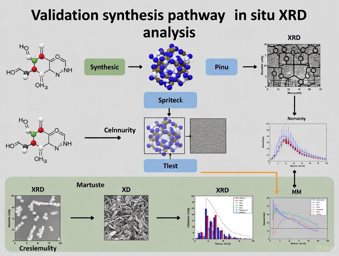
In Situ XRD Analysis: A Powerful Tool for Validating Material Synthesis Pathways in Biomedical Research
This article provides a comprehensive guide to in situ X-ray diffraction (XRD) analysis for validating material synthesis pathways, tailored for researchers and drug development professionals.
In Situ XRD Analysis: A Powerful Tool for Validating Material Synthesis Pathways in Biomedical Research
Abstract
This article provides a comprehensive guide to in situ X-ray diffraction (XRD) analysis for validating material synthesis pathways, tailored for researchers and drug development professionals. It covers the foundational principles of XRD and Bragg's Law, explores methodological setups for real-time monitoring of synthesis and catalytic processes, and offers practical troubleshooting for data interpretation. The content further examines how in situ XRD serves as a critical validation tool, comparing structural data with complementary techniques and computational modeling to confirm synthesis success and material performance, with a specific focus on applications in drug development and biomimetic material design.
Understanding In Situ XRD: Core Principles and Why It's Indispensable for Synthesis Validation
Bragg's Law and the Fundamental Principle of X-ray Diffraction
X-ray diffraction (XRD) is a powerful non-destructive analytical technique that has revolutionized our understanding of crystalline materials by providing unparalleled insights into their atomic and molecular structure [1]. This technique leverages the wave nature of X-rays, which have wavelengths comparable to the spacing between atoms in crystal structures (approximately 0.1–10 nm), allowing them to interact constructively with the periodic arrangement of atoms in crystalline materials [1]. The resulting diffraction pattern serves as a unique "fingerprint" for material identification and structural analysis, making XRD indispensable across scientific disciplines from materials science to pharmaceutical development [1] [2].
The fundamental principle governing XRD was formulated in 1913 by William Lawrence Bragg, who described the diffraction condition with remarkable simplicity [1] [3]. Bragg's Law establishes the mathematical relationship for constructive interference of X-rays scattered by parallel crystal planes through the equation:
nλ = 2d sin θ
Where:
- n = order of diffraction (integer: 1, 2, 3...)
- λ = X-ray wavelength, typically 1.5418 Å for copper Kα radiation
- d = interplanar spacing, the perpendicular distance between parallel crystal planes
- θ = Bragg angle, the angle between the incident X-ray beam and the crystal plane [1]
This elegant relationship enables researchers to calculate distances between crystal planes using measured diffraction angles, forming the foundation for determining crystal structures, lattice parameters, and various material properties [1]. The profound significance of Bragg's Law was demonstrated in landmark scientific achievements, most notably in determining the double helix structure of DNA through Rosalind Franklin's XRD work, which revealed key structural parameters including the 3.4 Å spacing between consecutive base pairs and the 34 Å helical repeat distance [1].
The XRD Instrument and Experimental Framework
Core Components of an X-ray Diffractometer
A modern X-ray diffractometer consists of several precision components working in coordination to measure diffraction patterns [1] [2]:
X-ray source: Generates monochromatic X-rays through electron bombardment of a metal target, most commonly copper (Cu Kα, λ = 1.5418 Å) or molybdenum (Mo Kα, λ = 0.71 Å) [1]. The X-ray tube operates at high voltage (typically 30–60 kV) and current (10–50 mA) to produce sufficient intensity for detection.
Incident beam optics: Conditions the X-ray beam using various optical elements including Soller slits for controlling beam divergence, monochromators for wavelength selection, and focusing mirrors for beam concentration [1].
Sample stage: Holds the specimen and allows precise positioning and rotation during measurement, providing accurate angular positioning that may include environmental controls for specialized analyses [1].
Detector system: Modern diffractometers employ position-sensitive detectors (PSDs) or area detectors that simultaneously collect data over a range of angles, significantly reducing measurement time while maintaining high resolution [1] [2].
Goniometer: A precision mechanical system controlling angular relationships between X-ray source, sample, and detector, with modern goniometers achieving angular accuracy better than 0.001° [1].
The instrument operates by directing X-rays at the sample while rotating both sample and detector according to θ-2θ geometry, ensuring the detector captures diffracted beams at the correct angle for constructive interference [1].
Key Research Reagent Solutions
Table 1: Essential Research Reagents and Materials for XRD Analysis
| Reagent/Material | Function | Application Examples |
|---|---|---|
| Copper (Cu) X-ray Tube | Produces characteristic Kα radiation (λ = 1.5418 Å) | Routine analysis of most materials [1] |
| Molybdenum (Mo) X-ray Tube | Produces shorter wavelength radiation (λ = 0.71 Å) | Samples with heavy elements or when higher resolution needed [1] |
| Crystalline Powder Standards | Reference materials for calibration and phase identification | Quality control, instrument calibration [2] |
| Silicon Zero-Diffraction Plate | Sample holder for powder analysis | Minimizes background signal during measurement [1] |
| ICDD Database (PDF-2) | Reference database for phase identification | Pattern matching for unknown material identification [2] |
XRD Pattern Interpretation and Analysis
Extracting Structural Information from XRD Patterns
An XRD pattern displays diffraction intensity versus diffraction angle (2θ), where each peak corresponds to a specific set of parallel crystal planes characterized by Miller indices (hkl) [1]. The diffraction pattern provides comprehensive structural information through various peak characteristics:
Peak position: The angular position directly relates to the d-spacing (interplanar spacing) through Bragg's law, enabling determination of lattice parameters, phase identification, and detection of structural changes due to composition, temperature, or pressure variations [1].
Peak intensity: The height or integrated area indicates the atomic arrangement within the crystal structure and the relative abundance of different phases, providing information about preferred orientation effects and enabling quantitative phase analysis [1].
Peak width: The breadth reveals crystal quality, including crystallite size and microstrain effects, with narrow peaks indicating large, well-formed crystals with minimal strain, while broad peaks suggest small crystallites or high levels of structural disorder [1] [3].
Peak shape: The detailed shape provides insights into crystal defects, stacking faults, and other structural imperfections, where asymmetric peak shapes often indicate compositional gradients or structural distortions [1].
The following diagram illustrates the fundamental relationship between atomic planes, Bragg's Law, and the resulting XRD pattern:
Diagram 1: Fundamental XRD Principle. This diagram illustrates the sequential relationship from atomic planes to measurable diffraction peaks governed by Bragg's Law.
Comparative Analysis of XRD Techniques
Table 2: Comparison of Primary XRD Techniques and Applications
| Technique | Sample Requirements | Structural Information Obtained | Data Collection Time | Limitations |
|---|---|---|---|---|
| Single Crystal XRD | High-quality single crystal (>0.1 mm) | Complete 3D atomic coordinates, bond lengths/angles, displacement parameters [1] | Hours to days | Requires large, well-formed single crystals [1] |
| Powder XRD | Polycrystalline powder (micron size) | Phase identification, lattice parameters, crystallite size, preferred orientation [1] [4] | Minutes to hours | Peak overlap limits structure solution complexity [4] |
| Thin Film XRD (GIXRD) | Thin film on substrate | Film phase, strain, texture, thickness, interface quality [2] [5] | Minutes to hours | Signal may be weak for very thin films [2] |
| In Situ/Operando XRD | Special cell for controlled environment | Real-time structural changes during synthesis, activation, reaction, deactivation [6] [7] | Minutes to days (time-resolved) | Complex experimental setup, potential lower data quality [6] |
In Situ XRD: Validating Synthesis Pathways in Real Time
Fundamental Principles and Methodologies
In situ X-ray diffraction has emerged as a transformative approach for validating synthesis pathways by enabling real-time characterization of catalysts and functional materials during their "lifetime" - under synthesis, activation, operation, and deactivation conditions [6] [7]. This methodology addresses a critical limitation of traditional ex situ characterization, where catalyst extraction from reactors after catalytic processes can cause significant alterations through exposure to atmospheric oxygen or other environmental factors, making determination of the active state impossible [7].
The terminology in this field distinguishes between:
- In situ XRD: Studies performed in non-ambient conditions (elevated temperature, pressure, specific gas environments)
- Operando XRD: Studies conducted under actual catalytic reaction conditions with simultaneous measurement of catalytic performance and structural properties [7]
The experimental framework for in situ XRD requires specialized reaction chambers or cells that allow control of temperature (typically from room temperature to 1000°C), pressure (from vacuum to several hundred bar), and gas environment while maintaining precise X-ray optical alignment [7]. These cells feature X-ray transparent windows (often beryllium or diamond) and integrated gas handling systems, with advanced setups incorporating mass spectrometry for simultaneous reaction product analysis [7].
Experimental Protocol for Catalyst Synthesis Validation
Objective: To monitor phase evolution during activation and operation of a Fischer-Tropsch synthesis (FTS) catalyst in real time [6].
Materials and Equipment:
- In situ XRD reactor cell with controlled temperature and gas environment
- Fe- or Co-based FTS catalyst precursor (typically oxide form)
- Syngas mixture (H₂:CO = 2:1)
- High-temperature X-ray diffractometer with position-sensitive detector
- Mass spectrometer for gas analysis (for operando studies)
Methodology:
- Sample Loading: Mount catalyst powder in the in situ reactor cell, ensuring uniform packing and contact with thermocouple for accurate temperature monitoring [6].
Baseline Measurement: Collect reference XRD pattern at room temperature in inert atmosphere (He or N₂) to establish baseline phase composition [6].
Temperature Programmed Activation: Heat the sample under reducing atmosphere (H₂ or syngas) while collecting sequential XRD patterns (2-5 minute intervals) through the critical activation temperature range (200-400°C) [6].
Reaction Monitoring: Maintain at operational temperature (220-250°C for FTS) under syngas flow, collecting XRD patterns continuously to track phase evolution during reaction [6] [7].
Data Integration: Correlate structural changes (phase transformations, lattice parameter shifts) with catalytic performance metrics (activity, selectivity) when mass spectrometry is incorporated [7].
The following workflow diagram illustrates the integrated process of in situ XRD analysis for synthesis validation:
Diagram 2: In Situ XRD Workflow. This diagram outlines the sequential process for validating catalyst synthesis pathways using in situ XRD analysis.
Advanced Applications and Future Directions
Machine Learning-Enhanced XRD Analysis
The integration of artificial intelligence and machine learning (ML) with XRD analysis represents a paradigm shift in materials characterization, particularly for handling the enormous datasets generated by high-throughput synthesis and in situ experiments [3]. ML approaches are being deployed to address longstanding challenges in XRD analysis:
Phase identification and classification: Supervised learning models can rapidly identify crystalline phases and mixtures in complex multiphase systems, significantly accelerating analysis compared to traditional database matching [3].
Crystal structure determination: Generative models like PXRDGen leverage diffusion models and neural networks to solve crystal structures from powder XRD data with unprecedented accuracy, achieving matching rates of 82% (1-sample) and 96% (20-samples) for valid compounds in the MP-20 dataset [4].
Pattern extraction from in situ studies: Unsupervised ML methods excel at identifying hidden patterns and trends in high-dimensional data from in situ and microscopic studies, enabling automated detection of phase transitions and intermediate states [3].
These AI-enhanced approaches are particularly valuable for analyzing the terabyte-scale datasets generated at synchrotron facilities and automated laboratories, where traditional analysis methods become impractical [3] [4]. However, researchers must exercise caution as ML methods are inherently physics-agnostic and require careful interpretation and validation against established physical principles [3].
Emerging Applications Across Scientific Disciplines
The application scope of XRD continues to expand into new scientific frontiers:
Pharmaceutical development: XRD is indispensable for polymorph screening, salt selection, and structure determination of active pharmaceutical ingredients (APIs), with in situ XRD enabling real-time monitoring of phase transformations during processing and formulation [1].
Energy materials research: From catalyst development for Fischer-Tropsch synthesis [6] to battery materials characterization, in situ XRD provides crucial insights into structural evolution under operational conditions.
Advanced materials discovery: XRD facilitates the structural characterization of novel materials systems including hybrid organic-inorganic semiconductors, metal-organic frameworks (MOFs), and nanostructured materials with tunable properties [3].
Extreme conditions science: Recent breakthroughs include the elucidation of liquid carbon structure using in situ XRD under extreme temperature and pressure conditions, demonstrating the technique's expanding capabilities [8].
The future of XRD analysis is trending toward more automated, miniaturized, and intelligent systems, with the global XRD market projected to exceed $1 billion by 2033, driven by innovations in robotics, AI integration, and laboratory-based 3D micro-beam XRD (Lab-3DμXRD) techniques [5]. These advances will further solidify XRD's role as an essential tool for validating synthesis pathways and accelerating materials discovery across scientific disciplines.
The pursuit of understanding material behavior under real-world conditions has driven a significant paradigm shift in analytical science, moving from traditional ex situ analysis toward advanced in situ and operando methodologies. This transition represents more than mere technical innovation; it constitutes a fundamental change in how researchers probe dynamic processes in functional materials. Where ex situ techniques provide valuable "before and after" snapshots, in situ and operando approaches offer a real-time cinematic view of material transformations as they occur. This comparative guide objectively examines the performance, capabilities, and limitations of these interconnected approaches, with particular focus on their application within validation synthesis pathway research using X-ray diffraction (XRD) analysis.
The critical distinction between these methodologies lies in their operational conditions. Ex situ analysis involves characterizing materials outside their operational environment after processes have occurred. In situ measurements observe materials under simulated reaction conditions (e.g., elevated temperature, applied voltage, presence of reactants), while operando techniques combine in situ observation with simultaneous measurement of functional activity or performance [9]. This evolution toward condition-relevant characterization has proven particularly transformative for investigating functional materials in energy storage, heterogeneous catalysis, and materials synthesis, where structure-property relationships are dictated by dynamic processes occurring under specific environmental stimuli.
Comparative Analysis: Technical Specifications and Performance Metrics
The selection between ex situ, in situ, and operando characterization strategies involves careful consideration of their respective capabilities, limitations, and information outputs. The table below provides a systematic comparison of these approaches across key analytical dimensions.
Table 1: Technical comparison of ex situ, in situ, and operando characterization approaches
| Analytical Dimension | Ex Situ | In Situ | Operando |
|---|---|---|---|
| Measurement Environment | Quenched/relaxed state, ambient conditions | Simulated reaction conditions | Actual working conditions |
| Temporal Resolution | Single time points (static) | Multiple time points (kinetics possible) | Real-time monitoring (full kinetics) |
| Structural Information | High resolution of initial/final states | Moderate resolution under conditions | Moderate resolution during operation |
| Chemical State Analysis | Post-process analysis, potential artifacts | Direct observation of intermediates | Direct correlation with activity |
| Technical Complexity | Low | Moderate to High | High |
| Reactor Design Requirements | Standard sample holders | Specialized environmental cells | Integrated performance monitoring |
| Data Interpretation | Straightforward, established protocols | Complex, requires environmental modeling | Highly complex, multi-parameter correlation |
Performance Analysis and Experimental Validation
Comparative studies across multiple material systems consistently demonstrate how the analytical approach significantly influences the mechanistic insights gained. In battery material research, a direct comparison between ex situ and operando XRD for studying lithium insertion in the fast ion conductor Li₀.₁₈Sr₀.₆₆Ti₀.₅Nb₀.₅O₃ revealed critical methodological distinctions. Ex situ analysis suggested a single-phase material existed throughout discharge, while operando XRD revealed a kinetically driven two-phase region during battery cycling below 1V that was not captured by quenched ex situ measurements [10]. This discrepancy highlights how ex situ approaches may miss transient intermediates that revert to more stable configurations when operational stresses are removed.
Similarly, in thermochemical energy storage research combining in situ XRD with thermogravimetry and differential scanning calorimetry revealed the dynamic redox behavior of metal oxide systems like Co₃O₄/CoO and Mn₂O₃/Mn₃O₄. While thermodynamic calculations predicted full reversibility for several metal oxide systems, in situ analysis demonstrated that only specific couples (Co₃O₄/CoO and Mn₂O₃/Mn₃O₄) exhibited complete reversibility, with other systems like PbO₂ showing no practical reversibility despite theoretical predictions [11]. These findings underscore how condition-relevant characterization provides essential validation for theoretical models.
Experimental Protocols: Methodologies for Advanced Characterization
Protocol for Operando XRD in Battery Materials
The investigation of dynamic phase transitions in energy storage materials represents a key application of operando methodologies. The following protocol details a representative experimental setup for operando XRD studies of lithium insertion materials, based on established methodologies [10]:
Cell Configuration: Utilize a specialized electrochemical cell with X-ray transparent windows (e.g., beryllium or Kapton windows) compatible with standard XRD instrumentation. For synchrotron studies, coin cells with modified housings provide sufficient photon transmission.
Electrode Preparation: Mix active material powder (e.g., Li₀.₁₈Sr₀.₆₆Ti₀.₅Nb₀.₅O₃) with conductive carbon additive and binder in an 80:10:10 weight ratio. Homogenize in N-methylpyrrolidone to form a slurry, which is then spread onto a current collector (18μm aluminum foil) and dried at 80°C under vacuum.
Data Collection Parameters: Employ synchrotron radiation (λ = 0.5-1.0 Å) or high-power laboratory X-ray source with incident beam geometry through transmission windows. Collect sequential diffraction patterns (30s-5min acquisition times) synchronized with electrochemical cycling.
Electrochemical Protocol: Apply constant current charge/discharge cycles between specified voltage limits (e.g., 0.4-3.0V vs. Li/Li+) with continuous potential monitoring. Current densities should be selected to approximate realistic operational conditions.
Data Analysis: Perform sequential Rietveld refinement of diffraction patterns to extract structural parameters (lattice constants, phase fractions) as a function of state of charge. Correlate structural evolution with electrochemical performance metrics in real time.
This methodology enabled the discovery of a 22(2)% reduction in the rate of unit cell expansion partway through the first discharge cycle in Li₀.₁₈Sr₀.₆₆Ti₀.₅Nb₀.₅O₃, indicating a change in lithium insertion mechanism that would be undetectable through ex situ approaches [10].
Protocol for In Situ XRD in Catalytic Materials
The investigation of catalyst behavior under operational conditions provides crucial insights for designing improved catalytic systems. The following protocol details methodology for in situ XRD studies of Fischer-Tropsch synthesis catalysts [6]:
Reactor Design: Utilize a dedicated in situ reaction chamber capable of maintaining controlled gas atmospheres (syngas: H₂/CO mixtures), elevated temperatures (200-350°C), and pressures (1-30 bar) while allowing X-ray transmission.
Catalyst Activation: Subject Fe- or Co-based catalysts to various activation protocols (H₂ reduction, CO treatment, or syngas pretreatment) while monitoring phase evolution via XRD. Temperature-programmed treatments provide insights into reduction mechanisms.
Data Collection During Reaction: Collect time-resolved diffraction patterns during exposure to reactive atmospheres. Utilize rapid detectors to capture transient phase evolution during initial activation and potential deactivation processes.
Promoter/Support Effects: Compare phase evolution of promoted catalysts (e.g., with K, Cu) versus reference materials to elucidate stabilization effects on active phases.
Correlation with Performance: When possible, integrate gas chromatography for simultaneous product analysis to establish structure-activity relationships under true operando conditions.
This approach has revealed how activation mode, promoters, and supports influence the phase evolution and ultimate performance of Fe- and Co-based Fischer-Tropsch catalysts, providing theoretical guidance for rational catalyst design [6].
Visualization: Experimental Workflows and Data Interpretation
The conceptual and practical relationships between characterization methodologies can be visualized through the following workflow:
Diagram 1: Progression from static to dynamic analysis
Essential Research Reagent Solutions
Successful implementation of advanced characterization methodologies requires specific research reagents and specialized components. The table below details key solutions for establishing robust in situ and operando characterization capabilities.
Table 2: Essential research reagent solutions for advanced characterization
| Reagent/Category | Function/Application | Specification Notes |
|---|---|---|
| Specialized Electrochemical Cells | Enable operando XRD of battery materials | X-ray transparent windows (Be, Kapton); compatible electrode configuration [10] |
| High-Temperature Reaction Chambers | In situ catalyst studies under operational conditions | Gas flow control; temperature capability to 1000°C; pressure rating to 30 bar [6] |
| Synchrotron-Grade X-Ray Sources | High brilliance for time-resolved studies | High photon flux; tunable wavelength; fast detector systems [10] |
| Reference Materials | Pattern calibration and instrument alignment | NIST-standard materials (e.g., Si, Al₂O₃) for quantitative analysis |
| Computational Analysis Tools | Automated phase identification and quantification | Machine learning platforms; Bayesian analysis methods [12] |
The methodological evolution from ex situ to in situ and operando analysis represents a critical advancement in materials characterization, particularly for validation of synthesis pathways. Each approach offers distinct advantages: ex situ provides high-resolution structural baseline information, in situ reveals dynamic structural evolution under simulated conditions, and operando directly correlates structural changes with functional performance in real-time. The research community's growing ability to bridge the information gaps between these approaches has enabled more accurate mechanistic understanding and accelerated the development of next-generation functional materials.
Future developments in this field will likely focus on addressing remaining technical challenges, including further optimization of reactor designs to better approximate real-world conditions [9], increased integration of multi-modal characterization approaches, and enhanced computational methods for interpreting complex time-resolved datasets [12]. As these methodologies continue to mature and become more accessible, they will undoubtedly play an increasingly central role in validating synthesis pathways and guiding the rational design of advanced materials across energy storage, catalytic, and functional material applications.
In situ X-ray diffraction (XRD) has emerged as a powerful non-destructive technique that enables researchers to monitor the evolution of a material's crystal structure in real time under controlled environments and external stimuli [1] [7]. Unlike conventional ex situ XRD, which provides only a static snapshot of a material's structure, in situ XRD allows for dynamic observation of structural transformations as they occur, providing unparalleled insights into reaction mechanisms, phase stability, and structural kinetics [7]. This capability is particularly valuable for validating synthesis pathways, where understanding intermediate phases and transformation sequences is crucial for rational materials design [13].
The fundamental principle of XRD remains grounded in Bragg's law (nλ = 2d sinθ), which describes the conditions under which constructive interference of X-rays occurs when scattered by crystalline planes [1] [14]. When applied in situ, this principle becomes a powerful tool for tracking how the key structural parameters—phase identity, crystallinity, and lattice dimensions—evolve during synthesis, activation, or operation of functional materials [7]. This guide examines these measurable properties through the lens of contemporary research applications, providing a comparative analysis of the insights gained through in situ XRD experimentation.
Key Structural Properties Accessible via In Situ XRD
Phase Identification and Transformation Pathways
Phase composition and transformation pathways represent the most direct application of in situ XRD, allowing researchers to identify crystalline intermediates and final products during synthesis or under operational conditions [7]. The technique captures the "fingerprint" diffraction patterns of crystalline phases at specific time or temperature intervals, enabling reconstruction of the entire reaction pathway [1] [13].
Table 1: In Situ XRD Applications in Phase Analysis Across Material Systems
| Material System | Phase Transformation Observed | Experimental Conditions | Key Findings | Citation |
|---|---|---|---|---|
| Fischer-Tropsch Co₃C catalysts | Stability of Co₃C phase under syngas atmosphere | H₂/CO = 2, 150-300°C, 0.2 MPa | Co₃C remains stable up to 300°C without decomposition | [15] |
| Additively manufactured Ti-6Al-4V | α' (martensite) → α + β (equilibrium) | Isothermal treatments, 400-700°C | Stepwise transformation through transitional α phase with asymmetrical lattice | [16] |
| Garnet-type Li₆.₅La₃Zr₁.₅Ta₀.₅O₁₂ | Formation via LLTO intermediate | Solid-state synthesis, ~1000°C | LLTO acts as structural template for final garnet phase | [13] |
| Al-Si-Mg casting alloy | Primary α-Al and eutectic Si formation | Solidification with ultrasonic processing | USMP refines α-Al grain size by 36% | [17] |
Recent studies have demonstrated the power of in situ XRD in elucidating complex phase evolution pathways that would be impossible to deduce from ex situ analysis alone. For instance, in the synthesis of garnet-type solid electrolyte LLZTO, quasi-in situ XRD revealed that the Ta-doped cubic phase forms through a specific intermediate (Li₅La₃Ta₂O₁₂) that shares structural homology with the target material [13]. Similarly, in Fischer-Tropsch catalysis research, in situ XRD has proven invaluable for establishing the stability windows of different catalyst phases under reaction conditions, directly linking structural features to catalytic performance [15].
Crystallinity and Microstructural Evolution
Crystallinity assessment through in situ XRD encompasses multiple aspects of material microstructure, including crystallite size, strain, and defect structure [1]. The technique tracks changes in diffraction peak characteristics—position, width, intensity, and shape—to quantify microstructural evolution during synthesis or processing [1].
Table 2: Crystallinity Parameters Accessible via In Situ XRD
| Parameter | XRD Manifestation | Extracted Information | Application Example |
|---|---|---|---|
| Crystallite Size | Peak broadening (Scherrer equation) | Grain growth, nucleation | Ultrasonic refinement of Al grains during solidification [17] |
| Microstrain | Peak broadening and shifting | Residual stress, defects | Stress relaxation in AM Ti-6Al-4V during heat treatment [16] |
| Crystallinity Degree | Sharp vs. diffuse scattering | Amorphous-crystalline transitions | Phase evolution in oxide catalysts during activation [7] |
| Preferred Orientation | Relative peak intensity changes | Texture development | Templated growth in complex oxide synthesis [13] |
The application of in situ XRD to microstructural analysis is well-illustrated by studies on additively manufactured Ti-6Al-4V, where researchers tracked the relaxation of internal strains and the decomposition of martensitic phase during post-build heat treatments [16]. By analyzing peak broadening and shifting, they determined that stress relaxation occurs between 25-400°C, while phase transformation proceeds in a stepwise manner between 550-750°C [16]. Similarly, in studies of aluminum alloy solidification, in situ synchrotron XRD has quantified how ultrasonic melt processing reduces primary α-Al grain size by 36% by slowing growth rates and promoting fragmentation [17].
Lattice Parameters and Unit Cell Dimensions
Precise determination of lattice parameters and their evolution under external stimuli represents another key application of in situ XRD. Changes in interplanar spacings (d-spacings), calculated from peak positions using Bragg's law, provide insights into thermal expansion, compositional changes, and structural transitions [1].
In catalyst research, in situ XRD has revealed how lattice parameters evolve during activation and reaction. For oxide and metal oxide catalysts, these measurements can track oxygen loss/uptake, reduction processes, and cation migration—all of which manifest as subtle changes in unit cell dimensions [7]. The high-temperature stability of Co₃C catalysts during Fischer-Tropsch synthesis was confirmed through the absence of significant lattice parameter changes until reaching critical temperature thresholds [15].
In metallurgy, in situ high-temperature XRD studies have quantified the lattice parameter evolution of both α-Al and Si phases during solidification of Al-Si-Mg alloys, enabling estimation of cooling rates and thermal expansion behavior [17]. Similarly, the transformation from martensite to equilibrium phases in Ti-6Al-4V manifests as a gradual shift in lattice parameters from compressed (a = 2.933 Å, c = 4.655 Å) to relaxed (a = 2.935 Å, c = 4.685 Å) values [16].
Experimental Protocols for In Situ XRD Analysis
Reactor Design and Environmental Control
The core challenge in in situ XRD experimentation lies in designing sample environments that simulate relevant process conditions while maintaining sufficient data quality [9]. Effective reactor design must balance several competing requirements: maintaining appropriate temperature, pressure, and atmosphere; allowing X-ray transmission to and from the sample; and ensuring representative mass transport conditions [9].
Table 3: Essential Research Reagent Solutions for In Situ XRD
| Reagent/Cell Component | Function | Technical Considerations |
|---|---|---|
| High-Temperature Reactors | Enable studies up to 1600°C | X-ray transparent windows (e.g., Be, SiO₂), uniform temperature zone, minimal thermal gradients |
| Gas/Liquid Flow Cells | Simulate reaction environments | Controlled atmosphere, laminar flow conditions, corrosion-resistant materials |
| Electrochemical Cells | Operando electrocatalyst studies | Working, counter, and reference electrode integration; ionically conductive but X-ray transparent electrolytes |
| Standard Reference Materials | Instrument calibration | Si, Al₂O₃, or other well-characterized materials for angle and line shape calibration |
For catalytic studies, in situ cells must allow precise control of gas composition and flow rates while maintaining temperature stability [7] [15]. For battery and electrocatalytic applications, specialized electrochemical cells incorporate electrode configurations and ion-conducting pathways while maintaining X-ray transparency [9]. A critical consideration is minimizing the mismatch between characterization conditions and real-world operating environments to ensure mechanistic insights remain relevant [9].
Data Collection and Analysis Methodologies
Successful in situ XRD experiments require careful planning of data collection strategies to capture relevant structural changes with appropriate temporal resolution. The specific methodology varies depending on the transformation kinetics—from rapid measurements capturing second-scale changes to slower experiments monitoring hour-long processes [13].
For time-resolved studies, protocols typically involve:
- Equilibration at initial conditions while collecting reference patterns
- Stepwise or continuous variation of stimulus (temperature, pressure, potential)
- Continuous or rapid-scan XRD data collection throughout the process
- Post-experiment calibration using reference standards if quantitative analysis is required
Data analysis workflows generally include:
- Phase identification via comparison with ICDD reference patterns [14]
- Rietveld refinement for quantitative phase analysis and lattice parameter extraction [1]
- Peak profile analysis for crystallite size and strain calculation [1] [16]
- Multivariate analysis for tracking complex multi-phase transformations [13]
The quasi-in situ XRD approach developed for studying garnet oxide synthesis exemplifies an innovative methodology that combines ultrafast high-temperature synthesis (UHS) with rapid cooling to "freeze" reaction intermediates, overcoming the temporal resolution limitations of conventional in situ XRD [13].
Comparative Data Presentation
Quantitative Comparison of Structural Parameters
Table 4: Comparative Quantitative Data from In Situ XRD Studies
| Material | Experimental Conditions | Measured Lattice Parameters | Crystallite Size (nm) | Phase Evolution Observations |
|---|---|---|---|---|
| Co₃C Catalyst [15] | FTS conditions, H₂/CO=2, 150-300°C | Stable Co₃C parameters throughout | Not reported | No phase changes observed up to 300°C |
| AM Ti-6Al-4V [16] | 5°C/min heating to 925°C | α': a=2.933→2.935Å, c=4.655→4.685Å | Not reported | α'→α transformation through αₜᵣ intermediate at 550-700°C |
| Al-Si-Mg Alloy [17] | Solidification, ~1°C/s cooling | Temperature-dependent a for α-Al and Si | 36% reduction with USMP | Primary α-Al at 616°C, eutectic at 573°C |
| Garnet LLZTO [13] | Solid-state synthesis, ~1000°C | Cubic LLZTO via LLTO intermediate | Not reported | Pathway: LiLa₂TaO₆→La₃TaO₇→LLTO→LLZTO |
Visualization of Experimental Workflows
The following diagram illustrates a generalized workflow for in situ XRD analysis of material synthesis pathways, integrating elements from multiple studies [16] [15] [13]:
In situ XRD analysis provides unparalleled capability for quantifying the key structural properties—phase identity, crystallinity, and lattice parameters—during active synthesis and operation of functional materials. The experimental data and comparative tables presented in this guide demonstrate how this technique enables researchers to move beyond static structural characterization to dynamic pathway validation. By implementing robust experimental protocols and careful data analysis methodologies, researchers can extract quantitative insights into transformation mechanisms, stability windows, and structure-property relationships across diverse material systems. As reactor designs become more sophisticated and detection systems more sensitive, in situ XRD will continue to expand its critical role in guiding rational materials design and synthesis optimization.
The Critical Role of Structural Validation in Drug Development and Biomimetic Design
Structural validation has become a cornerstone of modern drug development and biomimetic design, ensuring that therapeutic compounds and bio-inspired materials perform as intended. The ability to precisely characterize the atomic and micro-scale structure of active pharmaceutical ingredients (APIs), excipients, and biomimetic materials directly determines their efficacy, stability, and safety profiles. Structural validation techniques, particularly advanced X-ray analysis methods, provide the critical data needed to understand composition, polymorphic forms, and dynamic changes during manufacturing and application.
Within the broader thesis of validation synthesis pathway in situ XRD analysis research, this guide explores how integrated analytical approaches are revolutionizing quality control and design processes. The emergence of in-situ analysis represents a paradigm shift, enabling researchers to monitor structural changes in real-time under realistic conditions, rather than relying solely on static pre- and post-analysis. This capability is particularly valuable for tracking dynamic processes such as tablet dissolution, polymer crystallization during 3D printing, and biomimetic material performance under stress conditions. As this guide will demonstrate through comparative data and experimental protocols, the strategic selection of structural validation methodologies significantly impacts development timelines, regulatory success, and ultimately, the performance of final pharmaceutical and biomimetic products.
Core Structural Analysis Technologies: Principles and Applications
X-ray Diffraction (XRD) Techniques
X-ray diffraction techniques form the backbone of structural validation in pharmaceutical and materials science, each offering unique capabilities for different analytical needs.
Single Crystal X-ray Diffraction (SCXRD) provides the highest resolution structural data, enabling precise determination of molecular structures, atomic coordinates, and bond lengths. This technique has been fundamental to structure-based drug design, revealing critical details about drug-target interactions and facilitating the development of novel chemical entities with high selectivity and specificity toward therapeutic targets [18]. For example, SCXRD studies of human cytochrome P450 (CYP3A4) in complex with drugs like ketoconazole and erythromycin revealed unexpected binding stoichiometries and conformational changes that significantly advanced understanding of drug metabolism [18].
Powder X-ray Diffraction (PXRD) serves as a workhorse technique for analyzing materials that cannot be obtained as single crystals, including many pharmaceutical formulations and biomimetic materials. PXRD enables identification of crystalline phases, quantification of polymorphic content, and assessment of material stability [18] [19]. The technique is particularly valuable for quality control in pharmaceutical manufacturing, where it can detect unwanted polymorphs that might compromise product safety or efficacy. Recent advances have expanded PXRD applications to include amorphous solid dispersions (ASDs) through the pair distribution function (PDF), providing vital information about atomic arrangements in non-crystalline materials [18].
Synchrotron Radiation-based X-ray Microtomography (SR-µCT) represents a significant advancement over conventional laboratory-based µCT systems, offering dramatic improvements in spatial resolution and temporal performance [20]. This enables non-destructive 3D analysis of internal tablet morphology, coating thickness, porosity, and ingredient distribution at micrometer resolution. SR-µCT has proven particularly valuable for time-resolved monitoring of tablet dissolution processes, capturing dynamic events such as swelling in sustained-release formulations and API distribution changes [20].
Table 1: Comparative Analysis of X-ray Structural Validation Techniques
| Technique | Key Applications | Spatial/Temporal Resolution | Sample Requirements | Limitations |
|---|---|---|---|---|
| Single Crystal XRD (SCXRD) | Drug-target interaction studies, Absolute configuration determination, Biomimetic candidate characterization | Atomic resolution (static) | High-quality single crystals (>50-100 µm) | Requires crystallizable samples; Limited to static conditions |
| Powder XRD (PXRD) | Polymorph screening, Crystallinity assessment, Phase quantification, Stability testing | Bulk material analysis | Powder or polycrystalline material | Lower resolution than SCXRD; Peak overlap in complex mixtures |
| Synchrotron µCT (SR-µCT) | 3D morphology analysis, Coating thickness distribution, Dynamic dissolution monitoring | ~100 nm spatial resolution; 5-second temporal for dynamic studies | Intact tablets or formulations | Limited access to synchrotron facilities; Complex data processing |
| In-situ XRD | Real-time monitoring of manufacturing processes, Crystallization kinetics, Dynamic structural changes | Varies with setup; Seconds to minutes for time-resolved | Compatible with specialized cells/chambers | Requires specialized equipment; Data interpretation challenges |
Complementary and Emerging Techniques
While X-ray techniques provide comprehensive structural information, other analytical methods offer complementary insights crucial for complete structural validation.
Atomic Force Microscopy (AFM), particularly in liquid cell configurations, enables real-time nanoscale imaging of dynamic processes such as crystal growth, dissolution, and demineralization/remineralization cycles in biomimetic materials [21]. This technique has proven invaluable for studying enamel-like fluoridated hydroxyapatite (FHAp) crystals, revealing how fluoride incorporation influences crystal morphology, growth kinetics, and acid resistance [21].
Cellular Thermal Shift Assay (CETSA) has emerged as a powerful approach for validating direct drug-target engagement in physiologically relevant environments (intact cells and tissues), addressing the critical need for functional validation of pharmacological activity [22]. Recent advancements have combined CETSA with high-resolution mass spectrometry to quantify target engagement ex vivo and in vivo, bridging the gap between biochemical potency and cellular efficacy [22].
Artificial Intelligence and Machine Learning are revolutionizing structural analysis through accelerated pattern recognition and prediction. For powder X-ray diffraction, machine learning models now enable rapid prediction of space groups, cell parameters, and even atomic coordinates from diffraction patterns [23]. The development of benchmarks like SIMPOD (Simulated Powder X-ray Diffraction Open Database), containing 467,861 crystal structures and their simulated diffractograms, provides extensive training data for developing increasingly accurate models [23].
Experimental Protocols and Data Interpretation
Protocol for In-Situ Dissolution Monitoring via SR-µCT
The dynamic analysis of tablet dissolution behavior represents one of the most valuable applications of advanced structural validation techniques.
Sample Preparation:
- Select representative tablets from multiple production batches.
- For pantoprazole tablets used in one case study, ensure samples include both immediate-release and enteric-coated formulations for comparative analysis [20].
- Mount tablets using minimal supportive material to avoid interference with X-ray transmission.
Experimental Setup:
- Utilize a specially designed flow-through chamber that allows liquid from a 100 ml reservoir to circulate and renew during measurements [20].
- Maintain sink conditions throughout the experiment to mimic physiological relevance.
- Set synchrotron µCT parameters according to resolution requirements: high-resolution (0.5-2 µm voxel size) for detailed structural analysis or lower resolution for faster dynamic imaging [20].
- For rapid disintegration studies, employ tomogram recording times as short as 5 seconds to capture fast-evolving events [20].
Data Acquisition:
- Acquire baseline 3D scan before introducing dissolution medium.
- Initiate flow of physiologically relevant dissolution medium (typically pH-graded to simulate gastrointestinal transition).
- Collect sequential scans at predetermined intervals based on expected dissolution profile.
- For swelling-controlled release systems, extend acquisition to capture full hydration and expansion cycles.
Analysis and Interpretation:
- Reconstruct 3D volumes from projection data using filtered back-projection or iterative algorithms.
- Segment different components (API, excipients, pores) using grayscale thresholding or advanced machine learning approaches like U-Net convolutional neural networks [20].
- Quantify critical parameters including coating thickness variation, pore formation and evolution, API distribution changes, and swelling kinetics.
- Correlate structural changes with drug release profiles obtained through complementary analytical techniques.
Protocol for Biomimetic Material Performance Validation
Biomimetic materials inspired by natural structures like tortoiseshells, bamboo, and Rudraksha seeds require specialized validation approaches to confirm their enhanced performance characteristics [24] [25].
Sample Fabrication:
- Utilize additive manufacturing (3D printing) technologies to create complex bio-inspired architectures with precise control over internal structures [24] [25].
- Select materials matching target applications: Vero White Plus photopolymer for preliminary validation, specialized composites for functional prototypes [24].
- Implement hierarchical, graded, or hybrid structure characteristics inspired by natural models such as pomelo peel, horse hoof, spider-web, and sunflower [25].
Structural Characterization:
- Employ micro-CT scanning to verify internal architecture and identify manufacturing defects.
- Conduct powder XRD to determine crystalline structure, phase composition, and preferred orientation in mineralized biomimetic materials [21].
- For enamel-like hydroxyapatite crystals, calculate c-axis orientation ratio from XRD patterns by analyzing intensity ratios of (002) to (112) reflections [21].
Performance Validation:
- Subject samples to compressive, flexural, and impact loading using standardized mechanical testing platforms.
- Calculate Specific Energy Absorption (SEA) using the formula: SEA = Total Absorbed Energy / Mass, to quantify crashworthiness and impact resistance [24] [25].
- Compare performance against conventional designs; bio-inspired multi-cell tubes have demonstrated 35-107% improvement in SEA compared to traditional structures [25].
- For dental biomimetics, implement nanomechanical testing via AFM to measure hardness and modulus across different crystal planes, noting the anisotropy between basal (001) and prismatic (100) planes [21].
Table 2: Performance Comparison of Biomimetic Structural Designs
| Bio-inspired Structure | Natural Model | Key Performance Metrics | Improvement Over Conventional Designs |
|---|---|---|---|
| Multi-cell Tubes | Beetle Forewing | Specific Energy Absorption (SEA) | 9.79-107.68% increase in SEA [25] |
| Hexagonal Prismatic Tubes | Honeycomb | Energy Absorption (EA), Axial Compression | ~2.5x increase in SEA [25] |
| Foam-filled Structures | Cornstalk/Reed | Load-bearing Capacity, Energy Dissipation | Optimized stress distribution [25] |
| Functionally Graded Structures | Bamboo, Porcupine Quill | Buckling Strength, Stiffness | Buckling strength: 135.2 MPa (Hystrix structure) [25] |
| Sandwich Structures | Tortoiseshell | Flexural Strength, Impact Resistance | Enhanced multi-impact durability [25] |
Comparative Performance Data: In-Situ XRD Analysis
Quantitative Analysis of Method Performance
Recent studies have systematically evaluated the accuracy and applicability of different XRD quantification methods, providing valuable guidance for technique selection based on sample characteristics and accuracy requirements.
Method Comparison Protocol:
- Preparation of artificial mixtures using seven high-purity minerals (quartz, albite, calcite, dolomite, halite, montmorillonite, kaolinite) in randomly generated proportions [26].
- Measurement using Panalytical X'pert Pro X-ray powder diffractometer with Cu Kα radiation (λ = 1.5418 Å) [26].
- Application of three quantitative methods: Reference Intensity Ratio (RIR), Rietveld, and Full Pattern Summation (FPS) [26].
- Evaluation using absolute error (ΔAE), relative error (ΔRE), and root mean square error (RMSE) against known standard proportions [26].
Performance Findings:
- For samples without clay minerals, all three methods demonstrated comparable accuracy with minimal differences in error metrics [26].
- For clay-containing samples, significant differences emerged: FPS method showed widest applicability for sedimentary materials, while conventional Rietveld methods struggled with phases exhibiting disordered or unknown structures [26].
- The RIR method, while handy for rapid assessment, demonstrated lower analytical accuracy compared to both FPS and Rietveld methods [26].
Table 3: Accuracy Comparison of Quantitative XRD Methods for Mineral Analysis
| Quantitative Method | Principle | Best For | Limitations | Reported Accuracy |
|---|---|---|---|---|
| Reference Intensity Ratio (RIR) | Individual peak intensity using RIR values | Quick screening, Simple mixtures | Lower accuracy, Peak overlap issues | Varies significantly with clay content [26] |
| Rietveld Method | Whole-pattern fitting based on crystal structure | Non-clay samples with known structures | Struggles with disordered/unknown structures | High accuracy for non-clay samples [26] |
| Full Pattern Summation (FPS) | Summation of reference library patterns | Sediments, Clay-containing samples | Requires comprehensive reference library | Wide applicability, good for clay samples [26] |
In-Situ Monitoring of Manufacturing Processes
The integration of XRD analysis into manufacturing processes provides unprecedented insights into structural evolution during production.
Polymer Additive Manufacturing:
- Experimental Setup: Unique time-resolved XRD configuration for fixed material volume monitoring during material extrusion AM process [27].
- Key Findings: Processing temperature dominates crystal microstructure evolution compared to deposition velocity; lower temperatures just above melting point created favourable crystallization conditions [27].
- Correlation: Established between polymer crystallinity from printing parameters and resulting mechanical properties [27].
Pharmaceutical Formulation:
- Analysis Capabilities: Real-time monitoring of crystalline structure changes during manufacturing, polymorph conversion, and stability assessment [19].
- Technology: Benchtop systems like Malvern Panalytical's Aeris XRD provide specialized pharmaceutical analysis modes including solid state analysis, crystallinity assessment, and phase identification/quantification [19].
- Regulatory Value: Data utilized in submissions to FDA, EMA, and other regulatory agencies [19].
The Scientist's Toolkit: Essential Research Reagents and Materials
Successful structural validation requires carefully selected materials and reagents tailored to specific analytical needs.
Table 4: Essential Research Reagents and Materials for Structural Validation
| Reagent/Material | Function/Application | Examples/Specifications |
|---|---|---|
| High-Purity Minerals | Reference standards for quantitative XRD | Quartz, albite, calcite, dolomite, halite, montmorillonite, kaolinite [26] |
| Pharmaceutical Powders | API and excipient analysis | Polymorph screening, crystallinity assessment, stability testing [19] |
| Biomimetic Fabrication Materials | Creating bio-inspired structures | Vero White Plus photopolymer, Tango Black Plus, composite materials [24] [25] |
| Synchrotron Facilities | High-resolution µCT studies | Bright, coherent X-ray beams for phase contrast imaging, ~100 nm resolution [20] |
| Flow-Through Chambers | In-situ dissolution monitoring | 100 ml reservoir, sink condition maintenance, fluid circulation [20] |
| AFM Liquid Cells | Nanoscale in-situ observation | Real-time crystal growth/dissolution monitoring in physiological environments [21] |
| Computational Resources | Machine learning applications | SIMPOD database (467,861 crystal structures), Deep graph networks for virtual analogs [23] [22] |
Integrated Workflows and Pathway Analysis
Modern structural validation employs integrated workflows that combine multiple analytical techniques to provide comprehensive understanding of material behavior.
Figure 1: Integrated workflow for in-situ XRD analysis in drug development and biomimetic design, showing the continuous feedback loop between experimental, analytical, and application phases.
Figure 2: Computational pathway for predicting crystal structures from powder XRD data, highlighting the role of machine learning and structural databases in accelerating materials discovery.
The field of structural validation in drug development and biomimetic design is undergoing rapid transformation, driven by advances in in-situ analysis capabilities, computational methods, and multi-technique integration. The comparative data presented in this guide demonstrates that technique selection should be guided by specific material characteristics and validation requirements, rather than adopting one-size-fits-all approaches.
The emerging paradigm of validation synthesis pathway in situ XRD analysis research emphasizes continuous, real-time monitoring of structural changes during manufacturing and application, providing insights that were previously inaccessible through conventional post-hoc analysis. This approach, combined with machine learning-powered prediction and the systematic biomimetic design principles inspired by natural structures, enables more efficient development of optimized pharmaceutical formulations and high-performance materials.
As these technologies continue to evolve, the integration of advanced structural validation throughout the development pipeline will become increasingly essential for reducing attrition rates, accelerating time-to-market, and ensuring the efficacy and safety of final products. Researchers who strategically leverage these complementary analytical approaches will be best positioned to overcome the complex challenges in modern drug development and biomimetic material design.
The field of material characterization is undergoing a revolutionary transformation, driven by the synergistic advancement of synchrotron X-ray sources and photon-counting detector technologies. In situ X-ray diffraction (XRD) studies, which involve observing material changes under realistic reaction conditions, have particularly benefited from these developments. The emergence of fourth-generation synchrotron facilities like MAX IV in Sweden and the European Synchrotron Radiation Facility Extremely Brilliant Source (ESRF-EBS) in France has provided researchers with X-ray beams of unprecedented brightness and coherence [28]. Concurrently, breakthroughs in hybrid pixel detector technology have enabled single-photon counting capabilities even in the soft X-ray range, dramatically improving signal-to-noise ratios and temporal resolution [29]. This powerful combination is redefining what is possible in fields ranging from catalyst development to drug discovery, allowing scientists to probe molecular transformations with unprecedented spatial and temporal precision.
The integration of these technologies has been particularly transformative for structure-based drug design, where understanding the atomic-level interaction between drug candidates and their protein targets can significantly accelerate development timelines. Pharmaceutical companies like AstraZeneca have transitioned to a "synchrotron-only" model for X-ray data collection, leveraging the high throughput and remote accessibility of modern beamlines to deliver hundreds of unique protein-ligand complex structures annually [30]. Similarly, in materials science, the ability to track phase evolution in catalysts under operating conditions provides crucial insights for designing more efficient and stable materials [6] [31]. This guide examines the performance characteristics of modern synchrotron sources and detectors, providing experimental protocols and comparative data to help researchers select the optimal tools for their in situ investigations.
Comparative Analysis of Synchrotron Facility Capabilities
Technical Specifications of Major Biomedical Beamlines
Global synchrotron facilities offer specialized beamlines optimized for different experimental needs, with varying performance in beam energy, flux, and spot size. The table below summarizes the key characteristics of actively used beamlines for life science applications:
Table 1: Technical Specifications and Applications of Major Synchrotron Biomedical Beamlines
| Medical Beamline | Country | Energy Range | Beam Size (Max) | Flux Density | Featured Applications |
|---|---|---|---|---|---|
| ESRF (ID17) [28] | France | 25–185 keV | 150.0 mm × 7.0 mm | 2×10¹⁴ ph/s (at 33 keV) | Brain microsurgery, Mammography, Microbeam radiotherapy |
| Australian Synchrotron (IMBL) [28] | Australia | 25–250 keV | 50 cm × 4 cm | 3.39×10¹² ph/s (at 22.1 keV) | Lung function imaging, Bone measurement, Enhanced Mammography |
| Canadian Light Source (BMIT) [28] | Canada | 12.6–140 keV | 200 mm × 4 mm | 10¹² ph/s/mm² (pink beam) | Cytochemistry visualization, Strontium distribution detection |
| Elettra (SYRMEP) [28] | Italy | 8–40 keV | 160 mm × 5 mm | 2×10⁸ ph/s/mm² (at 20 keV) | Mammography, Synchrotron radiotherapy |
| SPring-8 (BL20B2) [28] | Japan | 5–113 keV | 300 mm × 20 mm | 3.6×10⁸ ph/s/mm² (at 40 keV) | Bone structure imaging, Neuron structure analysis |
The selection of an appropriate beamline depends heavily on the specific experimental requirements. High-energy applications such as deep tissue imaging or studying dense materials benefit from facilities like the Australian Synchrotron's IMBL, which offers energies up to 250 keV [28]. Conversely, high-flux sources like ESRF's ID17 beamline are particularly valuable for dynamic studies requiring rapid data acquisition, such as tracking catalyst phase transitions in real time [6] [28]. The trend toward fourth-generation sources emphasizes improved coherence and brightness, which enhances techniques like ptychographic imaging and enables the study of smaller crystals with weaker diffraction signals [29] [28].
Advanced Detector Technologies for Soft and Hard X-Rays
The evolution of detector technologies has been equally critical for expanding the capabilities of in situ studies. Recent innovations have addressed the longstanding challenge of detecting soft X-rays with single-photon counting efficiency:
Table 2: Performance Comparison of Advanced X-Ray Detector Technologies
| Detector Technology | Detection Principle | Energy Range | Key Advantages | Representative Applications |
|---|---|---|---|---|
| iLGAD-based Hybrid Detector [29] | Single Photon Counting | 550 eV and above | High QE (55% at 250 eV), No readout noise, High frame rate | Ptychography at Fe L₃-edge (707 eV), Spectromicroscopy |
| Thallium Bromide (TlBr) Detector [32] | Photoconductive | Wide range | High signal-to-noise, Minimum detectable limit: 130 nGy/s, High contrast imaging | Low-dose X-ray imaging, Medical diagnostics |
| Standard Hybrid Detectors (EIGER2) [29] | Single Photon Counting | >1.75 keV | High dynamic range, Radiation hard, Fine φ-slicing | Macromolecular crystallography, Time-resolved studies |
| Charge-Coupled Devices (CCDs) [29] | Charge Integrating | Soft X-ray range | High QE, Low electronic noise | Limited applications in soft X-ray ptychography |
The introduction of inverse Low Gain Avalanche Diode (iLGAD) sensors represents a particular breakthrough for soft X-ray experiments. By incorporating an internal charge multiplication layer, these detectors achieve sufficient signal amplification to distinguish single photons with energies as low as 550 eV from electronic noise [29]. This capability is crucial for experiments at the K-edges of light elements (C, O, N) and L-edges of 3d transition metals, which are fundamental for studying organic materials, biological systems, and magnetic materials [29]. The hybrid architecture of these detectors allows independent optimization of the sensor and readout electronics, providing flexibility for different experimental requirements without technological compromises [29].
Experimental Protocols for In Situ Studies
Protocol 1: Tracking Catalyst Phase Evolution During Synthesis
The application of in situ XRD to monitor catalyst formation provides crucial insights into nucleation kinetics and phase stability, enabling the rational design of optimized catalytic materials.
dot-1 Experimental Workflow for In Situ XRD-Guided Catalyst Synthesis
Step-by-Step Methodology:
- Reactor Preparation: Load precursor materials (e.g., nickel salts and phosphorus sources) in a hydrothermal reactor equipped with X-ray transparent windows [31].
- Temperature Program: Apply controlled temperature ramps from 150°C to 300°C while collecting XRD patterns at regular intervals (typically 1-2 minute acquisition times) [31].
- Real-Time Monitoring: Identify the formation of intermediate phases (e.g., Ni₂P) and monitor transitions to stable phases (e.g., Ni₁₂P₅) as temperature increases above 225°C [31].
- Kinetic Analysis: Calculate activation energies for nucleation (e.g., 91.0 kJ mol⁻¹ for Ni₂P), growth (62.3 kJ mol⁻¹), and phase transitions (115.5 kJ mol⁻¹) using data from multiple temperatures [31].
- Material Optimization: Correlate synthesis parameters with catalytic performance (e.g., oxygen evolution reaction activity) to identify optimal crystallite size and crystallinity [31].
Key Considerations: Crystallite size can be controlled by varying reaction time and temperature, with partly amorphous nanoparticles obtained through short reaction times. Absolute crystallinity quantification is essential as amorphous impurities significantly impact electrocatalytic performance, sometimes more than crystallite size alone [31].
Protocol 2: High-Throughput Protein-Ligand Complex Analysis
In pharmaceutical research, synchrotron-based macromolecular crystallography has become the standard method for determining protein-ligand structures to guide drug design.
dot-2 High-Throughput Protein-Ligand Structure Determination
Step-by-Step Methodology:
- Sample Preparation: Establish robust crystallization protocols for target proteins that tolerate ligand soaking [30].
- Ligand Introduction: Soak pre-grown protein crystals in solutions containing drug candidates (typically 24 hours) [30].
- Sample Shipping: Harvest and flash-cool crystals in liquid nitrogen, then ship to synchrotron facilities (2-day shipping allowance recommended) [30].
- High-Throughput Data Collection: Utilize automated sample changers and remote access capabilities to collect 120-160 datasets in a single 8-hour synchrotron shift [30].
- Rapid Structure Determination: Leverage automated data processing pipelines to solve structures within 24 hours of data collection [30].
- Design Iteration: Use structural insights on binding modes to guide synthetic chemistry efforts for improved drug candidates [30].
Key Considerations: The "synchrotron-only" model with remote data collection enables delivery of new structures within a working week from compound receipt. This workflow depends on reliable crystal shipping and seamless data transfer infrastructure between facilities and home institutions [30].
The Scientist's Toolkit: Essential Research Reagents and Materials
Successful in situ XRD studies require carefully selected materials and reagents optimized for specific applications. The following table details key solutions used in the experiments referenced throughout this guide:
Table 3: Essential Research Reagents and Materials for In Situ XRD Studies
| Research Reagent | Composition/Specification | Function in Experiment | Application Context |
|---|---|---|---|
| Fe- and Co-based Catalyst Precursors [6] | Metal salts (nitrates, chlorides), Promoters (K, Cu), Support materials (SiO₂, Al₂O₃) | Active phases for Fischer-Tropsch synthesis, Phase evolution studies | Catalysis research [6] |
| Nickel Phosphide Synthesis Precursors [31] | Nickel salts, Phosphorus sources (e.g., NaH₂PO₂), Solvents | Formation of Ni₂P and Ni₁₂P₅ nanoparticles under hydrothermal conditions | Electrocatalyst development [31] |
| Protein Crystallization Reagents [30] | Precipitants (PEGs, salts), Buffers, Additives | Generate diffraction-quality protein crystals for ligand binding studies | Drug discovery [30] |
| Fragment Libraries [30] | 500-1500 rule-of-three compliant compounds with high solubility | Identify initial binding motifs through crystallographic screening | Structure-based drug design [30] |
| iLGAD Sensors [29] | Inverse Low Gain Avalanche Diodes with customized doping profiles | Enable single-photon counting for soft X-rays (from 550 eV) | Spectromicroscopy, Ptychography [29] |
| Thallium Bromide Crystals [32] | Zone-refined TlBr single crystals (50 purification cycles) | High signal-to-noise X-ray detection with wide dynamic range | Low-dose imaging applications [32] |
The continuous advancement of synchrotron sources and detector technologies is fundamentally expanding the frontiers of in situ materials characterization. Fourth-generation synchrotrons with diffraction-limited storage rings are pushing the boundaries of brightness and coherence, while novel detector technologies like iLGAD sensors are opening previously inaccessible energy ranges to single-photon counting techniques [29] [28]. The parallel development of laboratory-scale techniques, such as the demonstration of three-dimensional XRD (3DXRD) using liquid-metal-jet sources, promises to make some advanced characterization capabilities more accessible to a broader research community [33].
Future progress will likely focus on increasing temporal resolution for studying ultrafast processes, improving detection limits for studying dilute systems, and enhancing data integration across multiple characterization techniques. These developments will further cement the role of in situ studies as an indispensable tool for advancing materials science, catalytic chemistry, and pharmaceutical development, enabling researchers to observe and understand molecular transformations under realistic conditions with unprecedented clarity.
Implementing In Situ XRD: Methodologies and Real-World Applications in Material Synthesis
In situ X-ray diffraction (XRD) has emerged as a foundational technique for elucidating the structural evolution of materials under realistic synthesis and operational conditions. Unlike conventional ex situ characterization, which risks altering material properties during transfer and measurement, in situ XRD provides real-time observation of a sample's response to external stimuli such as temperature, gas atmosphere, and electrochemical potential [34]. This capability is particularly vital for validating synthesis pathways, where understanding intermediate phases and transformation kinetics directly informs process optimization and material design. The technique's power lies in its ability to correlate dynamic structural changes with processing parameters, thereby closing the loop between synthesis conditions and final material properties.
This guide systematically compares the core components of in situ XRD experimentation—reaction cells, atmospheric control, and temperature management—to equip researchers with the practical knowledge needed to design robust experiments for synthesis pathway validation.
Comparative Analysis of In Situ Reaction Cell Designs
The reaction cell forms the core of any in situ XRD experiment, defining the environmental control and measurement capabilities. Designs vary significantly based on the applied stimuli and material system under investigation. The table below compares four specialized cell designs for different applications.
Table 1: Comparison of In Situ Reaction Cell Designs for XRD
| Cell Type | Primary Application | Key Features | Controlled Atmospheres | Typical Temperature Range | XRD Compatibility |
|---|---|---|---|---|---|
| High-Temperature Vacuum Cell [35] | Phase evolution in thin films (e.g., Nb:TiO₂) | Platinum thin film for in-situ temperature calibration, high-vacuum compatibility. | High vacuum, inert gas (Ar) | Up to ~1000°C (with calibration) | Grazing incidence, bulk-sensitive |
| Electrochemical Flow Cell [36] | Electrocatalyst studies (e.g., OER, CO2RR) | Adjustable aqueous electrolyte window, integrated flow system, 3-electrode setup. | Liquid electrolyte, gas product removal | Ambient (controlled by electrolyte) | Transmission, Fluorescence XAFS |
| Modular Electrochemical Cell [37] | Corrosion & hydrogen embrittlement | 3D-printed (chemically resistant resin), decoupled reference electrode, compact design. | Acidic/alkaline solutions, gas charging (H₂) | Up to ~80°C (coolant dependent) | BCDI, DFXM, Surface XRD |
| Capillary Flow Reactor [38] | Gas-solid reactions (e.g., iron ore reduction) | Quartz capillary tube, flow-gas furnace, integrated thermocouple. | 5% H₂/95% N₂, other reactive gases | RT to 1000°C (±10°C uncertainty) | Synchrotron XRD (SXRD) |
Selecting a Cell Design
The choice of cell is dictated by the synthesis pathway being validated. High-temperature vacuum cells are ideal for studying solid-state reactions, annealing effects, and phase stability in thin films and powders [35]. The electrochemical flow cell is specialized for probing structural changes in electrocatalysts during operation, such as oxygen evolution or carbon dioxide reduction reactions [36]. The modular electrochemical cell is tailored for investigating degradation processes like corrosion and hydrogen embrittlement under extreme environments [37]. Finally, the capillary flow reactor excels in studying gas-solid reaction kinetics, such as the reduction of metal oxides or catalytic transformations [38].
Experimental Protocols for Synthesis Pathway Validation
Detailed methodology is crucial for generating reproducible and reliable in situ XRD data. The following protocols are adapted from recent studies.
Protocol: Temperature Calibration for High-Vacuum Experiments
This protocol is essential for obtaining accurate temperature data during high-temperature synthesis or annealing in vacuum, where conventional thermocouple readings can be inaccurate [35].
- Sample Preparation: Deposit a thin, crystalline platinum film onto a standard substrate. Platinum is chosen for its well-characterized thermal lattice expansion behavior.
- Cell Setup: Place the sample in the high-vacuum chamber and establish a vacuum. Position the thermocouple in close proximity to the sample.
- Data Collection:
- Heat the sample to a series of set-point temperatures (e.g., 100°C, 200°C, etc.).
- At each set-point, acquire a high-resolution XRD pattern of the platinum thin film.
- Record the control temperature from the thermocouple.
- Data Analysis:
- For each temperature, determine the precise lattice parameter of platinum from the XRD peak shifts.
- Plot the measured lattice parameter against the thermocouple temperature and fit the data using platinum's known thermal expansion coefficient.
- This creates a calibration curve that correlates the true sample temperature (from lattice expansion) with the control thermocouple reading, achieving an uncertainty of less than 10 K [35].
Protocol: Tracking Phase Transformations in Reactive Gas Atmospheres
This protocol is used to validate the sequence of phase transformations during reactions, such as the reduction of iron ore with hydrogen [38].
- Sample Preparation: Load finely ground powder (particle size ~5-10 µm) into a quartz capillary reactor.
- Gas Control: Connect the capillary to a gas delivery system. Flow a controlled mixture of reactive gas (e.g., 5% H₂) and inert carrier gas (e.g., 95% N₂) through the sample at a constant rate (e.g., 0.04 ml/min).
- In Situ Measurement:
- Program a heating ramp (e.g., 10-20 °C/min) from room temperature to the target temperature (e.g., 1000°C).
- Continuously collect XRD patterns throughout the heating process and during any subsequent isothermal holds.
- Data Analysis:
- Identify the sequence of crystalline phases present at each temperature/time point (e.g., Fe₂O₃ → Fe₃O₄ → FeO → Fe).
- Calculate the relative abundance of each phase via Rietveld refinement or peak intensity analysis.
- Plot phase fractions versus temperature/time to determine onset temperatures and kinetics for each transformation step.
Protocol: Operando Monitoring of Electrocatalyst Evolution
This protocol couples electrochemical control with XRD to capture structural dynamics during electrocatalytic reactions [36].
- Cell Assembly: Integrate a working electrode (catalyst-coated carbon paper) with a custom-designed electrochemical cell featuring X-ray transparent windows (e.g., Kapton membrane).
- Electrochemical Control: Fill the cell with electrolyte and connect to a potentiostat. Apply a relevant potential or current program (e.g., linear sweep voltammetry, chronoamperometry) to drive the reaction (e.g., Oxygen Evolution Reaction).
- Simultaneous Data Acquisition:
- Simultaneously collect XRD patterns and electrochemical data (current, potential).
- For deeper insights, combine with X-ray absorption spectroscopy (XAS) to probe local electronic structure.
- Data Correlation:
- Correlate the appearance of new XRD peaks (signaling phase transitions) or shifts in existing peaks (indicating strain or intercalation) with specific electrochemical events.
- This directly links the catalyst's crystallographic structure to its operational activity and stability.
Workflow for Validating Synthesis Pathways
The following diagram illustrates the logical workflow for designing and executing an in situ XRD experiment to validate a material synthesis pathway.
Diagram 1: Experimental workflow for in situ XRD validation.
The Scientist's Toolkit: Key Reagents and Materials
Successful in situ experimentation relies on specialized materials and reagents tailored for operation under X-rays and in controlled environments.
Table 2: Essential Research Reagent Solutions for In Situ XRD
| Reagent/Material | Function in Experiment | Application Example |
|---|---|---|
| Kapton Polyimide Film | X-ray transparent window for reaction cells; chemically resistant and impermeable to moisture/oxygen. | Sealing electrochemical cells [36] [37] and gas reaction chambers. |
| Quartz Capillary Tubes | Micro-reactor for powder samples; withstands high temperatures and allows for gas flow. | Containing iron ore powder during hydrogen reduction studies [38]. |
| Platinum Thin Film | In-situ temperature calibration standard via thermal lattice expansion. | Calibrating sample temperature in high-vacuum, high-temperature XRD [35]. |
| Polyether Ether Ketone (PEEK) | Chemically inert, high-strength polymer for constructing cell bodies. | Fabricating electrochemical cell housings stable in pH 0-14 electrolytes [36]. |
| Beryllium Windows | Highly X-ray transparent material for detector and cell windows (requires careful handling due to toxicity). | Providing low-absorption windows for high-sensitivity diffraction measurements [39]. |
The strategic design of in situ experiments, centered on the selection and implementation of appropriate reaction cells, atmospheres, and temperature controls, is paramount for the accurate validation of material synthesis pathways. As demonstrated, specialized cells exist for high-temperature solid-state reactions, electrocatalytic processes, and gas-solid interactions, each providing unique control parameters. The integration of precise temperature calibration protocols and robust experimental workflows ensures that the collected XRD data truly reflects the material's behavior under realistic conditions. By leveraging this comparative guide and the associated protocols, researchers can make informed decisions to effectively probe and validate the complex structural dynamics that underpin material synthesis and performance.
Solid-state synthesis is a fundamental process for developing new materials, from pharmaceutical cocrystals to advanced battery components. Conventionally, the formation and transformation of crystalline phases during synthesis have been studied using ex situ methods, which require interrupting the process to analyze the final product. This approach provides only a snapshot of the reaction pathway and can miss critical transient intermediates and phase evolution dynamics [40]. In contrast, in situ X-ray diffraction (XRD) has emerged as a powerful analytical technique that enables researchers to monitor solid-state reactions in real time, providing unprecedented insight into reaction mechanisms, kinetics, and phase stability under actual synthesis conditions [41] [6].
The foundation of XRD lies in Bragg's law (nλ = 2d·sinθ), which describes the conditions under which X-rays constructively interfere when reflecting from crystal planes, producing characteristic diffraction patterns that serve as fingerprints for different crystalline phases [42] [43]. In situ XRD extends this fundamental principle to dynamic studies, allowing researchers to observe phase transitions as they occur during thermal treatment, mechanochemical synthesis, or electrochemical processes [44] [40]. This real-time monitoring capability has transformed materials characterization, enabling the direct observation of metastable intermediates, the quantification of transformation kinetics, and the validation of synthesis pathways across diverse research domains including pharmaceutical development, energy storage materials, and catalyst design [41] [6] [44].
Comparative Analysis of In Situ vs. Ex Situ XRD Monitoring
The fundamental difference between in situ and ex situ XRD monitoring approaches lies in their methodology and the quality of mechanistic information they provide. The table below summarizes the key distinctions:
Table 1: Comparison of In Situ vs. Ex Situ XRD Monitoring Approaches
| Feature | In Situ XRD Monitoring | Ex Situ XRD Monitoring |
|---|---|---|
| Temporal Resolution | Continuous real-time data collection during reaction | Discrete time points requiring process interruption |
| Phase Detection Capability | Captures transient intermediates and metastable phases | Often misses short-lived intermediates |
| Reaction Mechanism Insight | Direct observation of phase evolution pathways | Inferred mechanism from endpoint analysis |
| Data Artifacts | Minimizes relaxation, recrystallization, or moisture absorption artifacts | Prone to sample transformation before analysis |
| Experimental Complexity | Requires specialized equipment (e.g., reaction chambers, synchrotron access) | Standard laboratory XRD equipment sufficient |
| Kinetic Analysis | Enables direct quantification of transformation kinetics | Limited kinetic information with potential inaccuracies |
In situ XRD addresses a critical limitation of ex situ approaches: the inability to capture transient intermediates that may transform before analysis. As noted in mechanochemical synthesis studies, ex situ analysis can be "very poorly informative or even misleading as the samples transform before analysis due to relaxation, recrystallisation, desolvation or simply moisture absorption" [40]. This capability to observe previously undetectable intermediates has led to the discovery of new solid forms with potential commercial value, particularly in pharmaceutical development [40].
Furthermore, in situ monitoring provides superior insights into reaction kinetics and pathways. For instance, time-resolved in situ XRD revealed that mechanochemical cocrystallization of nicotinamide with suberic acid proceeds through a multi-step mechanism involving a short-lived 1:1 cocrystal intermediate that transforms into the final 2:1 cocrystal product—a pathway that had not been observed in previous ex situ studies of this well-characterized system [40].
Experimental Methodologies and Protocols
In Situ Monitoring of Mechanochemical Synthesis
Mechanochemical synthesis through grinding or milling represents an environmentally friendly "green" alternative to traditional solution-based crystallization methods [41] [40]. The protocol for in situ monitoring of these reactions has been successfully implemented for pharmaceutical cocrystal formation:
Table 2: Key Experimental Parameters for In Situ Mechanochemical XRD
| Parameter | Specification | Function/Rationale |
|---|---|---|
| Milling Vessel | Custom-designed PMMA jar with hemispherical steel ends | Minimizes background scattering while providing mechanical durability |
| X-ray Source | Synchrotron radiation (e.g., ESRF ID15B/ID31) | Provides sufficient intensity for poorly scattering organic materials |
| Reaction Scale | Reduced quantities (one-fourth standard amount) | Optimizes signal quality while maintaining representative reaction conditions |
| Data Collection | Time-resolved powder X-ray diffraction (TRIS-PXRD) | Captures phase evolution with appropriate temporal resolution |
| Liquid Additive | Catalytic amounts for liquid-assisted grinding (LAG) | Modifies reaction kinetics and enables selective phase formation |
A recent study demonstrated the power of this approach for the selective synthesis of multicomponent organic solids from 9-anthracenecarboxylic acid (ACA) and 4,4'-bipyridine (BPY). While conventional solution-based methods yielded mixtures of cocrystal (CC) and ionic cocrystal (ICC) forms regardless of solvent used, mechanochemical approaches with in situ monitoring enabled selective formation of either pure CC or ICC phases. Neat grinding or water-assisted grinding promoted complete transformation of the CC into ICC, with the reaction pathway clearly elucidated through time-resolved in situ PXRD [41].
In Situ Studies of Thermal Phase Transformations
In situ high-temperature XRD (HTXRD) provides critical insights into phase stability and thermal transformation pathways. The experimental setup typically employs an environmental chamber (e.g., Anton Paar HTK1200N) mounted on a diffractometer, allowing precise temperature control while collecting diffraction patterns [45]. A representative protocol for studying the phase transition of metastable amorphous aluminum oxide to crystalline polymorphs includes:
Sample Preparation: Metastable amorphous-AlOx@C nanocomposites synthesized via laser ablation synthesis in solution (LASiS) are deposited as uniform layers on the heating stage [45].
Temperature Programming: Samples are heated to target temperatures (e.g., 750-790°C) at controlled ramp rates (~50°C/min) for non-isothermal studies, or rapidly brought to specific temperatures for isothermal kinetics analysis [45].
Data Collection: Sequential XRD patterns are collected throughout the heating process, focusing on the growth of characteristic peaks for emerging crystalline phases (e.g., θ/γ-Al2O3 polymorphs) [45].
Kinetic Analysis: Phase transformation progress is quantified by integrating peak areas corresponding to the crystalline product. For the m-AlOx → θ/γ-Al2O3 transition, the contracting volume kinetics model provided the best fit, with an activation energy barrier of ~270±11 kJ/mol determined from Arrhenius plots [45].
This methodology has been successfully applied to study thermal stability in battery materials as well. For instance, in situ techniques including real-time X-ray analytical micro-furnace (RT-XAMF) and time-resolved XRD (TR-XRD) have elucidated the synthesis process and thermal stability of NaNiO2 cathode material for sodium-ion batteries, revealing phase transitions from rhombohedral to monoclinic structures and decomposition pathways [44].
Signaling Pathways and Phase Transition Workflows
The phase transition pathways observed during solid-state synthesis often involve complex multi-step mechanisms that can be visualized through workflow diagrams. The following diagrams illustrate common pathways elucidated through in situ XRD studies.
Figure 1: Mechanochemical Cocrystallization Pathways. Reaction pathways for ACA-BPY system showing selective formation of different solid forms depending on grinding conditions [41] [40].
Figure 2: Thermal Phase Transition Pathway. Solid-state transformation of metastable amorphous aluminum oxide to crystalline polymorphs showing volume contraction mechanism [45].
Essential Research Reagents and Materials
Successful in situ XRD monitoring requires specific reagents and materials tailored to the synthesis method and material system. The following table summarizes key research reagents and their functions based on the cited studies:
Table 3: Essential Research Reagents for In Situ XRD Studies of Solid-State Synthesis
| Reagent/Material | Function/Application | Representative Examples |
|---|---|---|
| 9-Anthracenecarboxylic Acid (ACA) | Hydrogen bond donor for cocrystal formation | Multicomponent organic solids with BPY [41] |
| 4,4'-Bipyridine (BPY) | Hydrogen bond acceptor with dual nitrogen sites | Cocrystal and ionic cocrystal formation with ACA [41] |
| Carbamazepine | Model pharmaceutical compound for cocrystal studies | Pharmaceutical cocrystal with saccharin [40] |
| Saccharin | Cocrystal former with active pharmaceutical ingredients | Mechanochemical cocrystal formation studies [40] |
| Nicotinamide | Vitamin B3 model compound with multiple hydrogen bonding sites | Stoichiometric-dependent cocrystal studies [40] |
| Suberic Acid | Dicarboxylic acid for multi-step cocrystal formation | Formation of 1:1 and 2:1 cocrystals with nicotinamide [40] |
| Na₂O₂ and NiO | Precursors for battery cathode materials | Solid-state synthesis of NaNiO₂ [44] |
| Aluminum Target | Source material for metastable oxide synthesis | Laser ablation synthesis of m-AlOx@C [45] |
| Organic Solvents | Liquid additives for assisted grinding | Methanol, THF for liquid-assisted grinding [41] |
Comparative Performance Data and Applications
The quantitative data derived from in situ XRD studies provides critical insights into reaction kinetics, phase stability, and transformation mechanisms across different material systems. The table below summarizes key performance metrics from recent studies:
Table 4: Quantitative Phase Transformation Data from In Situ XRD Studies
| Material System | Transformation Process | Kinetic Parameters | Experimental Conditions |
|---|---|---|---|
| ACA-BPY Cocrystals [41] | CC → ICC phase transformation | Complete transformation in <60 minutes | Neat grinding or water-assisted grinding |
| Nicotinamide-Suberic Acid [40] | 1:1 → 2:1 cocrystal transformation | Intermediate lifetime: ~3 minutes (LAG) ~40 minutes (neat grinding) | Liquid-assisted grinding (LAG) vs. neat grinding |
| m-AlOx → θ/γ-Al2O3 [45] | Amorphous to crystalline transition | Activation energy: 270±11 kJ/mol | High-temperature XRD (750-790°C) |
| NaNiO2 Synthesis [44] | Rhombohedral → monoclinic transition | Phase transition above 243°C | Real-time X-ray analytical micro-furnace |
| Na1-xNiO2 Decomposition [44] | Layered structure → rock salt phase (NiO) → metallic Ni | Decomposition temperature: ~300°C for Na0.91NiO2 ~280°C for Na0.5NiO2 | TR-XRD with electrolyte presence |
The applications of in situ XRD span diverse fields, each with specific analytical requirements:
Pharmaceutical Development: In situ monitoring enables the discovery of new cocrystal forms and polymorphs that can enhance drug solubility, bioavailability, and stability. The technique has revealed unexpectedly rapid mechanochemical reactions, with complete conversions occurring within minutes in some systems [40].
Battery Materials: Real-time XRD studies provide crucial information on synthesis pathways, structural evolution during charge/discharge cycles, and thermal stability of electrode materials. For all-solid-state batteries, in situ XRD monitors phase and lattice parameter changes in electrodes and electrolyte interfaces [42] [44].
Catalyst Characterization: In situ XRD elucidates phase evolution during catalyst activation and operation, guiding the design of more effective catalytic materials. Studies of Fischer-Tropsch synthesis catalysts have revealed how activation mode, promoters, and supports influence phase evolution and performance [6].
Energetic Materials: The kinetics of solid-state phase transitions in metastable materials informs the design of materials with tailored energy release profiles, with applications in propellants and explosives [45].
In situ XRD analysis has fundamentally transformed our understanding of solid-state synthesis pathways by providing real-time, mechanistic insights into phase formation and transitions. The technique's ability to capture transient intermediates and quantify transformation kinetics represents a significant advantage over conventional ex situ methods. As the field advances, integration with machine learning approaches for data analysis, development of more accessible synchrotron resources, and implementation of high-throughput experimental designs will further expand the capabilities of in situ XRD monitoring [43]. For researchers pursuing the rational design of functional materials, from pharmaceutical cocrystals to energy storage materials, in situ XRD has become an indispensable tool for validating synthesis pathways and accelerating materials discovery.
The development of advanced nanocomposites for applications in catalysis, energy storage, and biomedicine requires precise control over their structural properties. Validating the success of synthesis pathways has traditionally relied on ex situ characterization, which involves analyzing materials after the synthesis is complete. This approach, however, provides only a static snapshot and can miss critical transient phases and intermediate structures that form during the reaction process. In situ X-ray diffraction (XRD) analysis has emerged as a powerful alternative that enables researchers to monitor structural evolution in real-time under actual synthesis conditions [46] [7]. This guide compares the application of in situ XRD for validating two distinct nanocomposite systems: polycarbazole-copper oxide (PCz-CuO) for antibacterial applications and iron nickel selenide/reduced graphene oxide (FeNiSe/rGO) for electrocatalytic water splitting. By examining the experimental protocols, structural insights, and performance outcomes for these nanocomposites, we aim to demonstrate how in situ XRD provides unparalleled validation of synthesis pathways, ultimately leading to materials with enhanced functional properties.
Case Study 1:In SituValidation of PCz-CuO Nanocomposite Synthesis
Synthesis Methodology and Structural Features
The PCz-CuO nanocomposite was synthesized via in situ oxidative polymerization of carbazole monomer in the presence of pre-synthesized CuO nanoparticles [47]. The process involved adding both carbazole monomer and CuO nanoparticles to a round-bottom flask containing a 1:1 v/v mixture of methanol and water. Ferric chloride (FeCl₃) was used as the oxidizing agent, and the reaction proceeded for 6 hours at 30°C under ultrasonication. The resulting nanocomposite was filtered, washed, and dried under vacuum.
In situ XRD analysis would have been particularly valuable during the thermal treatment stages to monitor the crystallographic development of the composite structure. For the CuO nanoparticles themselves, which were synthesized via a precipitation method followed by calcination at 400°C for 2 hours, in situ XRD could have tracked the transformation of the copper hydroxide intermediate to phase-pure CuO [47].
Performance Comparison and Antibacterial Efficacy
The PCz-CuO nanocomposite demonstrated enhanced antibacterial performance compared to pure polycarbazole, as quantified through agar well diffusion assays against Gram-positive (Staphylococcus aureus) and Gram-negative (Escherichia coli) bacteria [47].
Table 1: Antibacterial Performance of PCz and PCz-CuO Nanocomposite
| Material | Inhibition Zone (E. coli) | Inhibition Zone (S. aureus) | Key Characteristics |
|---|---|---|---|
| Pristine PCz | 3.4–14.5 mm | 3.5–15.2 mm | Conducting polymer, moderate antibacterial activity |
| PCz-CuO Nanocomposite | 4.2–16.2 mm | 4.5–17.1 mm | Enhanced activity due to synergistic effects, CuO provides additional antibacterial mechanisms |
The superior performance of the nanocomposite is attributed to the synergistic effect between PCz and CuO nanoparticles. Molecular docking studies suggested that the enhanced antibacterial activity stems from effective inhibition of FabI and FabH enzymes, which are crucial for bacterial fatty acid synthesis [47].
Case Study 2:In SituValidation of FeNiSe/rGO Nanocomposite Synthesis
Synthesis Methodology and Structural Features
The FeNiSe/rGO nanocomposite was synthesized using a one-pot solvothermal method [48]. The process began with dispersing graphene oxide sheets in ethylene glycol using ultrasonic treatment. Nickel chloride hexahydrate, iron chloride hexahydrate, and selenium powder were then added to the solution, followed by the addition of hydrazine monohydrate as a reducing agent. The mixture was transferred to a Teflon-lined stainless-steel autoclave and heated at 180°C for 12 hours.
In situ XRD characterization confirmed the successful formation of the nanocomposite, showing distinct diffraction peaks for the FeNiSe phase while maintaining the characteristic features of reduced graphene oxide [48]. The incorporation of Fe into the NiSe lattice caused noticeable peak shifts and modified interplanar spacing, evidence of successful doping and formation of a hybrid material rather than a simple mixture of components.
Performance Comparison and Electrocatalytic Activity
The FeNiSe/rGO nanocomposite exhibited exceptional electrocatalytic performance for both hydrogen evolution reaction (HER) and oxygen evolution reaction (OER) in alkaline media [48].
Table 2: Electrocatalytic Performance of FeNiSe/rGO Nanocomposite
| Material | HER Overpotential @10 mA/cm² | OER Overpotential @10 mA/cm² | Overall Water Splitting Voltage | Key Characteristics |
|---|---|---|---|---|
| Benchmark Pt/C | Reference | - | - | High cost, limited abundance |
| Benchmark RuO₂ | - | Reference | - | High cost, limited abundance |
| FeNiSe/rGO | 140 mV | Competitive with RuO₂ | 1.5 V @10 mA/cm² | Bifunctional catalyst, excellent stability, cost-effective |
The enhanced performance is attributed to several factors: the inherent electrochemical activity of FeNiSe in alkaline conditions, the high electrical conductivity of rGO which prevents particle aggregation, and the modification of the electronic structure of NiSe through iron incorporation [48]. The rGO support also provides a high surface area and structural stability during prolonged operation.
Comparative Analysis: Synthesis Validation Insights
Methodological Comparison and XRD Applications
Table 3: Comparison of Synthesis Validation Approaches
| Aspect | PCz-CuO Nanocomposite | FeNiSe/rGO Nanocomposite |
|---|---|---|
| Synthesis Method | In situ oxidative polymerization | One-pot solvothermal |
| Key Characterization | FT-IR, XRD, FE-SEM, XPS, Molecular docking | XRD, FE-SEM, XPS, Raman, BET |
| XRD Role in Validation | Confirm CuO structure, composite formation | Verify crystal phase, Fe incorporation, hybrid structure |
| Critical Synthesis Step for In Situ XRD | Polymerization process, CuO calcination | Solvothermal reaction, phase formation |
| Advantages of In Situ XRD | Monitor polymerization effect on crystal structure | Track real-time phase evolution during solvothermal process |
The comparison reveals that while both nanocomposites benefit from XRD analysis, the specific validation challenges differ. For PCz-CuO, the primary concern is verifying the structural integrity of CuO nanoparticles during polymerization, whereas for FeNiSe/rGO, the focus is on confirming the successful incorporation of Fe into the crystal lattice and the formation of a well-integrated hybrid structure [47] [48].
2In SituXRD Experimental Protocol
The following diagram illustrates a generalized workflow for conducting in situ XRD analysis during nanocomposite synthesis:
Figure 1: Workflow for in situ XRD analysis during nanocomposite synthesis.
For successful in situ XRD experiments, researchers must consider several critical factors. Reaction cell design must allow for X-ray penetration while maintaining desired synthesis conditions (temperature, pressure) [7]. The time resolution of measurements should be appropriate to capture relevant phase transformations, which may require synchrotron sources for rapid processes. Additionally, data analysis approaches must account for potential complexities such as preferred orientation, amorphous content, and multiphase systems [49].
Essential Research Reagent Solutions
The following table details key reagents and materials used in the featured nanocomposite syntheses and in situ XRD validation:
Table 4: Essential Research Reagents for Nanocomposite Synthesis and Validation
| Reagent/Material | Function in Synthesis | Application in Characterization |
|---|---|---|
| Carbazole Monomer | Precursor for polycarbazole matrix | - |
| Copper Precursors | Source of CuO nanoparticles | XRD reference patterns for phase identification |
| Nickel/Iron Salts | Metal sources for FeNiSe | - |
| Selenium Powder | Chalcogen source for selenides | - |
| Graphene Oxide | 2D support material for hybrids | XRD for interlayer spacing measurement |
| Reducing Agents | Facilitate nanoparticle formation | - |
| Specialized XRD Cells | Enable synthesis under controlled conditions | Real-time structural analysis during reactions |
| Reference Materials | - | Calibration of XRD instruments |
The case studies of PCz-CuO and FeNiSe/rGO nanocomposites demonstrate the critical importance of robust synthesis validation using in situ XRD analysis. This technique provides real-time structural insights that are simply unattainable through conventional ex situ methods. For PCz-CuO, potential in situ XRD could validate the preservation of CuO crystal structure during polymerization, explaining the enhanced antibacterial performance. For FeNiSe/rGO, XRD confirmed the successful formation of the hybrid structure and iron incorporation, which directly correlates with exceptional electrocatalytic activity. As nanocomposite systems grow increasingly complex for applications in energy, medicine, and environmental technologies, in situ XRD will play an ever more crucial role in guiding synthesis optimization and ensuring reproducible material properties. Future developments in this field will likely focus on combining XRD with complementary techniques such as spectroscopy and microscopy for comprehensive multiscale characterization.
Understanding the dynamic behavior of catalysts under operating conditions is a fundamental challenge in heterogeneous catalysis. Traditional ex situ characterization methods, which analyze catalysts before and after reactions, provide a limited view because a catalyst's state can change dramatically under the influence of high temperature, pressure, and reactive environments [7]. X-ray diffraction (XRD) is a foundational technique for determining the bulk crystal structure, phase composition, and crystallite size of solid catalysts. The application of in situ XRD enables researchers to observe the structural and phase evolution of catalytic materials in real time, during their entire lifecycle—including synthesis, activation, operation, and deactivation [6] [7]. This capability is crucial for establishing valid structure-activity relationships, moving beyond static snapshots to a dynamic understanding of catalyst function. The field of heterogeneous catalysis research benefits substantially from this approach, as it allows for the direct observation of active phases and transient intermediates that are often impossible to capture post-reaction [7].
Core Concepts:In Situvs.OperandoAnalysis
In catalysis research, the terms in situ and operando have distinct meanings, though both involve studying catalysts under non-ambient conditions [7] [9].
- In situ XRD refers to measurements conducted under simulated reaction conditions (e.g., elevated temperature, specific gas atmospheres). It focuses on observing the structural changes of the catalyst material itself [7] [9].
- Operando XRD represents a more integrated approach, where the catalyst is studied under reaction conditions while its catalytic activity and selectivity are measured simultaneously. This dual capability directly links observed structural phases to catalytic performance metrics in real time [7] [9].
For oxide and metal oxide catalysts, which are the focus of this guide, in situ XRD is exceptionally powerful for investigating processes like the loss and uptake of oxygen, reduction-oxidation (redox) cycles, and solid-state reactions that occur during catalyst activation and operation [7].
Comparative Analysis: Application ofIn SituXRD Across Catalyst Systems
In situ XRD has been applied to elucidate the behavior of diverse catalyst systems. The table below provides a comparative overview of its insights for different catalytic materials.
Table 1: Comparative Application of In Situ XRD Across Catalyst Systems
| Catalyst System | Key Insights from In Situ XRD | Impact of Activation & Reaction Conditions |
|---|---|---|
| Fe-based FTS Catalysts [6] | Reveals phase evolution (e.g., to active iron carbides) during activation and reaction. Shows influence of activation mode (H₂ vs. CO/syngas) on phase composition. | Different activation gases lead to distinct phase pathways, directly impacting catalytic activity and stability. |
| Co-based FTS Catalysts [6] | Tracks the reduction of Co₃O₄ to metallic Co, which is the active phase for FTS. Elucidates the role of promoters and supports in this reduction process. | The degree of reduction to metallic Co is a critical performance determinant; affected by support interactions and promoters. |
| MnOx-ZrO₂ CO Oxidation [50] | Identifies stable phases under reaction and monitors solid solution decomposition during pre-treatment. | Reduction-oxidation pre-treatment decomposes solid solution, increases oxide dispersion, and enhances catalytic activity. |
Case Study: Phase Evolution in Fischer-Tropsch Synthesis (FTS) Catalysts
Fischer-Tropsch Synthesis is a key process for converting syngas into hydrocarbons, heavily reliant on Fe- and Co-based catalysts [6]. In situ XRD has been instrumental in mapping the complex phase transitions these catalysts undergo.
Fe-based Catalysts: The activation of iron oxide precursors (e.g., Fe₂O₃) can proceed via different pathways. Reduction in H₂ typically forms Fe₃O₄, while activation in CO or syngas can directly lead to the formation of various iron carbides (e.g., χ-Fe₅C₂, ε-Fe₂C), which are considered crucial active phases [6]. In situ XRD allows researchers to track these transformations in real time and understand how promoters and supports influence the stability and selectivity of these carbide phases.
Co-based Catalysts: The active phase for Co-based FTS is metallic cobalt. In situ XRD studies typically monitor the thermal reduction of Co₃O₄ precursors, which occurs in two steps: Co₃O₄ → CoO → Co. The technique helps quantify the extent of reduction and reveals how the choice of support (e.g., Al₂O₃, SiO₂, TiO₂) can inhibit reduction through strong metal-support interactions or the formation of irreducible mixed oxides like CoAl₂O₄ [6].
Case Study: Activation of MnOx-ZrO₂ for CO Oxidation
A study on a MnOx-ZrO₂ catalyst for CO oxidation showcases how operando XRD (combining XRD with mass spectrometry) can decode activation mechanisms [50]. The as-prepared catalyst contained Mn₃O₄, Mn₂O₃, and a (Mn,Zr)O₂ solid solution. During reduction-oxidation pre-treatment cycles, operando XRD revealed:
- Decomposition of the solid solution in a CO environment at 450°C, accompanied by the segregation of MnO.
- Re-oxidation in an oxygen-containing environment, which reformed Mn₂O₃ and Mn₃O₄ phases [50].
This structural evolution, specifically the decomposition of the solid solution and growth of oxide dispersion, was directly linked to a significant increase in catalytic activity for CO oxidation, demonstrating how pre-treatment can be optimized to engineer a more active catalyst structure [50].
Experimental Protocols: Methodologies forIn SituXRD Studies
The following workflow diagram illustrates a generalized experimental setup and process for an operando XRD study of a catalyst.
Diagram Title: Operando XRD Experimental Workflow
Detailed Methodology for anOperandoXRD Experiment
The experimental protocol for a typical operando XRD study involves several critical steps, as shown in the workflow above.
Catalyst Preparation and Reactor Loading: The catalyst powder is typically synthesized via standard methods like coprecipitation or impregnation, followed by calcination [50]. A thin layer of this powder is then loaded into a dedicated in situ reactor cell, which is equipped with X-ray transparent windows (often made of single-crystal sapphire or beryllium) and integrated with gas feed and mass spectrometry lines [50] [9].
Setting Reaction Conditions and Data Acquisition: The reactor cell is mounted in the XRD instrument. The desired gas mixture (e.g., CO/O₂/He for oxidation studies, H₂ for reduction, or syngas for FTS) is introduced at a controlled flow rate (e.g., 200 sccm [50]). The temperature is ramped according to the experimental plan, from room temperature up to reaction temperatures (e.g., 450°C [50]). Throughout this process, XRD patterns are collected continuously or at set intervals. Simultaneously, the effluent gas is analyzed by a mass spectrometer (MS) to monitor reactant consumption and product formation, enabling the direct correlation of structural changes with catalytic activity [50].
Data Analysis and Correlation: The acquired XRD patterns are analyzed using phase identification databases (e.g., ICDD PDF) and quantitative analysis methods like Rietveld refinement to track phase composition, crystallite size, and lattice parameters over time. This structural data is then plotted alongside the catalytic activity data (from MS) to establish direct links between the appearance or disappearance of specific phases and changes in catalyst performance [50] [7].
The Scientist's Toolkit: Essential Research Reagents and Materials
Successful in situ XRD studies require specialized equipment and materials. The following table details key components of the research toolkit.
Table 2: Essential Research Reagent Solutions and Materials for In Situ XRD
| Item Name | Function / Role in Experiment |
|---|---|
| High-Purity Gases (e.g., H₂, CO, O₂, Syngas, Inert Diluents) | Create controlled reactive atmospheres for catalyst activation, reaction, and regeneration within the reactor cell. |
| Dedicated In Situ Reactor Cell | A high-temperature/temperature-pressure chamber with X-ray transparent windows that maintains reaction conditions while allowing X-ray measurement. |
| Model Catalyst Powder | The material under investigation, often with well-defined composition (e.g., promoted Fe/Co catalysts, mixed oxides like MnOx-ZrO₂). |
| XRD Instrument with Non-Ambient Attachment | A diffractometer equipped with a reactor cell holder and capable of time-resolved data collection during temperature and gas programming. |
| Quantitative Phase Analysis Software | Software for performing Rietveld refinement on time-series XRD data to extract quantitative phase information and structural parameters. |
| Mass Spectrometer (MS) or Gas Chromatograph (GC) | Integrated analytical system for simultaneous measurement of gas-phase composition during XRD data collection, enabling operando analysis. |
Technical Considerations and Best Practices
The effective design and execution of in situ and operando experiments require careful consideration to avoid common pitfalls.
Reactor Design and Mass Transport: A significant challenge is the potential mismatch between the conditions in a specialized in situ reactor cell and those in a real-world catalytic reactor [9]. Many in situ cells use batch-type operation or planar electrodes, which can lead to poor mass transport of reactants and products compared to continuous-flow reactors or devices with gas diffusion electrodes [9]. This can create concentration gradients (e.g., pH shifts) at the catalyst surface, potentially obscuring the true intrinsic reaction kinetics and leading to misinterpretation of the data [9]. Best practices involve co-designing reactors to better approximate benchmarking conditions, for instance, by modifying zero-gap reactors with beam-transparent windows to allow characterization under more industrially relevant configurations [9].
Probe Path and Signal Optimization: The path length and material that an X-ray beam must traverse can significantly impact the signal-to-noise ratio. For experiments involving liquid electrolytes (e.g., in electrocatalysis), careful design is needed to minimize beam attenuation through the liquid while ensuring sufficient interaction with the catalyst surface to generate a strong signal [9]. Similarly, in techniques like grazing-incidence XRD (GIXRD), the beam's angle of incidence must be optimized to maximize the signal from the catalyst layer [9].
Table 3: Comparison of Catalyst Characterization Techniques Under Operating Conditions
| Technique | Primary Information | Key Application in Catalysis | Considerations for In Situ/Operando Use |
|---|---|---|---|
| In Situ/Operando XRD [7] | Bulk crystal structure, phase composition, crystallite size. | Tracking phase transformations during activation/reaction; identifying active phases. | Bulk-sensitive; less sensitive to surface species or highly dispersed phases (< 3-5 nm). |
| X-ray Absorption Spectroscopy (XAS) [9] | Local electronic structure and geometry around an element. | Determining oxidation state and coordination environment of active sites. | Element-specific; requires synchrotron source for best quality time-resolved data. |
| Vibrational Spectroscopy (IR, Raman) [9] | Molecular vibrations, identification of surface species and reaction intermediates. | Probing adsorbed reactants, intermediates, and reaction mechanisms. | Can be sensitive to reaction environment (e.g., water); surface-specific. |
| Electrochemical Mass Spectrometry (ECMS) [9] | Identification and quantification of volatile products/intermediates. | Linking applied potential to product formation rates in electrocatalysis. | Requires careful design to minimize delay between reaction event and detection. |
Poor Active Pharmaceutical Ingredient (API) stability in solid dose formulations presents a critical challenge, potentially leading to reduced efficacy, generation of toxic degradation products, lack of regulatory acceptance, and diminished shelf-life [51]. Simultaneously, the pharmaceutical industry faces the pervasive problem of poor aqueous solubility, which affects approximately 90% of developmental drugs and often results in low or variable oral bioavailability [52]. Amorphous Solid Dispersions (ASDs) have emerged as a prominent strategy to enhance solubility and bioavailability of poorly water-soluble drugs by stabilizing the amorphous API form within a carrier matrix [52]. Within this context, in situ X-ray diffraction (XRD) analysis serves as an essential characterization technique for probing structural evolution and solid-state stability under relevant processing and storage conditions, providing critical insights for validating synthesis pathways and formulation strategies.
This guide objectively compares the performance of various ASD carrier systems and alternative formulation approaches, focusing on their impact on API stability and dissolution enhancement. We present experimental data on protein-based, polymer-based, and nanocrystalline formulations, along with methodologies for characterizing their performance using advanced analytical techniques including in situ XRD.
API Instability: Root Causes and Formulation Implications
Primary Causes of API Degradation
Multiple factors can compromise API stability in solid dosage forms. The effect differs among APIs and depends on their chemical nature and other characteristics [51]:
- Environmental Factors: Exposure to heat, temperature fluctuations, moisture, oxygen, and light can accelerate degradation.
- Chemical Factors: Oxidation, exposure to extreme pH conditions, and interactions with other formulation components.
- Processing Factors: Compression forces during tableting, shear forces during mixing, and thermal stress during granulation or drying.
- Excipient-Related Factors: Direct chemical interactions, hygroscopicity of excipients, and impurities within excipients such as peroxides or reducing sugars.
Excipient-Induced Instability Mechanisms
Excipients play a paradoxical role in formulation stability—while they can stabilize APIs, they can also induce degradation through several mechanisms [51]:
- Direct Interaction/Reactivity: Incompatible excipients can react with APIs. For example, lactose can interact with amine groups via the Maillard reaction, while magnesium stearate is incompatible with acidic APIs like acetylsalicylic acid.
- Hygroscopicity: Excipients vary widely in water content; starch may contain up to 15% water while mannitol contains only 0.3%, creating different microenvironments that can promote hydrolytic degradation.
- Impurity-Mediated Degradation: Excipients may contain impurities including water, reducing sugars, aldehydes, and peroxides that promote API instability. Polyvinyl pyrrolidone (PVP)-based excipients like povidone and crospovidone often contain residual peroxide impurities that can vary among suppliers and induce oxidation [51].
Formulation Strategies Comparison: ASDs and Alternatives
Amorphous Solid Dispersions: Carrier System Performance
ASDs consist of an amorphous API molecularly dispersed in an excipient matrix that prevents crystallization through intermolecular interactions, anti-plasticization effects, and physical barriers to nucleation [52]. The choice of carrier significantly influences ASD performance.
Table 1: Comparison of Amorphous Solid Dispersion Carriers at 50% Drug Loading
| Carrier Type | Specific Carrier | Glass Transition Temperature | Dissolution Performance | Drug Loading Capacity | Key Advantages | Key Limitations |
|---|---|---|---|---|---|---|
| Protein-Based | β-lactoglobulin (BLG) | Highest among tested carriers | Fastest dissolution rate and highest drug solubility across pH 1.2, 4.5, and 6.5 | High (50% w/w demonstrated) | Diverse functional groups for API interaction; hydrophobic binding sites | Relatively novel approach with limited long-term stability data |
| Polymer-Based | HPMCAS | Intermediate | Moderate dissolution enhancement | Typically limited (<30% w/w for many APIs) | Well-established; sustains supersaturation | High carrier loadings can increase pill burden |
| Polymer-Based | Eudragit L100 | Lowest among tested carriers | Slowest dissolution | Typically limited (<30% w/w for many APIs) | pH-dependent release possible | Potential for drug-polymer incompatibilities |
Alternative Formulation Approaches
Beyond ASDs, several alternative strategies address solubility and stability challenges:
- Nanocrystalline Formulations: Reduce particle size to nanometer range to increase surface area and dissolution rate. RFX nanocrystals showed slower dissolution compared to BLG-ASDs in comparative studies [52].
- Nanoformed APIs: Controlled crystallization or precipitation in supercritical carbon dioxide (scCO₂) creates nano-sized API particles without organic solvents or excessive excipients, enabling smaller pills with high drug loading [53].
- Continuous Direct Compression (CDC): For low-dose APIs, CDC minimizes segregation risks by processing small powder quantities and avoiding large-scale powder transfers that cause de-mixing [54].
Case Study: Formulating Challenging APIs
Atorvastatin presents multiple stability challenges: sensitivity to heat, moisture, oxidation, light, and acids, alongside instability in its amorphous form. Successful formulation requires [51]:
- Using direct compression instead of wet granulation to address moisture and heat sensitivity
- Applying low compression force to negate heat sensitivity and amorphous instability
- Avoiding excipients containing peroxides (e.g., povidone, crospovidone)
- Addressing acid sensitivity through pH modification
For low-dose, moisture-sensitive APIs, a case study demonstrated successful formulation using particle-engineered mannitol (Parteck M 200) as a carrier. The API was premixed with 15% of the excipient before dilution with the rest of the formulation, enabling direct compression while maintaining content uniformity and stability [51].
Experimental Characterization and Analysis Protocols
In Situ XRD for Structural Analysis
In situ X-ray diffraction provides real-time monitoring of structural evolution under processing or storage conditions. However, experimental design critically influences data interpretation:
- Cell Geometry Considerations: Comparative in situ XRD studies of modified coin cells versus pouch cells reveal that coin cells with Kapton-tape windows can create "localized electrochemical dead zones" due to disrupted electron transport in the probed region [49]. Pouch cell configurations decouple electron and ion transport pathways from the X-ray beam direction, promoting more uniform electrochemical activity and accurate structural interpretation [49].
- Practical Implications: For pharmaceutical applications, this suggests that in situ XRD analysis of conducting formulations or those under stress conditions must account for cell geometry artifacts. Decoupling transport pathways from the beam direction enables more accurate monitoring of phase transformations during processing or storage.
ASD Preparation and Characterization Methods
Spray Drying Protocol for ASDs [52]:
- Solution Preparation: Dissolve carrier (BLG, HPMCAS, or EudL) and API (rifaximin) in appropriate solvents. For BLG-ASD, BLG dissolves in water (20 mg/mL) while RFX dissolves in ethanol (20 mg/mL). For polymer-based ASDs, dissolve all components in methanol.
- Spray Drying: Use a Büchi B-290 spray dryer in closed-loop configuration. For three-fluid nozzle (BLG-ASD), feed BLG solution through outer channel and RFX solution through inner channel at 1.8 mL/min. For two-fluid nozzle (polymer ASDs), feed solutions at 3 mL/min.
- Parameters: Set inlet temperature to 100°C, atomization air flow rate to 473 L/h, drying air flow rate to ~35 m³/h, achieving outlet temperature <65°C.
Nanocrystalline Formulation Protocol [52]:
- Milling: Combine API (2g RFX), stabilizer (400 mg poloxamer 407), and grinding media (40g glass beads blend) in a glass bottle.
- Wet Milling: Add 40 mL water and stir with magnetic stir bar at 400 rpm for 24 hours at room temperature.
- Purification: Separate milled particles from beads, centrifuge at 14.6K rpm for 12 minutes, discard supernatant, and resuspend particles in water. Repeat centrifugation and resuspension twice to remove excess stabilizer.
- Drying: Collect final RFX particles and dry overnight under ambient conditions.
Characterization Techniques:
- X-ray Powder Diffraction: Scan range 5-30° 2θ, scan speed 0.067° 2θ/s, step size 0.026° to confirm amorphous state or nanocrystalline nature.
- Modulated DSC: Determine glass transition temperatures and detect crystallinity.
- Dissolution Testing: Perform in multiple media (e.g., pH 1.2, 4.5, 6.5) to evaluate performance across physiological conditions.
The Scientist's Toolkit: Essential Research Reagents and Materials
Table 2: Essential Materials for ASD Development and Characterization
| Material/Technique | Function/Application | Specific Examples | Key Considerations |
|---|---|---|---|
| Protein Carriers | ASD co-former providing diverse interaction sites | β-lactoglobulin, whey protein isolate | Hydrophobic binding sites for hydrophobic compounds; enables high drug loading (50%) |
| Polymer Carriers | Traditional ASD matrix systems | HPMCAS, Eudragit L100 | May limit drug loading (<30%); can sustain supersaturation during dissolution |
| Direct Compression Excipients | Fillers for tableting processes | Mannitol (Parteck M), Microcrystalline Cellulose | Particle engineering enhances compressibility and content uniformity |
| Spray Dryer | ASD particle formation | Büchi B-290 with closed-loop configuration | Enables amorphous state preservation through controlled temperature profiles |
| Synchrotron XRD | High-resolution structural analysis | Synchrotron Radiation-based XRD | Provides faster scans, higher resolution than laboratory XRD; enables in situ monitoring |
| Modulated DSC | Thermal characterization | Glass transition determination | Detects amorphous content and physical stability of ASD systems |
| High-Performance Fillers | Content uniformity in low-dose formulations | Particle-engineered mannitol (Parteck M 200) | Large, structured surface area adsorbs API effectively, reducing segregation risk |
The comparative analysis presented herein demonstrates that protein-based ASDs, particularly those utilizing β-lactoglobulin, show superior dissolution performance and higher drug loading capacity compared to traditional polymer-based systems and nanocrystalline formulations [52]. However, the optimal formulation strategy remains API-dependent, requiring careful consideration of stability profiles, processing requirements, and characterization methodologies.
Successful formulation development must integrate robust characterization techniques like in situ XRD with appropriate experimental configurations to avoid analytical artifacts [49]. For low-dose APIs, continuous direct compression with engineered excipients presents a viable strategy to overcome content uniformity challenges while maintaining stability [54]. As formulation science advances, the integration of novel carrier systems like proteins with advanced characterization methodologies and continuous manufacturing platforms will provide comprehensive solutions to the intersecting challenges of API stability and solubility enhancement.
Overcoming Challenges: A Practical Guide to Optimizing In Situ XRD Data Quality
In in situ X-ray diffraction (XRD) analysis, where researchers investigate material behavior under dynamic conditions such as during Fischer-Tropsch synthesis catalysts, the quality of final analytical data is fundamentally constrained by the initial sample preparation [6]. The validation synthesis pathway for these advanced materials depends entirely on obtaining samples that accurately represent the bulk material and exhibit sufficient homogeneity for precise measurement. Without proper attention to these foundational steps, even the most sophisticated analytical instrumentation produces compromised data, leading to erroneous conclusions about material behavior, phase transitions, and catalytic performance [55] [56].
Sample preparation represents a critical yet often underestimated component of the analytical workflow, particularly for time-sensitive experiments where materials undergo transformation during analysis. The challenge intensifies when moving from heterogeneous bulk materials to the minute aliquots actually analyzed, with mass reductions spanning up to six orders of magnitude [57]. Within the context of in situ XRD research, where materials are subjected to controlled environments simulating real-world processes, ensuring that the initial sample properly represents the material system under investigation becomes paramount for validating synthetic pathways and understanding fundamental material behavior.
Theoretical Framework: Sampling Theory and Principles
The Theory of Sampling (TOS)
The Theory of Sampling (TOS) developed by Pierre Gy provides the scientific foundation for representative sampling, identifying eight potential sampling errors that can occur due to heterogeneity and inadequate sampling equipment design [57]. These errors manifest throughout the sampling, sub-sampling, preparation, and presentation processes, ultimately contributing to the Total Sampling Error (TSE) that affects analytical outcomes. The TOS establishes that all materials exhibit some degree of heterogeneity, making sampling protocols essential for reliable analysis [57].
A fundamental principle within TOS is the Fundamental Sampling Principle (FSP), which states that all increments in the lot must have an equal probability of being included in the final sample [57]. This "Golden Rule" of sampling ensures unbiased representation of the source material. For heterogeneous materials with irregular particle size distribution or compositional variations, collecting a sufficient number of small increments combined into a composite sample is necessary to accurately represent the total lot [57].
Sampling Design Strategies
Selecting an appropriate sampling design is essential for obtaining representative samples. Different scenarios require specific approaches as summarized in Table 1.
Table 1: Sampling Design Strategies and Their Applications
| Sampling Design | Key Characteristics | Recommended Applications |
|---|---|---|
| Simple Random Sampling | Uses random number generators; treats all subunits equally [55] | Homogeneous systems requiring statistically unbiased determinations [55] |
| Stratified Sampling | Separates system into non-overlapping homogeneous subpopulations (strata) [55] | Heterogeneous target populations that can be logically subdivided [55] |
| Systematic/Grid Sampling | Collects samples at regular spaced intervals (time or geometric) [55] | Determining analyte distribution across a system of interest [55] |
| Ranked Set Sampling | Two-phase method identifying and ranking field locations before selection [55] | Situations where field ranking costs are low compared to laboratory measurements [55] |
| Composite Sampling | Physically combines and mixes individual samples [55] | Reducing number of analyses needed; forming homogeneous representatives [55] |
| Judgmental Sampling | Relies on professional knowledge and opinion [55] | When prior system knowledge exists; limited statistical conclusions [55] |
Critical Pitfalls in Sample Preparation
Non-Representative Sampling
Non-representative sampling occurs when the collected sample does not accurately reflect the composition and characteristics of the source material, fundamentally compromising all subsequent analysis. The financial and operational consequences can be significant, potentially leading to incorrect product valuation, process optimization based on erroneous data, and flawed research conclusions [57]. A replication experiment conducted at Elkem Metal, Canada, quantified error distribution throughout the sampling and analysis process, revealing that 85% of the total sampling variance occurred before pulverization and laboratory analysis, with primary sampling alone contributing 35% of the total variance [57].
The most common cause of non-representative sampling violates the Fundamental Sampling Principle (FSP), where not all portions of the source material have equal probability of selection [57]. This frequently occurs when sampling from stationary lots like trucks, railcars, or stockpiles without proper increment extraction protocols. Additionally, insufficient sample mass relative to material heterogeneity creates representation problems, as the sample may not contain the full range of particle sizes and compositions present in the bulk material [55].
Inadequate Homogeneity
Sample homogeneity refers to the uniform distribution of analytes throughout the sample matrix, ensuring that any subsample taken for analysis accurately represents the whole [55]. In XRD analysis, homogeneity directly impacts diffraction pattern quality, peak intensities, and subsequent phase identification and quantification [58]. Inadequate homogenization manifests as inconsistent results between replicate analyses and preferential orientation effects that distort intensity measurements [56].
The particle size distribution significantly influences homogeneity, with finer powders generally providing better homogeneity due to more random crystallite orientation [59]. For XRD analysis, the granularity of powder is quantified by the value of μD (where μ represents the linear absorption coefficient and D denotes the average diameter of the crystal), with ideal particle sizes characterized by μD values less than 0.01 [59]. Achieving this optimal particle size requires proper grinding techniques and equipment selection based on material properties.
XRD-Specific Artifacts
Several analytical artifacts specific to XRD analysis originate from sample preparation deficiencies. Preferred orientation occurs when crystallites align non-randomly due to sample preparation or morphology, resulting in uneven peak intensities that distort phase quantification and structural analysis [56]. This artifact is particularly problematic for materials with platy or elongated crystal habits.
Peak broadening arises from instrumental factors, crystallite size effects, or microstrain introduced during sample preparation [56]. Mechanical grinding can induce surface strain and alter crystalline structure, while insufficient grinding may leave particles too large for proper analysis. Other XRD artifacts include fluorescence in samples containing elements with absorption edges close to the X-ray wavelength, specimen transparency in low-absorbing materials where X-rays penetrate deeply causing peak shifts, and surface roughness effects that distort peak shapes and intensities [56].
Table 2: Common XRD Artifacts and Mitigation Strategies
| Artifact | Impact on XRD Data | Mitigation Strategies |
|---|---|---|
| Preferred Orientation | Uneven peak intensities; distorts phase quantification [56] | Back-loading sample holders; side-loading techniques; sample rotation [56] |
| Peak Broadening | Broader peaks affecting crystallite size analysis [56] | Gentle grinding; annealing; use of standard reference materials [56] |
| Fluorescence | Increased background noise; reduced signal-to-noise ratio [56] | Switch X-ray source; monochromators; energy-dispersive detectors [56] |
| Surface Roughness | Distorted peak shapes and intensities [56] | Surface polishing; optimized incident angle; modeling corrections [56] |
| Specimen Transparency | Peak shifts and broadening [56] | Reduce sample thickness; thinner holders; absorption correction [56] |
Methodologies for Representative and Homogeneous Samples
Sample Collection Protocols
Implementing scientifically sound sample collection protocols forms the foundation for obtaining representative samples. The composite sampling approach, which combines and mixes multiple increments from throughout the lot, provides an effective method for achieving representative samples from heterogeneous materials [55]. The individual increments should be approximately equal in size, and when comparing multiple composite samples, the number of samples comprising each composite should be equal [55].
For dynamic sampling, collecting material while it is in motion (on a conveyor belt or in a pipe) generally provides better representation than sampling from stationary lots [57]. Several sampling devices facilitate proper collection, including:
- Sample thieves: Specialized tools for granulated solids, free-flowing powders, or liquids that feature pocketed or slotted rods for collection at defined depths [55]
- Corers: Depth-profiling devices that function as enlarged versions of apple corers for obtaining cross-sectional samples [55]
- Liquid sampling devices: Pipette-style devices and manual valve collection tubes for maintaining depth profiles in liquids [55]
Powder Sample Preparation for XRD
Proper powder preparation for XRD analysis requires meticulous attention to multiple steps to ensure homogeneity and avoid analytical artifacts. The following workflow outlines the critical steps for preparing representative powder samples for XRD analysis:
Grinding and particle size reduction represents the most critical step, with the sample ground into a fine powder to ensure numerous crystals in the exposed volume for accurate and reproducible diffraction data [59]. Different grinding equipment serves specific needs: vibratory mills process multiple samples simultaneously with capacities from 10g to 300g, while planetary mills offer alternative mechanisms for fine powder preparation [59]. The goal is achieving particle sizes typically in the micrometer range, with optimal XRD results obtained when μD < 0.01 [59].
Mounting techniques significantly impact XRD data quality. The prepared powder must be transformed into a specimen with an exceptionally flat surface to minimize diffraction line broadening [59]. Several mounting methods help achieve this:
- Back-loading: Filling the sample holder from the rear to minimize preferred orientation [56]
- Side-loading: Similar approach with different orientation to reduce preferential alignment [56]
- Front-pressing: Applying gentle pressure to create a flat surface without inducing orientation
- Sprinkle mounting: Distributing powder evenly across a prepared surface
Homogeneity Assessment and Validation
Validating sample homogeneity requires systematic testing protocols. Replicate analysis of multiple subsamples from different portions of the prepared material provides statistical measures of homogeneity through calculation of relative standard deviations. For XRD-specific validation, sample rotation during measurement helps average out orientation effects, with consistent diffraction patterns indicating adequate homogeneity [56].
The Replication Experiment methodology quantifies error contributions from each step in the sampling and analysis process [57]. This hierarchical approach identifies which stages introduce the most variability, allowing targeted process improvements. Results from such experiments demonstrate that the majority of total variance (85%) typically occurs during primary sampling and crushing phases, while pulverization and analysis contribute only 7.5% combined [57].
The Scientist's Toolkit: Essential Research Reagent Solutions
Table 3: Essential Equipment and Reagents for Representative Sample Preparation
| Tool/Reagent | Function | Application Notes |
|---|---|---|
| Vibratory Mills | Fine grinding of brittle materials to optimal particle sizes [59] | Processes multiple samples simultaneously; capacity 300g to 10g [59] |
| Planetary Mills | Alternative grinding mechanism for various sample types [59] | Suitable for diverse applications with different jar and ball materials [59] |
| Sample Thieves | Representative sampling from granular materials at defined depths [55] | Single-pocket for specific depths; multi-pocket for depth profiling [55] |
| Sieving Systems | Particle size classification and homogenization [59] | Ensures consistent particle size distribution; reduces segregation [59] |
| Back-Loading Sample Holders | XRD sample mounting minimizing preferred orientation [56] | Critical for accurate quantitative phase analysis [56] |
| Isotropic Materials (MgO, CaF₂) | Reference standards and orientation randomization [59] | Added to samples to mitigate selective orientation effects [59] |
| Mortar and Pestle | Initial particle size reduction and mixing [58] | Various materials (agate, porcelain) to prevent contamination [58] |
Impact on In Situ XRD Analysis and Data Interpretation
In in situ XRD studies of dynamic processes such as Fischer-Tropsch catalyst phase evolution, sample preparation quality directly influences the validity of conclusions about reaction mechanisms and material behavior [6]. Non-representative samples may miss critical intermediate phases or misrepresent phase distribution, leading to incorrect interpretation of reaction pathways. Similarly, inadequate homogeneity creates spatial inconsistencies in diffraction data, complicating the interpretation of time-dependent phase transitions.
The relationship between sampling errors and their impact on in situ XRD data interpretation follows a predictable pathway:
For validation synthesis pathways in materials research, where in situ XRD tracks structural evolution during synthesis, representative sampling ensures that the monitored region accurately reflects the entire material's behavior. This is particularly crucial for heterogeneous catalyst systems where active phase distribution determines overall performance [6]. Sample preparation artifacts can obscure genuine material behavior or create apparent effects that misinterpret the underlying chemistry, potentially leading to optimization of incorrect synthesis parameters.
Ensuring sample representativeness and homogeneity is not merely a preliminary step but a fundamental component of valid materials characterization, particularly for in situ XRD analysis of dynamic processes. The validation synthesis pathway for advanced materials depends on accurate structural characterization throughout synthesis and processing, which in turn relies on proper sample preparation protocols. By implementing scientifically sound sampling designs, appropriate preparation techniques, and rigorous homogeneity assessment, researchers can significantly enhance the reliability of their analytical results.
Future directions in sample preparation methodology include increased automation to reduce operator-dependent variability, development of standardized protocols for specific material classes, and improved real-time homogeneity assessment techniques. For researchers focusing on in situ analysis of dynamic processes, prioritizing sample preparation quality ensures that the substantial investment in advanced characterization instrumentation yields maximum return through reliable, interpretable, and reproducible data. The pathway to valid materials research begins long before the X-ray source is activated—it starts with representative and homogeneous samples that truly reflect the material system under investigation.
In-situ X-ray diffraction (XRD) analysis provides powerful insights into dynamic material changes during synthesis or catalytic reactions, but the complex experimental environments often introduce significant data artifacts that challenge interpretation. Researchers conducting validation of synthesis pathways frequently encounter three persistent categories of data imperfections: elevated background signals, systematic peak shifts, and abnormal intensity distributions. These artifacts arise from multiple sources, including complex sample environments, capillary effects, and the intrinsic limitations of in-situ reactor designs. High background signals frequently originate from scattering by non-crystalline sample cells, gases, or amorphous sample components, potentially obscuring weak diffraction signals critical for identifying minor phases or transient intermediates [9] [60]. Peak shifts present another common challenge, resulting from factors such as temperature fluctuations, sample displacement, or structural changes like lattice expansion/contraction during reaction processes [61] [60]. Abnormal intensities, whether from preferred orientation, texture effects, or anisotropic crystallite properties, can distort phase quantification and mislead structural interpretation [62] [60]. Effectively mitigating these issues requires both robust experimental design and sophisticated software correction capabilities, which vary significantly across available analysis platforms.
Comparative Analysis of XRD Software Solutions
Software Performance for Data Issue Mitigation
Table 1: Comparative capabilities of XRD analysis software for addressing common data issues
| Software Platform | High Background Correction | Peak Shift Analysis & Correction | Abnormal Intensity Handling | Specialized In-Situ Features |
|---|---|---|---|---|
| XRDWIN 2.0 [63] | Multiple background fitting routines; Interactive background selection | Parabolic, Gaussian, Pearson VII, Cauchy peak fitting; Absolute peak position or cross-correlation methods | Texture analysis; Pole figures; March-Dollase preferred orientation model | Residual stress mapping; Real-time pattern monitoring during reactions |
| FullProf Suite [64] | Polynomial coefficients; Fourier filtering; User-defined background models | Rietveld refinement; Profile matching; Strain analysis models | Multi-axial March-Dollase functions; Spherical harmonics for preferred orientation | Multi-pattern refinement; Combined analysis of multiple in-situ conditions |
| DIFFRAC.EVA [65] | Automated background subtraction; Compton scattering correction | Lattice parameter refinement with offset and sample height corrections; Pawley-like fitting | March-Dollase model; Spherical harmonics up to 6th degree; Amorphous phase quantification | Cluster analysis for large in-situ datasets; Non-ambient data analysis tools |
| XRD2DScan [62] | 2D background masking; Parasitic scattering removal | 2D peak position analysis; Strain mapping from 2D data | Anisotropy analysis; Crystallite size mapping from 2D patterns | Direct 2D data collection for textured samples; Fast texture experiments |
Quantitative Performance Metrics
Table 2: Quantitative performance metrics for software addressing XRD artifacts
| Software Platform | Background Reduction Efficiency | Peak Position Accuracy | Intensity Quantification Reliability | Processing Speed for Large Datasets |
|---|---|---|---|---|
| XRDWIN 2.0 [63] | High (Interactive fitting) | Very High (Multiple algorithms) | High (Texture correction) | Medium (Dedicated modules) |
| FullProf Suite [64] | Very High (Flexible models) | Highest (Rietveld method) | Very High (Comprehensive models) | Variable (Depends on refinement complexity) |
| DIFFRAC.EVA [65] | High (Automated + manual) | High (With corrections) | High (Multiple methods) | High (64-bit, multi-threading) |
| XRD2DScan [62] | Medium (2D specific) | High (2D analysis) | Very High (Anisotropy mapping) | Very High (Parallel processing) |
Experimental Validation Data
Table 3: Experimental results demonstrating software efficacy for data correction
| Study Context | Data Issue Addressed | Software Solution Applied | Improvement Achieved |
|---|---|---|---|
| Au/CeO₂ Catalytic Reaction Monitoring [60] | Subtle peak shifts from lattice parameter changes | Detailed difference pattern analysis | Detection of surface reconstruction and lattice swelling not visible with standard refinement |
| Hydrogen-Based Iron Ore Reduction [61] | Phase transformation tracking at high temperature/pressure | In-situ XRD with phase identification | Precise determination of metallic iron formation temperature shift (20°C lower at 3 bar) |
| FOX-7 Polymorphic Transition [66] | Temperature-induced phase transition identification | In-situ XRD with temperature control | Clear identification of α to β phase transition at 116°C correlating with impact desensitization |
| 2D Texture Analysis [62] | Intensity abnormalities from preferred orientation | XRD2DScan direct 2D data analysis | Streamlined assessment of materials texture and properties anisotropy |
Experimental Protocols for Data Validation
Standardized Workflow for In-Situ XRD Data Quality Assurance
Detailed Methodologies for Artifact Mitigation
Protocol for High Background Reduction
For reliable background subtraction in complex in-situ environments, implement a multi-step approach beginning with empty cell characterization. Collect diffraction patterns of the empty reaction cell under identical conditions (temperature, pressure, gas atmosphere) to be used in experimental runs. During data processing, apply consistent background modeling using appropriate mathematical functions (polynomial, spline, or Fourier filtering) that can accommodate the complex scattering from environmental components [64] [65]. For software-specific implementation, in DIFFRAC.EVA, utilize the automated background subtraction routine with manual adjustment points at regions known to contain no diffraction peaks [65]. In FullProf Suite, employ the polynomial coefficients or Fourier filtering background models with periodic refinement during Rietveld analysis [64]. For severe background issues, particularly in capillary or complex reactor setups, apply advanced masking algorithms like those in XRD2DScan to remove parasitic scattering contributions while preserving weak diffraction signals [62].
Protocol for Peak Shift Correction and Validation
Addressing peak shifts requires both instrumental correction and physical interpretation. Begin by implementing an internal standard methodology using certified reference materials (e.g., NIST Si or Al₂O₃) mixed with the sample or measured separately under identical conditions [60]. For software applications, utilize the lattice parameter refinement capabilities in DIFFRAC.EVA with offset and sample height corrections to account for instrumental contributions to peak shifts [65]. In XRDWIN 2.0, apply multiple peak-fitting routines (parabolic, Gaussian, Pearson VII, Cauchy) to accurately determine subtle peak position changes, particularly useful for tracking lattice parameter evolution during reactions [63]. For complex shift patterns, FullProf Suite's Rietveld refinement with strain analysis models can decouple instrumental, thermal, and structural contributions to observed shifts [64]. Experimental validation should include complementary techniques such as Raman spectroscopy, which can confirm lattice changes through frequency shifts, as demonstrated in ceria-based catalysts where both XRD peak shifts and Raman band shifts indicated Ce³⁺ formation without oxygen vacancy creation [60].
Protocol for Abnormal Intensity Normalization
Addressing abnormal intensity distributions requires identifying the root cause: preferred orientation, texture, or anisotropic crystallite effects. For standard powder samples with preferred orientation, implement the March-Dollase correction available in DIFFRAC.EVA, FullProf Suite, and XRDWIN 2.0 [63] [64] [65]. For strongly textured materials or those with large crystallites, transition to 2D XRD analysis using XRD2DScan, which enables direct characterization of anisotropy without powder averaging [62]. The protocol should include collection of sufficient data for reliable texture analysis—conventional point detector pole figures require measurement at multiple sample orientations, while 2D detectors can capture large portions of the pole figure simultaneously, significantly accelerating data collection [62]. For quantitative phase analysis in the presence of abnormal intensities, employ the reference intensity ratio (RIR) method with multiple peaks rather than single-peak intensity comparisons to minimize errors from preferred orientation effects [65].
The Scientist's Toolkit: Essential Research Reagents and Materials
Table 4: Essential materials and reagents for reliable in-situ XRD analysis
| Item | Function/Purpose | Application Example | Considerations |
|---|---|---|---|
| Certified Reference Materials (NIST Si, Al₂O₃) | Peak position calibration; Instrument alignment | Internal standard for tracking thermal expansion during heating experiments | Select materials with non-overlapping peaks; Chemically inert under experimental conditions |
| High-Purity Gases (He, O₂, H₂, reaction mixtures) | Controlled atmosphere for in-situ reactions; Background minimization | Hydrogen reduction studies of iron ores [61]; Catalytic reaction monitoring [60] | Ultra-high purity to prevent side reactions; Consider scattering cross-section for background |
| Specialized In-Situ Cells (Capillary reactors, heating stages, electrochemical cells) | Sample environment control during measurement | Temperature-induced phase transition studies [66]; Catalytic reaction monitoring [60] | Minimize parasitic scattering; Ensure thermal/chemical stability; Optimize for beam geometry |
| Capillary Tubes (Quartz, glass, Kapton) | Sample containment for in-situ experiments | Gas-solid reaction studies; Temperature-dependent measurements | Low background scattering; Chemical inertness; Temperature stability |
| Calibration Standards (LaB₆, Si) | Instrument performance verification; Resolution validation | Routine instrument quality control; Before/after in-situ experiment series | Certified line profile shapes; Well-characterized peak parameters |
Implementation Guide: Software Selection Framework
Software-Specific Implementation Protocols
FullProf Suite for Complex Refinement Scenarios
For research requiring advanced structural refinement, FullProf Suite provides comprehensive Rietveld analysis capabilities. Implementation begins with careful profile matching (Le Bail fit) to establish accurate starting parameters without structural assumptions [64]. For in-situ studies involving multiple patterns, utilize the multi-pattern capability to refine structural models against data collected across temperature, pressure, or reaction progress simultaneously. This approach ensures consistent structural parameters across the entire dataset while refining only those parameters expected to change during the experiment. For magnetic materials or complex incommensurate structures, FullProf Suite provides specialized refinement models that can accommodate satellite reflections and magnetic scattering contributions [64]. The software's ability to mix neutron and X-ray diffraction data further enhances validation opportunities for challenging structural problems.
DIFFRAC.EVA for High-Throughput In-Situ Studies
For time-resolved in-situ experiments generating large datasets, DIFFRAC.EVA offers efficient processing workflows and cluster analysis tools. Implement the software's workflow recording capability to ensure consistent data processing across all patterns in a series [65]. For automated phase identification and quantification, utilize the search/match module with multiple reference databases, taking advantage of the residual search feature to identify minor phases after accounting for major components. When dealing with amorphous content or nanocrystalline materials, apply the pair distribution function (PDF) analysis capability recently integrated into DIFFRAC.EVA, which provides local structure information complementary to traditional Bragg diffraction analysis [65]. For the most efficient processing of large in-situ datasets, leverage the software's 64-bit architecture and multi-threading capabilities, which significantly reduce computation time for batch processing operations.
Effectively mitigating common XRD data artifacts requires both thoughtful experimental design and selection of appropriate software tools matched to specific research needs. The comparative analysis presented here demonstrates that while all major software platforms address background, peak shift, and intensity abnormalities, their specialized capabilities make them particularly suited to different research scenarios. FullProf Suite excels in complex structural refinement scenarios, particularly for advanced materials with magnetic or incommensurate structures [64]. DIFFRAC.EVA provides optimized workflows for high-throughput studies and efficient analysis of large in-situ datasets [65]. XRD2DScan offers unique capabilities for textured and anisotropic materials through direct 2D data analysis [62], while XRDWIN 2.0 delivers specialized solutions for stress measurement and industrial applications [63]. By implementing the standardized protocols and validation methodologies outlined in this guide, researchers can significantly enhance the reliability of their in-situ XRD studies, ensuring that observed patterns reflect genuine material transformations rather than experimental artifacts. This approach ultimately strengthens the validation of synthesis pathways and provides more confident interpretation of dynamic material behavior under realistic conditions.
In situ X-ray diffraction (XRD) has emerged as a powerful characterization tool for real-time monitoring of structural changes in materials under operational conditions. Unlike conventional ex situ XRD, which provides only static snapshots, in situ techniques enable researchers to capture phase transitions, chemical transformations, and structural evolution as they occur [6]. This capability is particularly valuable for studying dynamic processes in fields ranging from heterogeneous catalysis to energy storage materials, where understanding structural dynamics under realistic conditions is essential for optimizing material performance and stability.
The effectiveness of in situ XRD analysis hinges on the careful optimization of instrumental parameters, primarily scan rate, step size, and counting time. These parameters collectively determine the temporal resolution, angular resolution, and signal-to-noise ratio of the measurements [9]. However, optimizing these parameters involves navigating inherent trade-offs between data quality and temporal resolution, making it crucial to align parameter selection with specific experimental objectives and material systems. This guide provides a comprehensive comparison of parameter optimization strategies across different application domains, supported by experimental data and detailed methodologies.
Fundamental XRD Parameters and Their Interrelationships
Core Parameter Definitions
- Scan Rate: The speed at which the detector or X-ray source moves through the angular range, typically expressed in degrees 2θ per minute. Faster scan rates enable better temporal resolution but may compromise data quality.
- Step Size: The angular increment between successive measurement points, measured in degrees 2θ. Smaller step sizes provide higher angular resolution and better definition of diffraction peaks but increase total acquisition time.
- Counting Time: The duration of data collection at each step, usually measured in seconds. Longer counting times improve signal-to-noise ratio but reduce temporal resolution for dynamic studies.
Parameter Interdependence and Trade-offs
The relationship between these parameters follows a fundamental equation: Total Scan Time = (Angular Range / Step Size) × Counting Time. This equation highlights the direct trade-off between temporal resolution and data quality. For dynamic processes with rapid transformations, researchers must prioritize temporal resolution by employing faster scan rates, which necessarily requires larger step sizes or shorter counting times [67]. Conversely, for processes with subtle structural changes or weak diffraction signals, longer counting times and smaller step sizes become necessary despite the penalty in temporal resolution.
Experimental Optimization Approaches Across Applications
Advanced Instrumentation for Enhanced Temporal Resolution
Recent advancements in X-ray source and detector technology have significantly improved the capability to study rapid dynamic processes. Laboratory-based systems featuring metal-jet X-ray sources can achieve high X-ray flux with brightness up to 3.0 × 10¹⁰ photons/(s·mm²·mrad²), while ellipsoidal mirrors with multilayer coatings produce quasi-parallel monochromatic light with a divergence of 0.6 mrad [67]. When coupled with high-efficiency detectors such as the Pilatus 3R 1M, these systems can capture full XRD spectra within 10 seconds, making them suitable for studying fast phase transitions in systems like lithium-ion batteries under extreme charge-discharge conditions [67].
Application-Specific Parameter Optimization
The optimal parameter configuration varies significantly depending on the specific application and material system under investigation. The following section compares parameter selection strategies across different research domains, highlighting how experimental objectives dictate parameter prioritization.
Table 1: Optimal XRD Parameters for Different Dynamic Processes
| Application Domain | Recommended Scan Rate | Optimal Step Size | Counting Time | Temporal Resolution | Key Considerations |
|---|---|---|---|---|---|
| Fischer-Tropsch Catalysis [6] [15] | Slow to Moderate (1-5°/min) | 0.01-0.02° | 1-5 seconds | Minutes to hours | Phase stability assessment; focus on identifying crystalline phases during activation and reaction |
| Battery Materials [67] | Very Fast (≥10 s/full pattern) | 0.02-0.05° | 0.1-1 second | Seconds | Rapid phase transitions during charge/discharge require maximum temporal resolution |
| General Phase Transformations | Moderate (2-10°/min) | 0.01-0.03° | 0.5-3 seconds | Minutes | Balance between identifying intermediate phases and capturing transformation kinetics |
| Weakly Diffracting Materials | Slow (0.5-2°/min) | 0.008-0.015° | 3-10 seconds | Hours | Maximize signal-to-noise ratio for low-crystallinity or dilute samples |
Reactor Design Considerations for Operando Studies
The design of experimental reactors for in situ XRD studies significantly influences parameter optimization and data quality. Effective reactor design must accommodate the specific requirements of the X-ray measurement while maintaining realistic reaction conditions. Key considerations include implementing optical windows transparent to X-rays, minimizing path lengths between reaction sites and detection systems, and ensuring efficient mass transport to avoid convolution of intrinsic kinetics with transport limitations [9]. For example, in differential electrochemical mass spectrometry (DEMS), depositing catalysts directly onto pervaporation membranes can eliminate long path lengths between catalyst surfaces and mass spectrometry probes, enabling detection of short-lived intermediates [9].
Case Studies: Parameter Optimization in Practice
Fischer-Tropsch Synthesis Catalysts
In situ XRD studies of Fischer-Tropsch synthesis (FTS) catalysts demonstrate the application of moderate scan rates and step sizes to capture phase evolution during reaction conditions. Research on single-phase Co₃C catalysts employed in situ XRD to investigate phase stability under syngas (H₂/CO = 2) atmosphere at temperatures ranging from 150°C to 300°C [15]. The experimental protocol involved:
- Step Size: 0.02° 2θ
- Counting Time: 2 seconds per step
- Angular Range: 30-50° 2θ (covering major Co₃C diffraction peaks)
- Total Pattern Acquisition Time: Approximately 20-30 minutes per scan
This parameter set enabled researchers to confirm the exceptional stability of the Co₃C phase, which showed no decomposition even at 300°C, attributed to its twinning structure [15]. The moderate temporal resolution was appropriate for FTS catalysis, where phase transformations typically occur over timescales of hours. The data revealed that Co₃C maintained structural integrity under reaction conditions while exhibiting CO conversion of 27% and C₅+ selectivity of 78.5% at 300°C, establishing structure-performance relationships that guided catalyst design.
Lithium-Ion Battery Electrodes
The investigation of lithium-ion battery materials represents the extreme of temporal resolution requirements, where rapid phase transitions occur during fast charging and discharging. Recent technological advances have enabled laboratory-based diffractometers to capture full XRD patterns within 10 seconds using:
- X-ray Source: Ga-In alloy metal-jet source providing high flux (3.0 × 10¹⁰ photons/(s·mm²·mrad²))
- Optics: Ellipsoidal mirror with multilayer coating producing quasi-parallel monochromatic light (divergence: 0.6 mrad)
- Detector: Pilatus 3R 1M with high efficiency and signal-to-noise ratio
- Scan Rate: 6° 2θ per second (for a 60° range)
- Step Size: 0.05° 2θ
- Counting Time: 0.1 seconds per step
This configuration provides sufficient temporal resolution to capture phase evolution even under extreme fast-charging conditions [67]. The data quality achieved with this laboratory system was comparable to synchrotron radiation XRD, making high-time-resolution studies accessible to more researchers. The optimized parameters enabled identification of metastable intermediate phases during lithium insertion and extraction, providing crucial insights into degradation mechanisms and informing the development of improved electrode materials.
Table 2: Research Reagent Solutions for In Situ XRD Experiments
| Reagent/Material | Function in Experimental Setup | Application Examples | Key Considerations |
|---|---|---|---|
| Co₃C Catalyst [15] | Active material under investigation | Fischer-Tropsch synthesis | Twinning structure enhances thermal stability up to 300°C |
| Ga-In Metal-Jet X-ray Source [67] | High-brightness X-ray generation | Fast time-resolution studies | Provides 3.0 × 10¹⁰ photons/(s·mm²·mrad²) brightness |
| Ellipsoidal Mirror with Multilayer Coating [67] | Produces quasi-parallel monochromatic light | Beam conditioning | Achieves 0.6 mrad divergence and 5.9 × 10⁻³ energy resolution |
| Pilatus 3R 1M Detector [67] | High-efficiency diffraction signal collection | Fast data acquisition | High signal-to-noise ratio for rapid measurements |
| Syngas (H₂/CO = 2) [15] | Reaction atmosphere for FTS studies | Catalyst testing | Creates realistic reaction environment for in situ studies |
| Zero-Gap Reactor with X-ray Windows [9] | Electrochemical cell for operando measurements | Battery and electrocatalyst studies | Enables characterization under industrially relevant conditions |
Experimental Protocols for Parameter Validation
Methodology for Fischer-Tropsch Catalyst Characterization
The experimental protocol for studying Fischer-Tropsch synthesis catalysts via in situ XRD involves several critical steps that ensure reliable data acquisition and interpretation:
Catalyst Preparation: Synthesize single-phase Co₃C catalyst using wet chemical method followed by carburization. Confirm phase purity using ex situ XRD (reference pattern: ICDD PDF #00-026-0450) [15].
In Situ Reactor Loading: Load catalyst powder into a high-temperature in situ XRD reactor chamber equipped with Be windows for X-ray transparency and gas flow capabilities.
Baseline Acquisition: Collect background pattern at room temperature under inert atmosphere (He or Ar) using parameters: step size 0.01°, counting time 2 s/step, scan range 20-60° 2θ.
Reaction Condition Implementation: Introduce syngas mixture (H₂/CO = 2) at specified flow rate (typically 10-20 mL/min) while gradually increasing temperature to desired reaction conditions (150-300°C).
Time-Resolved Data Collection: Acquire XRD patterns at predetermined intervals using optimized parameters: step size 0.02°, counting time 2 s/step, continuous scan mode at 2°/min [15].
Data Processing: Apply background subtraction, Kα₂ stripping, and smoothing algorithms. Identify phases by comparison with ICDD reference patterns for Co, CoO, Co₂C, and Co₃C.
Quantitative Analysis: Determine phase fractions using Rietveld refinement. Correlate phase evolution with catalytic performance data (CO conversion, product selectivity) obtained from simultaneous gas chromatography analysis.
Protocol for Battery Material Investigation
The methodology for studying lithium-ion battery materials under operando conditions requires specialized electrochemical cells and rapid data acquisition capabilities:
Electrode Preparation: Fabricate electrode sheets by casting active material slurry (e.g., LiFePO₄, graphite, or NMC) onto current collectors. For transmission geometry, use thin electrodes (<50 μm) to minimize X-ray absorption.
Operando Cell Assembly: Construct electrochemical cell with X-ray transparent windows (Be or Kapton). Incorporate reference and counter electrodes for three-electrode configuration when possible.
Electrochemical Protocol: Program appropriate charge/discharge cycles (constant current, constant voltage, or pulsed protocols) matching intended application conditions.
Rapid Data Acquisition: Implement fast XRD acquisition parameters: step size 0.05°, counting time 0.1 s/step, continuous scanning at 6°/s [67]. For a 60° angular range, this yields complete patterns every 10 seconds.
Multi-modal Correlation: Simultaneously record electrochemical parameters (voltage, current) synchronized with XRD data acquisition timestamps.
Phase Identification and Quantification: Use whole-pattern fitting or Rietveld refinement to identify crystalline phases and determine lattice parameters. Track phase fractions as function of state of charge.
Data Interpretation: Correlate structural evolution with electrochemical performance, identifying phase transitions that contribute to capacity fade or impedance growth.
Comparative Analysis of Parameter Optimization Strategies
Cross-Domain Parameter Selection Trends
Analysis of parameter optimization across different application domains reveals distinct trends based on process dynamics and material characteristics. Catalytic systems such as Fischer-Tropsch synthesis typically employ moderate temporal resolution (minutes to hours) with emphasis on identifying and quantifying multiple crystalline phases [6] [15]. In contrast, energy storage materials require significantly faster data acquisition (seconds) to capture rapid phase transitions during charge-discharge cycles [67]. These differences highlight how parameter optimization must align with the characteristic timescales of the dynamic processes under investigation.
The selection of step size demonstrates particular sensitivity to material properties, with weakly diffracting or multiphase systems requiring smaller step sizes (0.008-0.015°) for adequate phase identification and quantification. Materials with strong diffraction signals and well-separated peaks can tolerate larger step sizes (0.03-0.05°) without significant loss of structural information. This trade-off becomes particularly important when studying nanoscale or defect-rich materials where diffraction peaks are naturally broadened.
Impact of Technological Advancements on Parameter Optimization
Recent technological innovations have substantially expanded the parameter space accessible for in situ XRD studies. The development of high-brightness laboratory X-ray sources, such as Ga-In metal-jet systems, has enabled faster scanning rates without compromising data quality [67]. Similarly, advanced detectors like the Pilatus series offer high quantum efficiency with minimal readout noise, permitting shorter counting times while maintaining adequate signal-to-noise ratios. These advancements are particularly beneficial for studying rapid dynamic processes that were previously accessible only through synchrotron-based techniques.
The integration of computational methods with experimental parameter optimization represents another significant advancement. Machine learning approaches are increasingly being employed to optimize scan strategies by identifying minimal data acquisition requirements for specific scientific questions. These approaches can significantly enhance measurement efficiency by focusing data collection on critical angular ranges or time points, thereby maximizing information content while minimizing total experiment duration.
Optimizing scan rate, step size, and counting time for in situ XRD studies of dynamic processes requires careful consideration of multiple competing factors, including temporal resolution, angular resolution, and signal-to-noise requirements. The optimal parameter combination is highly dependent on specific application objectives, with catalytic studies typically prioritizing phase identification capability while battery research emphasizes temporal resolution. Technological advancements in X-ray sources, optics, and detectors continue to expand the accessible parameter space, enabling increasingly sophisticated studies of material dynamics under realistic operating conditions. By applying the systematic optimization strategies and experimental protocols outlined in this guide, researchers can extract maximum insight from in situ XRD investigations across diverse material systems and applications.
Strategies for Resolving Severe Peak Overlap in Complex Multi-Phase Systems
In the field of materials science and pharmaceutical development, X-ray diffraction (XRD) serves as a cornerstone technique for the identification and quantification of crystalline phases. However, a significant challenge persists in the form of severe peak overlap in complex multi-phase systems, which can obscure critical structural information and compromise the accuracy of phase analysis. This challenge is particularly acute in the context of validation synthesis pathway in situ XRD analysis research, where precise phase identification and quantification are essential for understanding reaction mechanisms and kinetics.
The complexity arises from several factors: the presence of multiple phases with similar crystal structures, the occurrence of polymorphs with identical chemical composition but different crystal packing, and the inherent limitations of conventional XRD analysis when dealing with degraded or incomplete data. These challenges necessitate the development and comparison of advanced strategies that can deconvolute overlapping diffraction patterns to reveal underlying structural information.
This article provides a comprehensive comparison of modern approaches for resolving severe peak overlap, with a focus on their underlying principles, experimental requirements, and performance characteristics. By objectively evaluating traditional refinement techniques against emerging machine learning methodologies, we aim to equip researchers with the knowledge to select appropriate strategies for their specific multi-phase analysis challenges.
Fundamentals of Peak Overlap in Multi-Phase Systems
Peak overlap in XRD patterns occurs when Bragg reflections from different crystalline phases coincide at similar diffraction angles (2θ). This phenomenon is mathematically described by the Bragg's Law (nλ = 2d sinθ), where different crystal structures with similar d-spacings will produce peaks at nearly identical positions. In complex multi-phase systems, the probability of peak overlap increases significantly with the number of constituent phases and the symmetry relationships between their crystal structures.
The fundamental challenge in phase identification lies in distinguishing between materials that may share the same elemental composition but exhibit different crystal structures and properties. For instance, titanium dioxide polymorphs anatase and rutile both contain the same titanium and oxygen atoms in identical ratios, but their atomic arrangements differ substantially, resulting in different physical properties [68]. Similarly, calcium carbonate can exist as calcite or aragonite, phases with identical chemical composition but vastly different crystal structures and material properties [68].
In pharmaceutical development, this challenge extends to polymorph identification, where different crystalline forms of the same active pharmaceutical ingredient can dramatically affect drug bioavailability, stability, and processing characteristics. The ability to accurately identify and quantify these distinct phases amidst severe peak overlap is therefore critical for successful drug development and regulatory approval.
Traditional Resolution Techniques and Their Limitations
Reference Intensity Ratio and Whole Pattern Fitting Methods
Traditional approaches for phase quantification in multi-phase systems primarily rely on mathematical deconvolution of overlapping peaks. The two most established methods are the Reference Intensity Ratio (RIR) and Whole Pattern Fitting (WPF) techniques, both of which require high-quality reference patterns from databases such as the International Centre for Diffraction Data (ICDD) [69].
The RIR method performs quantification iteratively on groups of peaks, with the quality of fitted results displayed in a difference plot. This approach calculates weight percentages based on the relative intensities of the strongest peaks for each phase present. In contrast, the WPF method employs Rietveld refinement techniques to completely fit a simulated diffraction pattern to an experimental pattern. This method first optimizes composition parameters before refining more granular diffraction-related parameters such as lattice constants and site occupancy [69].
Quantitative studies evaluating these methods have revealed significant limitations in dealing with severe peak overlap. When analyzing mixtures of calcite, anatase, and rutile with known compositions, both methods demonstrated reasonable accuracy at higher concentrations (60 wt% and 30 wt%), but deviated from actual concentrations by more than 10% at 10 wt%, approaching the XRD detection limit of approximately 3-5 wt% [69]. The precision of both methods inversely correlates with concentration, with improving precision as concentrations increase.
Table 1: Performance Comparison of Traditional XRD Quantification Methods
| Method | Principle | Accuracy at 60 wt% | Accuracy at 30 wt% | Accuracy at 10 wt% | Detection Limit |
|---|---|---|---|---|---|
| RIR | Iterative fitting of peak groups | High (<5% error) | Moderate (<10% error) | Low (>10% error) | ~3-5 wt% |
| WPF | Whole pattern Rietveld refinement | High (<5% error) | Moderate (<10% error) | Low (>10% error) | ~3-5 wt% |
Data Preparation and Processing Requirements
The effectiveness of traditional techniques heavily depends on meticulous data preparation and processing. Proper assembly of two-dimensional diffraction patterns from raw diffraction data is crucial to obtaining reconstructions of the highest possible consistency [70]. This process involves multiple challenging steps: identifying and removing saturated pixels, eliminating anomalous values from cosmic ray events, normalizing recordings to synchrotron beam current, and scaling averages from different exposure times.
The Automated Merging Program (AMP) has been developed to address these challenges through quantitative analysis of CCD chip characteristics, including dark current calculation and CCD noise variation assessment [70]. AMP implements weighted averages and weighted normalizations based on error propagation rules, calculating normalization corrections based on pixels common to arrays being averaged. This represents a significant improvement over manual per-dataset assembly procedures that often produce user-dependent variations in assembled Fourier intensities.
Despite these advancements, traditional methods remain constrained by fundamental limitations in dealing with severe peak overlap, particularly in systems with numerous phases or those containing polymorphic compounds with nearly identical diffraction patterns.
Emerging Deep Learning Approaches
Convolutional Neural Networks for Phase Identification
Recent advances in deep learning have introduced transformative approaches for handling severe peak overlap in multi-phase systems. Convolutional Neural Networks (CNNs) have demonstrated remarkable capabilities in phase identification by treating XRD patterns as one-dimensional images and learning underlying features from large datasets of synthetic patterns [71].
This approach fundamentally differs from traditional rule-based analysis. Rather than relying on crystallography principles and practical logics to separate independent peaks, CNNs learn features that are scarcely understandable by traditional logic. Researchers have developed models trained on 1,785,405 synthetic XRD patterns prepared by combinatorically mixing simulated powder XRD patterns of 170 inorganic compounds in the Sr-Li-Al-O quaternary system [71].
These CNN models achieve nearly perfect accuracy (approximately 100%) for phase identification and 86% accuracy for three-step-phase-fraction quantification when tested with real experimental XRD data [71]. Remarkably, these models can complete in less than a second tasks that would require several hours for an expert with decade of experience using conventional analysis tools.
Table 2: Deep Learning Performance in XRD Phase Identification
| Model Architecture | Training Dataset Size | Phase Identification Accuracy | Phase Fraction Quantification Accuracy | Processing Time |
|---|---|---|---|---|
| CNN_2 | 1,785,405 synthetic patterns | ~100% | 86% (3-step) | <1 second |
| CNN_3 | 1,785,405 synthetic patterns | ~100% | 86% (3-step) | <1 second |
| CrystalNet | Materials Project data | 93.4% SSIM similarity | N/A | Variable |
End-to-End Structure Determination with CrystalNet
Beyond phase identification, more sophisticated deep learning approaches have been developed for end-to-end structure determination from powder diffraction data. CrystalNet represents a breakthrough as a variational query-based multi-branch deep neural network architecture that takes powder XRD patterns and chemical composition information as input and outputs a continuous function related to the 3D electron density distribution [72].
This model employs a Cartesian mapped electron density (CMED) representation that maps electron density from the crystallographic coordinate system to a Cartesian coordinate system. This distortion places the electron density on a universal basis, enabling seamless training on structures from different crystal systems with different unit cell parameters [72]. The actual electron density distribution can be recovered through inverse mapping, with discrete molecular structure decoded from this electron distribution if needed.
When evaluated on theoretically simulated data for cubic and trigonal crystal systems, CrystalNet achieves up to 93.4% average similarity (measured by structural similarity index) with ground truth on unseen materials [72]. The system demonstrates successful reconstruction even with degraded and incomplete input data, showing particular strength with high-symmetry structures where reconstruction is generally successful with only XRD data and no compositional information.
Experimental Protocols and Methodologies
Sample Preparation and Data Collection
Proper sample preparation is critical for obtaining high-quality XRD data capable of resolving severe peak overlap. For powder samples, the preferred method involves packing specimens into glass capillaries with approximately 100μm inner diameter, as glass has low X-ray absorbance and is not crystalline, thus avoiding interference with the X-ray pattern [68]. This approach minimizes preferred orientation effects that can exacerbate peak overlap challenges.
For multi-phase systems containing polymorphs, careful attention must be paid to representative sampling and particle size reduction without inducing phase transformations through excessive grinding. The optimal particle size for XRD analysis typically ranges between 1-10 micrometers to ensure adequate powder averaging while maintaining crystallinity.
Data collection parameters significantly impact resolution capability. Higher intensity X-ray sources, such as rotating anodes, provide faster turnaround time and enable analysis of much smaller particles [68]. For complex multi-phase systems with severe overlap, longer counting times and smaller step sizes are recommended to improve signal-to-noise ratio and better define peak shapes and positions.
Deep Learning Implementation Protocol
Implementing deep learning approaches for phase identification requires careful attention to dataset preparation and model training. The following protocol outlines the key steps for applying CNN-based phase identification:
Training Data Generation: Simulate plausible powder XRD patterns for all potential phases in the compositional system of interest. For the Sr-Li-Al-O system, researchers simulated patterns for 170 inorganic compounds [71].
Dataset Creation: Combinatorically mix simulated powder XRD patterns to create a comprehensive training dataset. The referenced study prepared 1,785,405 synthetic XRD patterns through this method [71].
Model Architecture Selection: Implement convolutional neural networks with optimized architectures. The referenced models included CNN2 and CNN3 architectures with varying kernel sizes and pooling schemes [71].
Training and Validation: Train models using the synthetic dataset while withholding a subset for validation. The referenced models achieved nearly 100% validation accuracy [71].
Experimental Validation: Test trained models with real experimental XRD patterns to evaluate real-world performance. The referenced study used 100 real experimental XRD patterns measured in the lab [71].
For structure determination using CrystalNet, the implementation differs significantly:
Input Preparation: Provide powder XRD patterns along with partial chemical composition information.
Coordinate Querying: The model processes queried coordinates through multiple branches and fuses them into a shared representation.
Density Prediction: The charge density regressor outputs predicted charge densities at each queried coordinate.
Reconstruction: Generate 3D CMED maps at any desired resolution through multiple queries [72].
Comparative Analysis of Resolution Strategies
Performance Metrics and Limitations
Table 3: Comprehensive Comparison of Peak Overlap Resolution Strategies
| Strategy | Best Use Cases | Accuracy Range | Limitations | Resource Requirements |
|---|---|---|---|---|
| RIR Method | Systems with limited phases and good reference patterns | 5-10% uncertainty in quantification [69] | Limited to ~3-5 wt% detection limit; struggles with severe overlap [69] | Moderate computational requirements; extensive reference database needed |
| WPF Method | Systems with known crystal structures | 5-10% uncertainty in quantification [69] | Similar detection limits as RIR; requires high-quality structural models [69] | High computational requirements for refinement; expert knowledge essential |
| CNN Phase ID | High-throughput screening of complex multi-phase systems | ~100% phase ID; 86% 3-step quantification [71] | Requires extensive training dataset; limited to known phases in training set [71] | Significant upfront training investment; lower ongoing computational cost |
| CrystalNet | Structure determination from powder data with partial information | 93.4% SSIM similarity with ground truth [72] | Performance varies by crystal system; lower accuracy for low-symmetry structures [72] | High computational requirements for training and inference |
Integration Pathways for Enhanced Resolution
The most effective approach for addressing severe peak overlap often involves integrating multiple strategies rather than relying on a single methodology. A hybrid workflow that combines traditional refinement with deep learning validation offers significant advantages for complex multi-phase systems.
For validation synthesis pathway in situ XRD analysis, we recommend an integrated approach beginning with traditional RIR or WPF analysis to establish baseline phase identification and quantification. This should be followed by CNN-based phase identification to validate results and identify potential misclassifications, particularly for polymorphic systems. For completely unknown structures or systems with exceptionally severe overlap, CrystalNet or similar end-to-end deep learning approaches can provide structural insights that might be impossible through traditional methods alone.
This integrated methodology is particularly valuable for pharmaceutical development, where polymorphic purity is critical and regulatory requirements demand robust characterization methods. The combination of approaches mitigates the limitations of individual techniques while leveraging their respective strengths.
The Scientist's Toolkit: Essential Research Reagents and Materials
Table 4: Essential Research Reagents and Materials for Multi-Phase XRD Analysis
| Item | Function | Specific Application Notes |
|---|---|---|
| ICDD Database | Reference patterns for phase identification | Contains over 350,000 references; updated annually [68] |
| Glass Capillaries | Sample containment for powder analysis | Low X-ray absorbance; non-crystalline; ~100μm inner diameter ideal [68] |
| High-Intensity X-ray Source | Enhanced signal-to-noise for complex patterns | Rotating anode sources enable faster analysis of smaller particles [68] |
| AMP Software | Automated merging of Fourier intensities | Improves consistency in assembled patterns; reduces user-dependent variations [70] |
| CNN Training Datasets | Synthetic XRD patterns for machine learning | Should comprehensively cover compositional space of interest [71] |
| Rietveld Refinement Software | Whole pattern fitting analysis | Enables WPF quantification; requires structural models for all phases [69] |
Workflow and Signaling Pathways
The following diagram illustrates the integrated workflow for resolving severe peak overlap in complex multi-phase systems, combining traditional and deep learning approaches:
The resolution of severe peak overlap in complex multi-phase systems remains a significant challenge in XRD analysis, particularly for validation synthesis pathway research in pharmaceutical development. Traditional methods including RIR and WPF provide reliable quantification for systems with limited phases and adequate reference patterns but struggle with detection limits around 3-5 wt% and severe peak overlap scenarios.
Emerging deep learning approaches represent a paradigm shift in addressing these challenges. CNN-based phase identification offers nearly perfect accuracy with processing times under one second, while end-to-end systems like CrystalNet demonstrate remarkable capability in determining complete structures from powder data with only partial chemical information. These approaches come with their own requirements, particularly regarding training data comprehensiveness and computational resources.
For researchers facing severe peak overlap challenges, an integrated approach leveraging both traditional and deep learning methodologies provides the most robust solution. This combined pathway maximizes the strengths of each technique while mitigating their respective limitations, offering comprehensive phase identification and quantification even in the most complex multi-phase systems encountered in pharmaceutical development and materials science research.
X-ray diffraction (XRD) is a cornerstone technique for determining the atomic and molecular structure of crystalline materials. The analysis of XRD data bridges the gap between raw diffraction patterns and comprehensive material characterization. This guide objectively compares the primary data analysis methods, from fundamental peak identification to the advanced Rietveld refinement method, providing researchers with a clear framework for validating synthesis pathways, particularly in in situ studies.
Foundational Principles of XRD Data Analysis
When a monochromatic X-ray beam interacts with a crystalline material, the atoms within the crystal planes scatter the X-rays. Under specific conditions, this scattering results in constructive interference, producing a unique diffraction pattern that serves as a fingerprint of the material's internal structure [14]. This process is governed by Bragg's Law:
nλ = 2d sinθ
Where:
- n is an integer representing the order of diffraction.
- λ is the wavelength of the incident X-rays.
- d is the spacing between the atomic planes in the crystal.
- θ is the angle of incidence of the X-rays [43] [14].
The resulting pattern, typically a plot of scattered X-ray intensity versus the diffraction angle (2θ), contains peaks whose positions and intensities are characteristic of the material. The fundamental goal of XRD data analysis is to extract meaningful information about phase composition, crystal structure, and microstructural features from this pattern [43].
Comparative Analysis of XRD Data Analysis Techniques
The choice of analysis technique depends on the complexity of the material and the specific information required. The table below compares the primary methods used in XRD data analysis.
Table 1: Comparison of Primary XRD Data Analysis Techniques
| Technique | Primary Function | Key Applications | Complexity & Data Requirements | Key Advantages | Inherent Limitations |
|---|---|---|---|---|---|
| Peak Identification (Qualitative Analysis) | Identifying crystalline phases present in a sample by matching peak positions and intensities to reference databases [14] [73]. | - Rapid phase identification [14].- Quality control of raw materials [73].- Patent filing support [73]. | Low Complexity. Requires an experimental pattern and access to a reference database (e.g., ICDD PDF) [14]. | - Fast and straightforward [14].- Non-destructive [14].- High confidence for single-phase or simple mixtures. | - Limited for complex mixtures with peak overlap [73].- Not quantitative.- Susceptible to preferred orientation errors [73]. |
| Quantitative Phase Analysis (QPA) | Determining the abundance (weight fraction) of different crystalline phases in a mixture [73]. | - Polymorphic purity assessment of APIs [74] [73].- Quantifying components in clinker, ceramics, and alloys [43]. | Medium Complexity. Requires standard patterns or calibration curves for each phase [73]. | - Direct quantification of crystalline phases.- Can be based on single-peak or whole-pattern methods. | - Accuracy affected by micro-absorption, preferred orientation, and amorphous content [73].- Requires careful calibration. |
| The Rietveld Refinement Method | A full-pattern fitting method for detailed crystal structure refinement and quantitative analysis [75] [73]. | - Accurate lattice parameter determination [75].- Quantitative analysis of complex multi-phase mixtures [73].- Microstructural analysis (crystallite size, microstrain) [43]. | High Complexity. Requires a structural model for each phase (e.g., from single-crystal XRD) as a starting point [73]. | - Highest accuracy for QPA [73].- Extracts multiple structural and microstructural parameters simultaneously.- Mitigates peak overlap issues. | - Computationally intensive.- Requires expert knowledge for model building and validation.- Results depend on the quality of the initial model. |
| Machine Learning (ML) / Deep Learning Analysis | Automated phase identification, classification, and crystal symmetry determination from XRD patterns [12] [43]. | - High-throughput screening for new materials [43].- Autonomous phase identification [12].- Crystal system classification [12]. | Medium to High Complexity. Requires large, curated datasets for model training [12] [43]. | - Extremely high speed for classifying known phases [12].- Can quantify prediction uncertainty (Bayesian methods) [12].- Reduces need for expert intervention. | - "Black box" nature can lack interpretability [12].- Performance depends on training data quality and diversity [12] [43].- Limited generalizability to unseen crystal systems. |
Experimental Protocols for Key Techniques
Protocol for Qualitative Phase Identification via Peak Matching
This is the most common starting point for XRD analysis.
- Sample Preparation: Grind the sample into a fine powder (particle size < 10 µm) to ensure a large number of randomly oriented crystallites. Load the powder into a holder to create a flat, uniform surface, minimizing "preferred orientation" that can alter relative peak intensities [14] [73].
- Data Collection: Use a powder diffractometer with a Cu Kα X-ray source (λ = 1.54178 Å). Typical operating conditions are 40 kV voltage and 100 mA current. Scan a 2θ range from 5° to 80° with a step size of 0.02° and a scan rate of 5° per minute [76].
- Data Processing: Perform basic processing on the collected pattern, including background subtraction and Kα2 stripping.
- Peak Identification: Identify the 2θ position and relative intensity of the top diffraction peaks.
- Database Search & Matching: Compare the processed pattern against a reference database such as the International Centre for Diffraction Data (ICDD) Powder Diffraction File (PDF). A match is confirmed when the peak positions and relative intensities of the sample align with a reference pattern within a tolerance of ±0.2° in 2θ for the most intense peaks [14] [73].
Protocol for Quantitative Analysis via Rietveld Refinement
This protocol is used for extracting precise quantitative and structural data.
- Prerequisite - Structural Model: Obtain a starting structural model for each phase expected in the sample. This is ideally derived from single-crystal XRD data, which provides atomic coordinates, site occupancies, and thermal parameters [73].
- Data Collection: Collect a high-quality, high-resolution XRD pattern with good counting statistics, often requiring longer scan times than qualitative analysis.
- Initialization: In Rietveld refinement software (e.g., TOPAS), input the experimental XRD data and the structural model(s). Initialize the unit cell parameters, phase scale factors, and instrumental parameters (e.g., zero-point error, peak shape function).
- Whole-Pattern Fitting: The software calculates a theoretical diffraction pattern based on the structural models and refines the model parameters by minimizing the difference between the calculated and observed patterns across the entire 2θ range [75] [73].
- Sequential Refinement: Refine parameters in a logical sequence:
- Background: Fit a polynomial function to the background.
- Scale Factors: Refine the scale factor for each phase, which correlates directly to its weight fraction in the mixture.
- Lattice Parameters: Refine the unit cell dimensions (a, b, c, α, β, γ).
- Peak Profile: Refine parameters governing peak shape and width, which can be used to calculate crystallite size and microstrain [43].
- Atomic Parameters: In advanced stages, refine atomic positions and occupancies.
- Validation: Continuously monitor the agreement indices (e.g., R-pattern, R-weighted pattern) and the goodness-of-fit. The refinement is considered complete when parameter shifts are negligible and the fit between calculated and observed patterns is visually and statistically satisfactory [73].
Workflow Integration for Validated Synthesis Pathway Analysis
The following diagram illustrates how different XRD data analysis techniques can be integrated into a coherent workflow, from material synthesis to final validation, crucial for in situ studies.
The Scientist's Toolkit: Essential Research Reagents and Materials
Successful XRD analysis, especially in regulated environments like drug development, relies on specific materials and computational tools.
Table 2: Essential Reagents, Materials, and Software for XRD Analysis
| Item / Solution | Function / Application |
|---|---|
| High-Purity Reference Materials | Certified pure materials (e.g., Silicon powder standard NIST 640c) used for instrument calibration and as internal standards for quantitative analysis to ensure data accuracy [73]. |
| ICDD Powder Diffraction File (PDF) | The primary database of reference XRD patterns used as a fingerprint library for phase identification via peak matching [14]. |
| Rietveld Refinement Software | Specialized software (e.g., TOPAS, GSAS-II, FullProf) that performs whole-pattern fitting to extract quantitative, structural, and microstructural parameters from XRD data [75] [73]. |
| Single-Crystal XRD Model | A structural model derived from single-crystal XRD data, providing the atomic-level starting parameters mandatory for a successful Rietveld refinement [74] [73]. |
| Computational Resources | High-performance computing (HPC) resources are increasingly needed for running complex Rietveld refinements and for training and deploying machine learning models for XRD analysis [12] [77]. |
The Future of XRD Analysis: Integrating Machine Learning
Machine learning (ML) is fundamentally disrupting traditional XRD analysis workflows. Deep learning models, such as convolutional neural networks (CNNs), are now used for automated crystal symmetry classification and phase identification directly from XRD patterns [12] [43]. For instance, a Bayesian-VGGNet model has achieved 84% accuracy on simulated spectra and 75% on external experimental data, while simultaneously estimating prediction uncertainty—a critical feature for reliable autonomous analysis [12].
These ML models are particularly powerful in high-throughput and in situ experiments, where they can screen thousands of patterns to identify phases of interest in real-time. Furthermore, ML techniques are being integrated with crystal structure prediction (CSP) algorithms, using experimental XRD data to guide the search for the most probable atomic structures, thereby accelerating the solution of complex crystal structures [77]. Future developments will likely focus on improving model interpretability and integrating physical constraints to ensure predictions align with fundamental crystallographic principles [12] [43].
Corroborating Evidence: Validating In Situ XRD Results with Complementary Techniques
Correlative microscopy addresses a fundamental challenge in materials science: the inherent limitations of using a single characterization technique. X-ray diffraction (XRD) excels at identifying crystal structures and phases but lacks spatial resolution, while electron microscopy (SEM/TEM) provides high-resolution structural and compositional data from specific, yet potentially non-representative, micro-regions [78] [79] [80]. By integrating these techniques, researchers can bridge the gap between atomic-scale structure, micro-scale morphology, and bulk-averaged properties, validating synthesis pathways with unprecedented confidence [81] [49]. This guide compares the performance of these techniques individually and in correlation, providing the experimental data and protocols to implement this powerful approach.
Individual Technique Comparison
The synergy of correlative analysis stems from the complementary strengths and weaknesses of each individual technique. The table below provides a quantitative comparison.
Table 1: Key Characteristics of XRD, SEM, and TEM
| Characteristic | X-Ray Diffraction (XRD) | Scanning Electron Microscopy (SEM) | Transmission Electron Microscopy (TEM) |
|---|---|---|---|
| Primary Information | Crystal structure, phase identification, crystallite size, strain [78] | Surface topography, chemical composition (with EDS), phase distribution [78] [79] | Internal structure, atomic-scale defects, crystallography (SAED), nanoscale composition [78] [79] |
| Typical Resolution | Bulk-average (no spatial resolution) | 1 nm - 20 nm [78] [79] | < 0.1 nm (atomic scale) [78] |
| Analysis Volume/Area | Several mm² (bulk powder or solid) | Up to several cm² [79] | Typically < 100 µm² [79] |
| Sample Preparation | Minimal (powder or solid surface) | Moderate (often requires conductive coating) [78] | Complex and time-intensive (requires electron-transparent thin sections <100 nm) [78] [79] |
| Key Limitations | No direct spatial information; limited for amorphous materials [78] | Limited surface information only; resolution limited by beam interaction volume [78] | Very small area analyzed; complex sample prep can introduce artifacts [79] |
Correlative Workflow and Experimental Protocols
The power of correlation is realized through a structured workflow that ensures data from different instruments can be accurately aligned and interpreted. The following diagram and protocols outline this process.
Experimental Protocol for Correlative XRD-SEM-TEM Analysis
This protocol is adapted from studies on complex materials like high-strength steels and cementitious composites [79] [80].
Sample Preparation:
- Bulk Sample: Begin with a representative bulk sample.
- XRD Sub-Sample: A portion is gently ground into a fine powder for XRD to ensure random crystal orientation and accurate phase identification [78].
- SEM/TEM Sub-Sample: A separate portion is prepared for electron microscopy. This involves:
Data Acquisition:
- XRD Analysis: Perform powder XRD on the prepared powder. Refine the data using the Rietveld method to quantify phase fractions and determine crystallite size via the Warren-Averbach method [78] [82].
- SEM Analysis: Image the polished cross-section.
- Use Backscattered Electron (BSE) imaging for compositional contrast to identify different phases [79].
- Employ Energy-Dispersive X-ray Spectroscopy (EDS) to create elemental maps and quantify local chemistry [80].
- Use Electron Backscatter Diffraction (EBSD) to map crystal orientations and phases [79].
- TEM Lamella Preparation: Using features identified in SEM as a guide, extract a site-specific thin lamella via Focused Ion Beam (FIB) milling [79].
- TEM Analysis: Perform:
- Selected Area Electron Diffraction (SAED) to confirm crystal structure from nanoscale regions, directly validating XRD phase identification [78].
- High-Resolution TEM (HRTEM) to image atomic lattices and defects.
- STEM-EDS for nanoscale elemental mapping.
Data Correlation and Analysis:
- Spatial Alignment: Use the FIB lift-out process or fiduciary markers to trace the TEM lamella back to its original location in the SEM and bulk XRD sample [83].
- Phase Validation: Correlate the phases identified by bulk XRD with the BSE/EDS/EBSD data from SEM and the SAED patterns from TEM. This confirms that the micro/nano-features observed are responsible for the bulk diffraction pattern.
- Quantitative Integration: Combine crystallite size from XRD line broadening with direct grain size measurements from SEM and TEM images to create a comprehensive size distribution profile from nano- to micro-scale [82].
Table 2: Essential Research Reagent Solutions for Correlative Studies
| Item | Function/Application |
|---|---|
| Diamond Polishing Suspensions (1 µm, 3 µm) | Final polishing of samples for SEM/EDS to achieve a scratch-free, flat surface for high-quality imaging and analysis [79]. |
| Conductive Carbon Tape/Coating | Mounting and grounding non-conductive samples in the SEM to prevent charge accumulation [78]. |
| Focused Ion Beam (FIB) System | Site-specific extraction of electron-transparent lamellae from regions of interest identified by SEM for subsequent TEM analysis [79]. |
| Silicon Nitride/Silicon Oxide TEM Windows | Support film for holding powder samples or FIB lamellae during TEM observation. |
| Image Analysis Software (e.g., ImageJ, Relate) | Used for particle size measurement, denoising EDS maps, and correlating (aligning) images from different modalities [83] [79]. |
Case Studies and Performance Data
Case Study: Mitigating Artifacts in Battery Electrode Analysis
In situ XRD is crucial for probing structural evolution in battery electrodes, but the cell design can introduce significant artifacts. A 2025 study on lithium-ion batteries demonstrated this critical issue [49].
- Problem: In situ XRD using modified coin cells with Kapton windows led to Localized Electrochemical Dead Zones (LEDZs) in the X-ray probed region. This was caused by disrupted electron/ion transport pathways, resulting in a distorted view of the electrode's structural changes [49].
- Correlative Solution: Researchers compared coin cell data with data from a pouch cell configuration, which decouples the transport pathways from the X-ray beam.
- Validation: The pouch cell geometry promoted uniform electrochemical activity, eliminating the LEDZ artifact. This proved that accurate structural interpretation depends on the correlative approach that considers electrochemistry alongside XRD data, validating the true synthesis pathway of the electrode material under operation [49].
Case Study: Quantitative Phase Analysis in Cementitious Materials
A study on ultra-high-performance cement paste developed a framework to quantify microphases using SEM-EDS, validated by XRD and simulation software [80].
- Workflow:
- XRD provided qualitative identification of crystalline compounds present.
- SEM-EDS mapping at high resolution (0.74 µm/pixel) over a 100 µm field of view generated quantitative chemical "hypermaps."
- Elemental atomic ratios (e.g., Si/Ca, Al/Ca) were calculated from EDS data to segment and quantify different microphases.
- Result: The EDS-based quantification of hydrated and unreacted phases showed good agreement with independent simulations (VCCTL), demonstrating that correlative SEM/XRD can reliably quantify complex, multi-phase microstructures [80].
Performance Data: Grain Size Distribution in Metallic Powders
A direct comparison of XRD and TEM for measuring grain size in milled metallic powders highlights the necessity of a correlative approach [82].
Table 3: Comparison of Grain Size (nm) by XRD and TEM [82]
| Material System | Milling Time | XRD Result (70% of grains < X nm) | TEM Result (70% of grains < X nm) | Agreement |
|---|---|---|---|---|
| Cu-15at%Al | 180 ks | 7.5 nm | 7.5 nm | Excellent |
| 360 ks | 12 nm | 12 nm | Excellent | |
| 864 ks | 20 nm | ~20 nm (curve narrower) | Good | |
| Cu-20at%Ni | 180 ks | 35 nm | ~35 nm (bimodal distribution) | Good |
| 360 ks | 22 nm | 22 nm | Excellent | |
| 864 ks | 16 nm | 16 nm | Excellent |
The data shows that while XRD and TEM results can be highly consistent, TEM provides the additional context of a direct visual image and can reveal complexities (like bimodal distributions) that might be obscured in the volume-averaged XRD data [82]. This confirms that TEM is essential for validating and providing context for XRD-based grain size measurements.
In the fields of materials science and pharmaceutical development, a comprehensive understanding of a material's chemical identity requires insights into its elemental composition, chemical bonding, and crystal structure. No single analytical technique can provide this complete picture. X-ray Photoelectron Spectroscopy (XPS), Fourier-Transform Infrared (FT-IR) Spectroscopy, and Raman Spectroscopy are powerful surface and molecular analysis tools, but they interrogate different aspects of a material's chemistry.
When these techniques are integrated, particularly within research frameworks that include in situ X-ray Diffraction (XRD) for structural validation, they provide a powerful, multi-modal approach to linking molecular structure with surface chemistry. This guide objectively compares the performance of XPS, FT-IR, and Raman spectroscopy and details how their complementary data can be synthesized to validate material synthesis pathways.
Each technique is based on a distinct physical principle, which dictates the type of information it gathers and its appropriate applications.
XPS is a surface-sensitive technique that uses the photoelectric effect. It irradiates a sample with X-rays, causing the ejection of core electrons. The measured kinetic energy of these electrons allows for the calculation of their binding energy, which is unique to each element and its chemical state [84] [85]. This makes XPS exceptional for determining elemental composition, empirical formulas, and chemical states of the top 1-10 nm of a material [85] [86] [87].
FT-IR Spectroscopy measures the absorption of infrared light by a sample. The absorbed frequencies correspond to the vibrational energies of specific chemical bonds, providing a fingerprint of the functional groups present (e.g., C=O, O-H, N-H) [88].
Raman Spectroscopy relies on the inelastic scattering of monochromatic light. It detects the subtle shifts in energy when photons interact with molecular vibrations, offering insights into molecular structure and crystal phases. It is particularly sensitive to symmetrical bonds and carbon-based frameworks [88].
The table below provides a direct comparison of these core techniques.
Table 1: Core Technique Comparison: XPS, FT-IR, and Raman Spectroscopy
| Feature | XPS | FT-IR | Raman |
|---|---|---|---|
| Primary Information | Elemental identity, chemical state, empirical formula | Functional groups, molecular fingerprints | Molecular structure, crystal phases, symmetries |
| Analysis Depth | ~1-10 nm (highly surface-sensitive) [86] | 0.5-2 µm (transmission); surface-sensitive with ATR | 0.5-2 µm (bulk-sensitive) |
| Spatial Resolution | 10-200 µm [84] | 10-50 µm | Sub-micrometer [88] |
| Sample Environment | Ultra-high vacuum (UHV) required [84] [85] | Ambient air, vacuum, or controlled atmosphere | Ambient air, vacuum, or through glass containers |
| Key Strength | Quantitative surface chemistry, oxidation states | Sensitive to polar bonds (C=O, O-H) | Excellent for aqueous samples, carbon structures |
| Primary Limitation | Requires UHV; can damage sensitive materials [84] | Strong water absorption can interfere | Fluorescence can mask weak signals |
Complementary Roles in Integrated Analysis
The synergy between these techniques is best understood not as redundancy, but as a multi-faceted interrogation strategy. They are like different senses, each perceiving a different aspect of the same object [89].
The Synergy Between XPS, FT-IR, and Raman
A practical example of this integration is found in the study of the organic ion pair pyridinium hydrogen squarate ([C5H6N]+[C4O4H]–). In this work:
- FT-IR and Raman spectroscopy were used to identify and characterize intramolecular hydrogen bonds (O–H···O and N–H···O) within the structure by revealing specific vibrational modes [90].
- XPS supported these findings by providing direct evidence of the chemical states inferred from the vibrational data. The C 1s spectrum was deconvoluted into components assignable to specific fragments like C=C, C=O–H, and C=N–C. Furthermore, the N 1s spectrum showed a clear peak for the protonated nitrogen (C=N+–H) at 400.1 eV, directly confirming the ionic nature of the pair [90].
- UV-Vis spectroscopy complemented the analysis by revealing strong absorbance in the 230-255 nm range, associated with n→π∗ and π→π∗ electronic transitions influenced by the hydrogen-bonded structure [90].
This case demonstrates how vibrational spectroscopies (FT-IR, Raman) can propose structural features that electron spectroscopy (XPS) can directly confirm on a quantitative, elemental level.
The Validating Role of In Situ XRD
Within a thesis on validating synthesis pathways, in situ XRD provides the critical structural context for the chemical data obtained from XPS, FT-IR, and Raman. While the spectroscopic techniques probe chemistry and bonding, in situ XRD tracks real-time changes in long-range order and crystal structure during synthesis or under operational conditions [6] [91].
For instance, in situ XRD has been crucial for:
- Catalyst Development: Monitoring the phase evolution of Fe- and Co-based Fischer-Tropsch synthesis catalysts during activation and reaction, linking performance to specific crystalline phases [6].
- Battery Material Synthesis: Studying the formation of superdense lithium-graphite intercalation compounds (like C₂Li) under high pressure and temperature, which is difficult to characterize post-synthesis [91].
- Polymorph Screening: Identifying and controlling the polymorphic composition of Active Pharmaceutical Ingredients (APIs), where different crystal structures can significantly alter a drug's bioavailability and stability [92].
The integration of spectroscopic data with in situ XRD creates a powerful feedback loop: spectroscopy suggests chemical mechanisms, while in situ XRD confirms the resulting structural transformations.
Experimental Protocols for Integrated Analysis
To obtain reliable and correlatable data, a structured experimental workflow is essential.
Workflow for Multi-Technique Characterization
The following diagram outlines a logical workflow for integrating these techniques to validate a material's synthesis and properties.
Detailed Methodologies
Protocol 1: XPS Analysis of Surface Chemistry and Composition
- Sample Preparation: Samples are typically analyzed as solid surfaces. Electrically insulating samples may require charge compensation with an electron flood gun [85] [87]. Analysis must be conducted under ultra-high vacuum (<10⁻⁹ Torr) [84] [85].
- Data Acquisition: A broad survey scan (0-1100 eV binding energy) is first collected to identify all elements present. High-resolution narrow scans are then performed on key elemental peaks (e.g., C 1s, O 1s, N 1s) with a pass energy of 20-50 eV for high resolution [85].
- Data Analysis: Elemental quantification is achieved by measuring the area under each peak, corrected with relative sensitivity factors (RSF) [84] [85]. Chemical state identification is performed by analyzing precise binding energy shifts (e.g., a C 1s peak for C-C is at ~284.8 eV, while C-O is at ~286.5 eV) [85] [86].
Protocol 2: FT-IR Spectroscopy for Functional Group Identification
- Sample Preparation: Solids can be prepared as KBr pellets, thin films, or analyzed via Attenuated Total Reflectance (ATR) which requires minimal preparation. Liquids can be analyzed between salt plates [88].
- Data Acquisition: Spectra are collected over a mid-infrared range (typically 4000-400 cm⁻¹). A background spectrum is collected first and automatically subtracted from the sample spectrum [88].
- Data Analysis: Identify functional groups by correlating absorption bands to known vibrational frequencies (e.g., O-H stretch ~3200-3600 cm⁻¹, C=O stretch ~1700-1750 cm⁻¹).
Protocol 3: Raman Spectroscopy for Molecular Structure and Polymorphs
- Sample Preparation: Minimal preparation is needed. Solids, liquids, and gases can be analyzed directly, often through glass containers. Dark-colored samples can suffer from laser-induced heating [88].
- Data Acquisition: A monochromatic laser source (e.g., 532 nm, 785 nm) focuses on the sample, and the scattered light is analyzed by a spectrometer. A sufficiently high laser power is used to achieve a good signal-to-noise ratio while avoiding sample damage [88].
- Data Analysis: Identify Raman shifts (in cm⁻¹) from the laser line. These shifts correspond to vibrational modes and provide a molecular fingerprint. The technique is excellent for distinguishing carbon allotropes and polymorphic forms [88].
Essential Research Reagent Solutions
The following table details key materials and reagents commonly used in experiments involving these characterization techniques.
Table 2: Essential Reagents and Materials for Spectroscopy and in situ Analysis
| Reagent/Material | Function/Application | Technical Notes |
|---|---|---|
| Squaric Acid (C₄O₄H₂) | Reactant for synthesizing organic ion pairs (e.g., pyridinium hydrogen squarate) for spectroscopic study [90]. | Serves as a model compound for studying hydrogen bonding and charge transfer via FT-IR, Raman, and XPS [90]. |
| Lithium Metal (Li) | Reactant for in situ XRD studies of lithium-graphite intercalation compounds (e.g., C₂Li) under high pressure [91]. | Handling requires an inert atmosphere (argon glovebox) to prevent oxidation and reaction with moisture [91]. |
| Potassium Bromide (KBr) | Matrix for preparing solid samples for FT-IR transmission analysis [88]. | Must be optically pure and dried to avoid absorbing moisture, which creates interfering IR absorption bands. |
| Nonaqueous Electrolytes (e.g., LiPF₆ in EC/DEC) | Medium for the electrochemical pre-lithiation of graphite to form C₆Li for subsequent in situ XRD studies [91]. | Must be thoroughly removed from the sample before high-pressure/high-temperature treatment to avoid side reactions [91]. |
| Argon (Ar) Gas | Source for ion guns used in XPS depth profiling to sputter and etch sample surfaces [85] [87]. | Enables depth-dependent compositional analysis by sequentially removing surface layers. |
XPS, FT-IR, and Raman spectroscopy are not competing techniques but are complementary pillars of modern materials characterization. XPS provides unparalleled quantitative data on surface elemental composition and chemical state, while FT-IR and Raman offer deep insights into molecular structure, functional groups, and bulk crystal phases. When these spectroscopic datasets are integrated and validated against the structural benchmark provided by in situ XRD, researchers can achieve a definitive link between a material's chemistry and its structure. This multi-modal approach is indispensable for deconvoluting complex synthesis pathways, optimizing materials for drug development, and building robust structure-property relationships in advanced research.
Benchmarking performance through the correlation of structural data with functional testing is a fundamental methodology in advanced materials research. This approach enables scientists to move beyond simple performance observation to understanding the underlying structural mechanisms that dictate material behavior in electrochemical and biological environments. The integration of advanced characterization techniques, particularly in-situ X-ray diffraction (XRD), with functional testing provides a powerful validation pathway for material synthesis and application development. For researchers and drug development professionals, this correlation is essential for rational material design, allowing for the optimization of key properties such as catalytic activity, corrosion resistance, and antibacterial efficacy based on structural insights rather than empirical observation alone.
The validation of synthesis pathways through in-situ structural analysis represents a paradigm shift in materials development, offering real-time monitoring of phase transformations, crystal growth, and structural evolution under operational conditions. This guide provides a comprehensive comparison of benchmarking methodologies, experimental protocols, and data correlation techniques that bridge the gap between material structure and function, with particular emphasis on applications in electrochemical systems and antibacterial materials development.
Experimental Methodologies for Structural and Functional Correlation
Structural Characterization Techniques
X-ray diffraction (XRD) serves as the cornerstone technique for crystalline material characterization, providing essential information about phase composition, crystal structure, crystallite size, and strain. For benchmarking applications, two primary quantification methods are employed: Reference Intensity Ratio (RIR) and Whole Pattern Fitting (WPF). The RIR method utilizes iterative fitting of selected peak groups and compares relative intensities to reference patterns, while WPF employs Rietveld refinement techniques to fit complete experimental patterns, optimizing composition before refining structural parameters like lattice constants and site occupancy [69]. Both methods demonstrate reasonable accuracy at concentrations above 10 wt%, with precision improving significantly at higher concentrations (30-60 wt%), though they approach detection limits near 3-5 wt% [69].
Advanced in-situ XRD electrochemical cells have been developed to overcome traditional limitations of strong X-ray attenuation in electrolyte solutions. A novel design utilizing backside illumination of a thin metal film working electrode through a polyimide foil enables high-intensity signal detection without the electrochemical limitations of thin-layer cells. This configuration allows the X-rays to penetrate only the polymer foil and sputtered metal layer, resulting in high-intensity signals from the electrolyte/electrode interface while maintaining unrestricted electrochemical performance [93]. This technical advancement facilitates quantitative measurements during electrochemical processes, such as monitoring the cathodic deposition of Cu₂O thin layers with excellent agreement between diffracted X-ray peak intensities and calculated thicknesses [93].
Electrochemical Performance Benchmarking
Electrochemical benchmarking evaluates material performance through standardized testing protocols that measure activity, selectivity, and stability under relevant operating conditions. For electrocatalytic applications such as hydrogen peroxide (H₂O₂) production, performance is benchmarked through the two-electron oxygen reduction reaction (2e- ORR) pathway, where key metrics include selectivity, faradaic efficiency, and overpotential [94]. The associative mechanism involves adsorbed O₂ coupling with an electron and proton to form the *OOH intermediate, which further reacts to produce H₂O₂, competing with the 4e- pathway that produces water [94]. The adsorption mode of O₂ molecules on the catalyst surface critically determines the reaction pathway, making structural characterization essential for understanding performance differences.
The Magnéli-phase Ti₄O₇ reactive electrochemical membrane (REM) system demonstrates comprehensive electrochemical benchmarking for wastewater treatment applications. Standardized testing evaluates degradation efficiency of contaminants like sulfamethoxazole (SMX) under controlled parameters including current density (0.008-0.8 mA cm⁻²), initial pH (3-11), electrolyte composition and concentration (0.01-0.2 mol L⁻¹ Na₂SO₄, NaCl, or NaNO₃), and presence of interfering substances like humic acid (0-80 mg L⁻¹) [95]. Performance is quantified through degradation percentage (99.7% achieved under optimal conditions), energy consumption, and identification of reactive oxygen species through quenching experiments and electron paramagnetic resonance analysis [95].
Antibacterial Performance Benchmarking
Antibacterial efficacy benchmarking employs standardized microbiological assays to quantify material toxicity to bacterial strains. The minimum inhibitory concentration (MIC) and time-kill assays provide quantitative measures of antibacterial activity, while materials are further characterized for their ion release profiles and reactive oxygen species (ROS) generation capabilities. For metal-based antibacterial materials, surface characterization and elemental mapping correlate structural features with antibacterial mechanisms.
Studies on magnesium hydroxide nanoparticles (Mg(OH)₂ NPs) demonstrate comprehensive antibacterial benchmarking against sulfate-reducing bacteria (SRB). Antibacterial efficacy is quantified through bacterial counts measured in log₁₀ colony forming units (CFUs)/mL after treatment with NPs of varying sizes (20.3 nm and 29.6 nm) and concentrations (0-10.0 mg/mL) under optimized treatment conditions (time, temperature) [96]. Mechanism investigation includes evaluation of cellular damage via electron microscopy, measurement of intracellular lactate dehydrogenase activity, and quantification of oxidative stress markers (catalase activity, H₂O₂ content) [96]. The smallest NPs (20.3 nm) at the highest concentration (10.0 mg/mL) reduced SRB counts from 8.77 ± 0.18 to 6.48 ± 0.13 log₁₀ CFUs/mL within 6 hours, demonstrating the critical relationship between structural parameters and antibacterial efficacy [96].
Copper-bearing stainless steel fabricated via selective laser melting (SLM) provides another benchmarking case study, where antibacterial efficacy is correlated with copper content (4-8 wt%) and microstructure. Antibacterial performance is quantified through percentage reduction of bacterial colonies, with SLMed Fe-Cr-Ni-Cu alloys achieving 97.6% to 98.5% improvement due to continuous Cu⁺ ion release from ε-Cu precipitates [97]. Structural characterization identifies the formation of ε-Cu phases within the γ-FeNiCrCu matrix, with increasing Cu content leading to altered oxide-to-metal ratios in the passive film composition, compromising integrity and enhancing copper ion release [97].
Comparative Analysis of Benchmarking Data
Table 1: Benchmarking Data for Electrochemical Materials
| Material | Structural Features | Electrochemical Performance | Testing Conditions | Key Findings |
|---|---|---|---|---|
| Magnéli-phase Ti₄O₇ REM [95] | Magnéli-phase titanium oxide with oxygen vacancy defects | 99.7% SMX degradation; Hydroxyl radicals dominant | 0.08 mA cm⁻², pH 7, 0.1 mol L⁻¹ Na₂SO₄, 2 hours | Combined excellent conductivity with corrosion resistance; Low energy consumption |
| Carbon-based Electrocatalysts [94] | Tunable nanostructures (1D, 2D, porous); Heteroatom doping | H₂O₂ production via 2e- ORR pathway; High selectivity | Acidic/alkaline conditions; Varying potentials | Structure-performance relationship guided catalyst design |
| Electrodeposited Cu₂O Thin Films [93] | Crystalline Cu₂O layers on copper substrate | Electrodeposition monitoring | In-situ XRD electrochemical cell | Quantitative correlation between XRD peak intensity and layer thickness |
Table 2: Benchmarking Data for Antibacterial Materials
| Material | Structural Features | Antibacterial Performance | Testing Organisms | Key Mechanisms |
|---|---|---|---|---|
| Mg(OH)₂ Nanoparticles [96] | Nanoplatelet morphology; 20.3 nm vs 29.6 nm size | Reduction from 8.77±0.18 to 6.48±0.13 log₁₀ CFUs/mL | Sulfate-reducing bacteria (SRB) | Oxidative stress; Cell membrane damage; ROS generation |
| Cu-bearing Stainless Steel [97] | ε-Cu precipitates in γ-FeNiCrCu matrix; 4-8 wt% Cu | 97.6-98.5% improvement in antibacterial efficacy | Standard bacterial strains | Continuous Cu⁺ ion release; Compromised passive film |
| S-Triazino-Benzimidazoles [98] | Fluorophenyl substituents on triazine ring | Enhanced bactericidal activity | E. coli, P. aeruginosa, S. aureus, S. typhimurium | Electrochemical oxidation; Electrodeposition on electrodes |
Table 3: Structural Characterization Techniques for Benchmarking
| Technique | Structural Information | Quantification Capabilities | Limitations | Application Examples |
|---|---|---|---|---|
| X-ray Diffraction (XRD) [69] | Crystal structure, phase composition, crystallite size | RIR and WPF methods accurate >10 wt%; Detection limit 3-5 wt% | Limited to crystalline materials; Poor surface sensitivity | Phase quantification in mixtures of calcite, anatase, rutile |
| In-situ XRD Electrochemical Cell [93] | Real-time structural evolution during electrochemical processes | Quantitative layer thickness measurement | Specialized equipment required | Electrodeposition of Cu₂O thin films on copper |
| Electron Microscopy (SEM/TEM) [96] | Morphology, particle size, elemental distribution | Size distribution analysis; Elemental mapping | Sample preparation complexity; Vacuum requirements | Mg(OH)₂ nanoplatelet characterization (20.3 nm, 29.6 nm) |
Correlation Workflows and Data Interpretation
Integrated Structural and Functional Analysis Framework
The correlation between structural data and functional performance follows a systematic workflow that integrates characterization results with electrochemical and biological testing outcomes. This framework begins with comprehensive structural fingerprinting of materials using techniques such as XRD, electron microscopy, and surface analysis to establish baseline structural parameters including crystalline phase, particle size, morphology, and elemental distribution. These structural metrics are then correlated with functional performance data through statistical analysis and pattern recognition to identify structure-property relationships.
For electrochemical materials, the correlation workflow links structural descriptors (crystal structure, defect density, surface area) with performance metrics (activity, selectivity, stability) to identify key structural features that enhance performance. In the Ti₄O₇ REM system, the Magnéli-phase structure with its oxygen vacancy defects and metallic-like conductivity correlates directly with enhanced electrochemical activity and hydroxyl radical generation [95]. Similarly, for carbon-based electrocatalysts, heteroatom doping and tailored nanostructures correlate with improved H₂O₂ production selectivity through the 2e- ORR pathway [94].
For antibacterial materials, the correlation workflow connects structural parameters (particle size, composition, phase distribution) with antibacterial efficacy (MIC, kill rates, ROS generation) and cytotoxicity profiles. In Mg(OH)₂ nanoparticles, the smaller particle size (20.3 nm) correlates with significantly enhanced antibacterial activity compared to larger particles (29.6 nm), attributed to increased surface area and improved contact with bacterial cells [96]. For copper-bearing stainless steel, the presence and distribution of ε-Cu precipitates directly correlate with antibacterial efficacy through enhanced Cu⁺ ion release [97].
In-Situ Analysis for Synthesis Pathway Validation
In-situ characterization techniques provide direct validation of synthesis pathways by monitoring structural evolution in real-time under relevant processing conditions. The specialized electrochemical cell for in-situ GI-XRD measurements enables quantitative analysis of electrode surfaces during electrochemical operations without the limitations of traditional thin-layer cells [93]. This approach allows researchers to directly correlate electrochemical signals with structural changes, providing unprecedented insight into reaction mechanisms and degradation pathways.
For antibacterial material development, time-resolved characterization captures dynamic interactions between materials and biological systems. The analysis of Mg(OH)₂ nanoparticles demonstrates how structural parameters (size, concentration) correlate with temporal antibacterial efficacy and mechanistic pathways (oxidative stress, membrane damage) [96]. Similarly, for S-triazino-benzimidazole compounds, electrochemical behavior correlates with antibacterial activity, suggesting redox-activated mechanisms [98].
Essential Research Reagent Solutions
Table 4: Essential Research Reagents and Materials for Benchmarking Studies
| Reagent/Material | Function in Research | Application Examples | Key Considerations |
|---|---|---|---|
| Magnéli-phase Ti₄O₇ [95] | Reactive electrochemical membrane anode | Wastewater treatment; Contaminant degradation | High conductivity; Corrosion resistance; Oxygen vacancy defects |
| Carbon Nanomaterials [94] | Metal-free electrocatalysts | H₂O₂ production; Oxygen reduction reaction | Tunable structure; Heteroatom doping; Defect engineering |
| Mg(OH)₂ Nanoparticles [96] | Antibacterial agents | SRB inhibition; Water treatment | Size-dependent activity; ROS generation; Low cytotoxicity |
| Copper-bearing Stainless Steel [97] | Antimicrobial surfaces | Medical devices; Biomedical implants | ε-Cu precipitates; Selective laser melting fabrication |
| S-Triazino-Benzimidazoles [98] | Redox-active antibacterial compounds | Antimicrobial agents; Electroactive coatings | Structure-activity relationship; Fluorine substituent effects |
| XRD Reference Materials [69] | Phase quantification standards | Calcite, anatase, rutile mixtures | ICDD database patterns; RIR and WPF quantification |
The integration of structural characterization with functional performance benchmarking provides an essential framework for validating synthesis pathways and advancing materials development. Through systematic correlation of structural data from techniques like XRD with electrochemical and antibacterial testing results, researchers can establish definitive structure-property relationships that guide rational material design. The comparative analysis presented in this guide demonstrates how standardized benchmarking protocols, coupled with advanced in-situ characterization methods, enable meaningful performance comparisons across material systems.
For electrochemical applications, the critical structural parameters include crystalline phase, defect density, and surface characteristics, which directly influence activity, selectivity, and stability. For antibacterial materials, particle size, phase distribution, and compositional homogeneity emerge as key structural factors controlling efficacy and mechanism. The continued advancement of in-situ characterization techniques, particularly for real-time monitoring of synthesis pathways and operational performance, will further enhance our ability to correlate structural evolution with functional behavior, accelerating the development of next-generation materials for electrochemical and biomedical applications.
The convergence of computational chemistry and materials characterization is revolutionizing the process of scientific validation in modern research. Computational validation represents a paradigm shift, enabling researchers to cross-verify experimental findings through multiple independent computational techniques, thereby enhancing the reliability of scientific conclusions. This approach is particularly crucial in fields like drug development and materials science, where understanding molecular structure and interactions forms the basis of discovery.
The integration of Molecular Docking and Deep Learning (DL) with X-ray Diffraction (XRD) pattern interpretation creates a powerful synergistic framework. Traditionally, XRD has served as a fundamental technique for determining the atomic and molecular structure of crystals. Meanwhile, molecular docking predicts how small molecules interact with target proteins. Deep learning now bridges these domains, offering unprecedented capabilities in analyzing complex datasets from both experimental and computational sources. This triad enables researchers to build robust validation pathways, moving from structural characterization to functional prediction with quantified confidence. This guide examines the performance, protocols, and practical applications of these integrated methodologies, providing a comparative analysis for research professionals.
Comparative Analysis of Integrated Approaches
The combination of molecular docking, deep learning, and XRD analysis is not a one-size-fits-all solution. Different integration strategies offer distinct advantages and are suited to particular research scenarios. The table below objectively compares the performance and characteristics of three primary approaches documented in recent literature.
Table 1: Performance Comparison of Integrated Computational Validation Approaches
| Integration Approach | Primary Application Scope | Reported Accuracy/Performance | Key Advantages | Limitations & Challenges |
|---|---|---|---|---|
| Deep Learning-Enhanced Docking for Target Validation [99] [100] | Drug discovery: Identifying novel enzyme inhibitors (e.g., TACE). | Docking scores and stable MD simulations (300 ns) confirming binding [99]. DL models outperform traditional scoring functions [100]. | Identifies novel chemical scaffolds; Accelerates virtual screening of large libraries; Provides superior binding affinity predictions. | Limited direct linkage to experimental structural validation (e.g., XRD); High computational cost for MD simulations. |
| DL-Based XRD for Autonomous Phase Identification [12] [3] | Materials science: Classifying crystal structures and space groups from XRD spectra. | 84% accuracy on simulated spectra, 75% on external experimental data [12]. Effective in high-throughput screening. | High-throughput automation; Quantified prediction uncertainty; Reduces expert dependency for initial phase ID. | "Black box" interpretability issues; Requires large, high-quality datasets for training; Performance depends on data diversity. |
| Sequential XRD & Docking for Experimental Compound Validation [101] [102] | Validating synthesized novel compounds (e.g., azo compounds, benzonitrile derivatives). | Strong binding energies and multiple hydrogen bonds confirmed via docking with proteins (e.g., 1HNY, 1PGG) [102]. | Direct experimental validation of synthesized materials; Links synthesized structure directly to bioactivity. | Lacks AI-driven efficiency; Primarily correlative rather than predictive; Slower and less scalable for discovery. |
Experimental Protocols for Key Methodologies
Deep Learning-Driven Molecular Docking and Dynamics Workflow
This protocol, used to identify novel TACE inhibitors, integrates deep learning with rigorous simulation techniques [99].
Ligand-Based Virtual Screening with a Graph Convolutional Network (GCN):
- Dataset Curation: A dataset of known active and decoy molecules for the target (e.g., from DUD-E database) is assembled. Molecular structures are represented as SMILES strings.
- Model Training: A GCNN model (e.g.,
GraphConvMolfrom DeepChem) is trained. The model treats atoms as nodes and bonds as edges in a graph, using message-passing layers to learn hierarchical molecular features directly from structure to predict activity. - Screening: The trained model screens a large, diverse chemical library (e.g., Enamine library) to predict compounds with high potential inhibitory activity.
Structure-Based Molecular Docking:
- Protein Preparation: The 3D crystal structure of the target protein (e.g., TACE from PDB: 2OI0) is obtained. The structure undergoes energy minimization, loop refinement, and protonation at physiological pH.
- Binding Site Definition: The active binding pocket is defined using residues known to coordinate the native ligand or catalytic site.
- Docking Execution: The top hits from the GCNN screening are docked into the binding site using docking software. Compounds are ranked based on their docking scores.
Validation with Molecular Dynamics (MD) Simulations:
- System Setup: The top-ranking docked complexes are solvated in an explicit water model and neutralized with ions.
- Production Run: Extended MD simulations (e.g., 300 ns) are performed to evaluate the stability of the protein-ligand complex.
- Analysis: Root-mean-square deviation (RMSD), root-mean-square fluctuation (RMSF), and hydrogen bonding profiles are analyzed to confirm stable binding, comparing the novel hits to a reference inhibitor.
Deep Learning for Autonomous XRD Analysis
This protocol addresses the challenge of automating crystal structure identification from XRD patterns, enhancing throughput and objectivity [12].
Dataset Construction and Synthesis:
- Data Collection: Crystal structures are sourced from databases (e.g., ICSD, Materials Project). Their corresponding XRD patterns are simulated.
- Data Augmentation (Template Element Replacement): To combat data scarcity, a TER strategy is used. This involves replacing elements within a known crystal template (e.g., perovskite ABX₃) to generate a vast and diverse library of "virtual" but physically plausible structures and their XRD spectra.
- Dataset Blending: The final training dataset (SYN) is created by blending these virtual structure spectral data (VSS) with real structure spectral data (RSS) to improve model generalizability to experimental data.
Model Training and Uncertainty Quantification:
- Architecture Selection: A specialized deep learning model, such as a Bayesian-VGGNet, is designed for spectrum classification.
- Training: The model is trained on the SYN dataset to classify XRD patterns into crystal systems or space groups.
- Uncertainty Estimation: The Bayesian framework allows the model to estimate its own prediction uncertainty, which is crucial for reliable autonomous characterization.
Validation and Interpretation:
- Testing: The model's performance is evaluated on a held-out set of real experimental XRD spectra.
- Interpretability: Tools like SHAP (SHapley Additive exPlanations) are used to post-hoc interpret the model's predictions, identifying which features of the XRD spectrum were most influential in the classification decision.
Successful implementation of these integrated computational workflows relies on a suite of software tools, databases, and chemical resources.
Table 2: Key Research Reagent Solutions for Computational Validation
| Resource Name | Type | Primary Function in Workflow |
|---|---|---|
| Enamine Compound Library [99] | Chemical Library | A diverse collection of synthetically accessible small molecules for virtual screening and lead identification. |
| DUD-E Database [99] | Benchmarking Dataset | Provides curated sets of known active and decoy molecules for training and benchmarking machine learning models. |
| PDBbind [100] | Database | A comprehensive database of protein-ligand complexes with binding affinity data for training scoring functions. |
| ICSD & COD [12] [3] | Crystallographic Database | Primary sources for experimentally determined crystal structures used for training XRD analysis models and validation. |
| DeepChem [99] | Software Library | An open-source toolkit providing implementations of deep learning models (e.g., GraphConvMol) for drug discovery and quantum chemistry. |
| GNINA / AtomNet [100] | Software Tool | Deep learning-based molecular docking suites that use CNNs to significantly improve binding pose and affinity prediction. |
| RDKit [99] | Cheminformatics Toolkit | An open-source collection of tools for cheminformatics and machine learning, used for descriptor calculation and molecular manipulation. |
| Bayesian-VGGNet [12] | Deep Learning Model | A specialized CNN architecture capable of classifying XRD patterns while quantifying prediction uncertainty. |
| SHAP [12] | Interpretation Tool | A method for interpreting the output of any machine learning model, explaining the contribution of input features (e.g., XRD peaks) to the final prediction. |
Establishing Structure-Property Relationships for Predictive Material Design
In materials science and drug development, the fundamental principle that a material's properties are dictated by its internal structure forms the cornerstone of predictive material design. Establishing quantitative structure-property relationships (SPRs) enables researchers to move beyond trial-and-error approaches, allowing for the targeted design of materials with predefined characteristics. The validation of synthesis pathways through in situ X-ray diffraction (XRD) analysis has emerged as a powerful research paradigm, providing real-time insights into the dynamic structural evolution of materials during synthesis and processing. This guide objectively compares the performance of three dominant methodological approaches—experimental, computational, and machine learning—for establishing these critical relationships, with a specific focus on the role of in situ XRD as a validation tool.
Methodological Comparison for Establishing Structure-Property Relationships
The establishment of reliable SPRs relies on a triad of complementary approaches: experimental characterization, computational modeling, and data-driven machine learning. The table below compares their performance, applications, and outputs, which is critical for selecting the appropriate methodology for a given research goal.
Table 1: Performance Comparison of Methodologies for Establishing Structure-Property Relationships
| Methodology | Key Function | Applications | Typical Outputs | Relative Speed | Key Strengths |
|---|---|---|---|---|---|
| Experimental In Situ XRD | Real-time monitoring of crystal structure evolution during synthesis or processing [27] | Polymer AM process optimization [27]; Catalyst phase transition analysis [6] | Time-resolved diffraction patterns; Crystallinity metrics; Phase identification | Slow | Provides direct, empirical validation of structural changes under realistic conditions |
| Computational Crystal Plasticity | Predicts mechanical response based on microstructural features using physics-based models [103] | Design of dual-phase steels; Prediction of damage initiation [103] | Stress-strain curves; Damage indicator maps; Sobol sensitivity indices [103] | Medium | Isolates influence of specific microstructural parameters (e.g., martensite fraction, grain size) |
| Machine Learning (ML) | Learns complex, non-linear patterns between structural descriptors and properties from large datasets [104] [105] | Crystal system classification [104]; Property prediction (e.g., band gap, solubility) [105] | Classification of crystal systems/space groups [104]; Quantitative property predictions | Fast (after training) | High-throughput screening of vast chemical spaces; Identifies non-intuitive correlations |
Table 2: Quantitative Data from Representative Studies
| Study Focus | Key Parameter Analyzed | Quantitative Result | Impact on Property |
|---|---|---|---|
| Polymer Material Extrusion AM [27] | Processing Temperature | Lower temperature just above melting point favoured crystallization | Dominant influence on crystal microstructure and final mechanical properties |
| DP800 Steel [103] | Martensite Fraction | Stronger impact on damage tolerance than martensite strength | Governed overall damage tolerance of the material |
| DP800 Steel [103] | Martensite Elongation | Dominant parameter for martensite fracture | Controlled fracture behavior in the hard phase |
| Deep Learning XRD Classifier [104] | Crystal System Classification Accuracy | High performance on experimental data (exact accuracy not stated) [104] | Enabled automated, high-throughput analysis of large XRD datasets |
Experimental Protocols forIn SituXRD Analysis
1In SituXRD for Material Extrusion Additive Manufacturing
Objective: To capture the nucleation and crystallization sequences of a polymer during a continuous deposition process [27].
Detailed Protocol:
- Experimental Setup: A unique time-resolved synchrotron XRD setup is configured to monitor a fixed volume of material throughout the extrusion and deposition process. The sample environment mimics the thermal conditions of the 3D printer [27].
- Data Acquisition: Time-resolved wide-angle X-ray scattering (WAXS) patterns are collected at a high frame rate (e.g., 10 Hz) during the printing process. This captures the evolution of diffraction rings, which correspond to the development of crystalline structures [27].
- Thermal Simulation: A complementary numerical model is run to simulate the thermal history (temperature profile over time and location) of the printed filament. This model is calibrated against thermal measurements [27].
- Data Correlation: The acquired XRD patterns are analyzed to identify the emergence of Bragg peaks, their position, and intensity. This structural timeline is directly correlated with the simulated thermal profile to understand the temperature dependence of crystallization [27].
- Ex Situ Validation: Printed samples are subjected to post-process characterization, including tensile testing and microscopy, to validate the correlation between the observed crystallinity (from XRD) and the resulting mechanical properties [27].
2In SituXRD for Fischer–Tropsch Synthesis Catalysts
Objective: To characterize the phase transitions of Fe- and Co-based catalysts during activation, reaction, and deactivation [6].
Detailed Protocol:
- Reactor Cell Setup: A catalytic reactor cell compatible with in situ XRD is loaded with the catalyst powder. The cell allows for controlled gas flow (e.g., syngas: CO + H₂), pressure, and temperature [6].
- Activation Phase: The catalyst is heated under a specific gas atmosphere (e.g., H₂ or CO). XRD patterns are continuously collected to monitor the reduction of precursor phases (e.g., Fe₂O₃ to Fe₃O₄ to metallic Fe or carbides) [6].
- Reaction Phase: The gas is switched to syngas at reaction pressure and temperature. Time-resolved XRD tracks the stability of active phases (e.g., iron carbides) and the potential formation of deactivated phases (e.g., coke or oxides) [6].
- Data Analysis: Diffraction patterns are refined using Rietveld analysis to quantify the abundance of different crystalline phases over time. The phase composition is then correlated with simultaneous measurements of catalytic activity and selectivity [6].
Diagram Title: Workflow for In Situ XRD-Based Material Analysis
The Scientist's Toolkit: Essential Research Reagents & Materials
Table 3: Key Reagent Solutions and Materials for In Situ XRD Research
| Item | Function / Relevance | Example Use-Case |
|---|---|---|
| Synchrotron X-ray Source | Provides high-intensity, monochromatic X-rays necessary for time-resolved studies of rapid structural changes [27]. | Capturing crystallization kinetics during polymer additive manufacturing [27]. |
| In Situ Reactor Cell | A controlled environment chamber that allows samples to be subjected to realistic process conditions (heat, pressure, gas atmosphere) during XRD measurement [6]. | Studying phase evolution of Fischer-Tropsch catalysts under reaction conditions [6]. |
| Crystalline Material Samples | The subject of analysis. Must possess a long-range periodic atomic arrangement to produce a sharp diffraction pattern for structural analysis [1]. | Metal-organic frameworks (MOFs), pharmaceutical polymorphs, catalyst powders, engineered polymers. |
| Computational Modeling Software | Enables crystal plasticity simulations or thermal modeling to interpret experimental data and predict properties [103]. | Quantifying the influence of martensite fraction on damage in DP steels [103]. |
| Deep Learning Models (e.g., CNN) | Automated, high-throughput classification of crystal systems and space groups from XRD patterns, surpassing human analytical speed [104]. | Screening large materials databases or real-time analysis of in situ XRD data streams [104]. |
| Explainable AI (XAI) Tools (e.g., SHAP, LIME) | Provides interpretability for machine learning models by identifying which structural features most impact a target property [106]. | Extracting human-interpretable structure-property relationships from black-box models [106]. |
No single methodology holds a monopoly on establishing structure-property relationships. The most powerful insights arise from a correlative investigation that integrates experimental, computational, and machine learning approaches [27]. Experimental in situ XRD provides the foundational, empirical validation of structural evolution. Computational models offer deep, physics-based insights into microstructural drivers of properties, while machine learning enables the rapid exploration of vast compositional and processing spaces. The future of predictive material design lies in frameworks that seamlessly combine these approaches, using in situ XRD to validate synthesis pathways and computational tools to accelerate the discovery of next-generation materials and pharmaceuticals.
Conclusion
In situ XRD analysis has emerged as a transformative technique for the real-time validation of material synthesis pathways, providing unparalleled insights into phase transitions, structural evolution, and stability under relevant conditions. By mastering its foundational principles, methodological applications, and optimization strategies, researchers can reliably deconvolute complex synthesis pathways. The convergence of in situ XRD with complementary characterization tools and advanced computational methods like deep learning creates a powerful, multi-faceted validation framework. This approach is poised to accelerate the development of novel biomimetic drugs, stable pharmaceutical formulations, and advanced functional materials, ultimately shortening the path from laboratory discovery to clinical application. Future directions will likely see increased automation and the integration of AI for end-to-end structure solution from diffraction data.
References
- 1. XRD Explained: Principles, Analysis & Applications [https://www.technologynetworks.com/analysis/articles/x-ray-diffraction-xrd-xrd-principle-xrd-analysis-and-applications-404260]
- 2. X-ray Diffraction (XRD) - Overview [https://www.malvernpanalytical.com/en/products/technology/xray-analysis/x-ray-diffraction]
- 3. Machine learning in X-ray diffraction for materials discovery ... [https://www.sciencedirect.com/science/article/abs/pii/S2590238525003157]
- 4. Powder diffraction crystal structure determination using ... [https://www.nature.com/articles/s41467-025-62708-8]
- 5. X-ray Diffraction: Principles, Applications of XRD Analysis [https://gnr.it/blog/x-ray-diffraction-principles-techniques-and-applications-of-xrd-analysis/]
- 6. Recent advances in the application of in situ X-ray ... [https://www.sciencedirect.com/science/article/pii/S2950155524000892]
- 7. In Situ X-ray Diffraction as a Basic Tool to Study Oxide and ... [https://www.mdpi.com/2073-4344/13/11/1421]
- 8. The structure of liquid carbon elucidated by in situ X-ray ... [https://www.nature.com/articles/s41586-025-09035-6]
- 9. Best practices for in-situ and operando techniques within ... [https://www.nature.com/articles/s41467-025-57563-6]
- 10. Comparative analysis of ex-situ and operando X-ray ... [https://www.sciencedirect.com/science/article/abs/pii/S0378775315304031]
- 11. Combining in-situ X-ray diffraction with thermogravimetry ... [https://www.sciencedirect.com/science/article/abs/pii/S0038092X17304152]
- 12. Interpretable X-ray diffraction spectra analysis using ... [https://www.nature.com/articles/s41524-025-01743-x]
- 13. Unveiling phase evolution of complex oxides toward ... [https://pmc.ncbi.nlm.nih.gov/articles/PMC12292902/]
- 14. XRD in Materials Analysis [https://www.labmanager.com/xrd-in-materials-analysis-34316]
- 15. In Situ XRD Study on Stability and Performance of Co3C ... [https://www.mdpi.com/2073-4344/14/8/483?]
- 16. In-situ XRD study on the effects of stress relaxation and ... [https://www.sciencedirect.com/science/article/abs/pii/S0921509321008030]
- 17. In-Situ Synchrotron X-Ray Diffraction of Ultrasonic ... [https://link.springer.com/article/10.1007/s11661-025-07939-6]
- 18. Using X-ray Diffraction Techniques for Biomimetic Drug ... [https://pmc.ncbi.nlm.nih.gov/articles/PMC7838816/]
- 19. XRD Solution for Pharmaceutical Powder Analysis [https://www.malvernpanalytical.com/en/learn/knowledge-center/insights/aeris-xrd-the-smart-solution-for-pharmaceutical-solid-state-and-powder-analysis]
- 20. Static and in-situ synchrotron X-ray microtomography ... [https://www.sciencedirect.com/science/article/abs/pii/S0378517325006660]
- 21. Biomimetic Enamel-like Crystals: A Versatile Platform for ... [https://pmc.ncbi.nlm.nih.gov/articles/PMC12442002/]
- 22. Top 5 Drug Discovery Trends 2025 Driving Breakthroughs [https://www.pelagobio.com/cetsa-drug-discovery-resources/blog/drug-discovery-trends-2025/]
- 23. A new benchmark for machine learning applied to powder ... [https://www.nature.com/articles/s41597-025-05534-3]
- 24. Biomimicking Nature-Inspired Design Structures—An ... [https://pmc.ncbi.nlm.nih.gov/articles/PMC9680522/]
- 25. A review of recent research on bionic structural ... [https://www.sciencedirect.com/science/article/pii/S1359836825005827]
- 26. Comparison of Quantitative X-ray Diffraction Mineral ... [https://www.mdpi.com/2075-163X/13/4/566]
- 27. In situ X-ray diffraction and thermal simulation of material ... [https://www.sciencedirect.com/science/article/pii/S0264127524006300]
- 28. Application of Synchrotron Radiation in Fundamental ... [https://pmc.ncbi.nlm.nih.gov/articles/PMC12189727/]
- 29. Single-photon counting pixel detector for soft X-rays [https://www.nature.com/articles/s42005-025-02240-9]
- 30. the impact of synchrotron radiation on structure-based drug ... [https://journals.iucr.org/s/issues/2025/02/00/he5682/]
- 31. In situ X-ray diffraction guided synthesis of Ni 2 P ... [https://pubs.rsc.org/en/content/articlelanding/2025/ta/d5ta02072g]
- 32. Wide dynamic range X-ray detector utilizing spectroscopic ... [https://www.sciencedirect.com/science/article/pii/S1738573325003857]
- 33. Taking three-dimensional x-ray diffraction (3DXRD) from ... [https://www.excillum.com/taking-three-dimensional-x-ray-diffraction-3dxrd-from-the-synchrotron-to-the-laboratory-scale/]
- 34. In-situ measurement [https://rigaku.com/resources/techniques/in-situ-measurement]
- 35. Research article In-situ temperature calibration for high ... [https://www.sciencedirect.com/science/article/pii/S092583882503405X]
- 36. (IUCr) Design and application of an electrochemical cell for ... [https://journals.iucr.org/s/issues/2025/05/00/art5003/]
- 37. Multipurpose in situ cell design for 3D X-ray imaging ... [https://arxiv.org/html/2506.04543]
- 38. In Situ SXRD Study of Phase Transformations and ... [https://link.springer.com/article/10.1007/s11663-025-03725-2]
- 39. Using In-Situ Laboratory and Synchrotron-Based X-ray ... [https://www.mdpi.com/2410-3896/5/4/75]
- 40. Mechanochemical synthesis of pharmaceutical cocrystals monitored ... [https://esrf.fr/home/UsersAndScience/Publications/Highlights/highlights-2014/SOM/SOM07.html]
- 41. Sustainable mechanochemical approach for the selective synthesis of... [https://pubs.rsc.org/en/content/articlehtml/2025/ce/d5ce00663e]
- 42. A classic case of in - situ for all- XRD - solid batteries - Dandong... state [https://www.tongdaxrd.com/news/a-classic-case-of-in-situ-xrd-for-all-solid-state-batteries]
- 43. X-ray Diffraction Data Analysis by Machine Learning ... [https://www.mdpi.com/2076-3417/13/17/9992]
- 44. RT-XAMF and TR-XRD studies of solid-state synthesis ... [https://www.sciencedirect.com/science/article/pii/S0272884222008902]
- 45. Kinetic analyses for solid-state phase transition of ... [https://www.nature.com/articles/s41598-025-90586-z]
- 46. In-situ/Operando X-ray Powder Diffraction - IMSERC [https://imserc.northwestern.edu/services-crystallography-in-situ.html]
- 47. In situ synthesis and characterization of polycarbazole–copper ... [https://pmc.ncbi.nlm.nih.gov/articles/PMC12376980/]
- 48. In-situ solvothermal synthesis of FeNiSe/rGO electrocatalyst ... [https://pmc.ncbi.nlm.nih.gov/articles/PMC12518709/]
- 49. Unveiling structural artifacts in in situ XRD analysis [https://www.sciencedirect.com/science/article/abs/pii/S2095495625006497]
- 50. ZrO 2 catalyst in CO oxidation: Operando XRD study [https://www.sciencedirect.com/science/article/abs/pii/S0167577X22003147]
- 51. Active Pharmaceutical Ingredient (API) Stability in Solid ... [https://www.sigmaaldrich.com/US/en/technical-documents/technical-article/pharmaceutical-and-biopharmaceutical-manufacturing/solid-formulation-strategies/api-stability-solid-dose-formulation?srsltid=AfmBOooXjo9vp-D6cBKe8q_aZB8D_BGjOFDVVtdDtko33kzJymCSMms9]
- 52. A Comparative Study between A Protein Based ... [https://pmc.ncbi.nlm.nih.gov/articles/PMC9865404/]
- 53. Nanoformed API A Superior Alternative To Solid Dispersions [https://www.drugdeliveryleader.com/doc/nanoformed-api-a-superior-alternative-to-solid-dispersions-0001]
- 54. The Comparison of Two Challenging Low Dose APIs in a ... [https://pmc.ncbi.nlm.nih.gov/articles/PMC7151305/]
- 55. Putting the Sample into Sample Preparation [https://www.chromatographyonline.com/view/putting-the-sample-into-sample-preparation]
- 56. Limitations and Artifacts in XRD [https://atomfair.com/semiconductor-material-primer/article.php?id=G23-460]
- 57. Representative Sampling of Bulk Materials - Why is it ... [https://mark-wedell.com/mw-jawo-sampling/why-representative-sampling/]
- 58. XRD Sample Preparation: Best Practices for Different ... [https://www.drawellanalytical.com/xrd-sample-preparation-best-practices-for-different-sample-forms/]
- 59. Preparing XRD Samples: A Comprehensive Guide [https://www.linkedin.com/pulse/preparing-xrd-samples-comprehensive-guide-kintek-solution-s2cjc]
- 60. In situ diffraction monitoring of nanocrystals structure ... [https://www.nature.com/articles/s41598-023-28557-5]
- 61. Investigation of structural changes during hydrogen-based ... [https://www.sciencedirect.com/science/article/pii/S2213343725050092]
- 62. Achieve deeper X-ray diffraction insights with XRD2Dscan [https://www.malvernpanalytical.com/en/learn/knowledge-center/insights/x-ray-diffraction-insights-with-xrd2dscan]
- 63. X-ray Diffraction Software [https://www.protoxrd.com/products/xrd-software]
- 64. FullProf Suite [https://www.ill.eu/sites/fullprof/]
- 65. DIFFRAC.EVA [https://www.bruker.com/en/products-and-solutions/diffractometers-and-x-ray-microscopes/x-ray-diffractometers/diffrac-suite-software/diffrac-eva.html]
- 66. Phase-transition-mediated impact desensitization of FOX-7 ... [https://pubs.rsc.org/pt-br/content/articlepdf/2025/cp/d5cp02471d]
- 67. Laboratory-based X-ray diffractometer with fast time ... [https://www.sciopen.com/article/10.26599/EMD.2025.9370057]
- 68. X-ray Diffraction–Solving Problems with Phase Analysis [https://www.mccrone.com/mm/xray-diffraction-phase-analysis-solutions/]
- 69. Validating Phase Quantification of Crystalline Materials ... [https://www.mccrone.com/mm/validating-phase-quantification-of-crystalline-materials-using-x-ray-diffraction/]
- 70. Data preparation and evaluation techniques for x-ray ... [https://pmc.ncbi.nlm.nih.gov/articles/PMC3076089/]
- 71. A deep-learning technique for phase identification in ... [https://www.nature.com/articles/s41467-019-13749-3]
- 72. Towards end-to-end structure determination from x-ray ... [https://www.nature.com/articles/s41524-024-01401-8]
- 73. Current Applications of Powder X-Ray Diffraction in Drug ... [https://www.americanpharmaceuticalreview.com/Featured-Articles/155545-Current-Applications-of-Powder-X-Ray-Diff-raction-in-Drug-Discovery-and-Development/]
- 74. Advances in Quantitative Analytical Methods for Solid Drugs [https://www.mdpi.com/2073-4352/15/1/38]
- 75. Recent developments in X-ray diffraction/scattering ... [https://pmc.ncbi.nlm.nih.gov/articles/PMC10493554/]
- 76. In situ synthesis and characterization of polycarbazole–copper ... [https://pubs.rsc.org/en/content/articlehtml/2025/ra/d5ra02065d]
- 77. Powder X-ray diffraction assisted evolutionary algorithm for ... [https://pubs.rsc.org/en/content/articlehtml/2025/dd/d4dd00269e]
- 78. 14.2 Characterization Techniques: XRD, SEM, and TEM [https://fiveable.me/inorganic-chemistry-i/unit-14/characterization-techniques-xrd-sem-tem/study-guide/GvUUHChzjuB3xSAO]
- 79. Scanning Electron Microscopy versus Transmission Electron ... [https://pmc.ncbi.nlm.nih.gov/articles/PMC8112914/]
- 80. Performance Evaluation of the Quantification of Cement ... [https://link.springer.com/chapter/10.1007/978-3-031-69626-8_29]
- 81. Correlative transmission electron and soft x-ray microscopy ... [https://www.sciencedirect.com/science/article/pii/S1748013225001045]
- 82. Comparison of grain size distributions obtained by XRD ... [https://www.sciencedirect.com/science/article/abs/pii/S0965977399004109]
- 83. A step by step guide for correlative data analysis [https://www.oxinst.com/blogs/a-step-by-step-guide-for-correlative-data-analysis]
- 84. X-ray photoelectron spectroscopy [https://en.wikipedia.org/wiki/X-ray_photoelectron_spectroscopy]
- 85. X-Ray Photoelectron Spectroscopy (XPS; aka Electron ... [https://serc.carleton.edu/msu_nanotech/methods/xps.html]
- 86. Comprehensive Guide to Understanding X-ray ... [https://www.brighton-science.com/blog/using-an-xps-to-understand-surfaces-critical-to-adhesion]
- 87. X-Ray Photoelectron Spectroscopy | Learning Center [https://www.thermofisher.com/us/en/home/materials-science/learning-center/surface-analysis.html]
- 88. FTIR vs. Raman Spectroscopy: Selecting the Right ... [https://sfr.ca/blogs/news/ftir-vs-raman-spectroscopy-selecting-the-right-analytical-technique]
- 89. XPS/EDS vs FTIR/Raman [closed] [https://chemistry.stackexchange.com/questions/138462/xps-eds-vs-ftir-raman]
- 90. Spectroscopic (FT–IR, Raman, UV–Vis, XPS) features for ... [https://www.sciencedirect.com/science/article/abs/pii/S0022286025031114]
- 91. In situ X-ray diffraction studies on nominal composition of C ... [https://www.nature.com/articles/s41598-024-77744-5]
- 92. Active Pharmaceutical Ingredients - XRD Analysis [https://www.thermofisher.com/blog/materials/x-ray-diffraction-analysis-of-active-pharmaceutical-ingredients/]
- 93. A versatile electrochemical cell for in-situ GI-XRD ... [https://www.sciencedirect.com/science/article/pii/S1572665724005691]
- 94. Innovative engineering strategies and mechanistic insights for ... [https://pubs.rsc.org/en/content/articlehtml/2025/mh/d5mh00221d]
- 95. Abstract - RSC Publishing [https://pubs.rsc.org/en/content/articlehtml/2025/ra/d5ra06264k?page=search]
- 96. Understanding the mechanisms behind the antibacterial ... [https://www.nature.com/articles/s41598-024-72516-7]
- 97. In-situ surface modification and bulk alloying of anti- ... [https://www.sciencedirect.com/science/article/pii/S0264127525005301]
- 98. A Correlation between the Electrochemical Behavior, and ... [https://link.springer.com/article/10.1134/S1023193524040025]
- 99. Integration of Deep Learning with Molecular Docking and ... [https://www.mdpi.com/2673-9879/5/4/55]
- 100. Harnessing Deep Learning and Generative AI for ... [https://www.intechopen.com/online-first/1232081]
- 101. Computational and molecular docking analysis of a novel ... [https://pubmed.ncbi.nlm.nih.gov/41193856/]
- 102. 4-[(4-hydroxybenzylidene)hydrazinyl]benzonitrile - ADS [https://ui.adsabs.harvard.edu/abs/2025JMoSt132540911R/abstract]
- 103. Exploring structure–property relations in dual phase steels ... [https://www.sciencedirect.com/science/article/pii/S0264127525012146]
- 104. Automated classification of big X-ray diffraction data using ... [https://www.nature.com/articles/s41524-023-01164-8]
- 105. A Comprehensive Review of Machine-Learning ... [https://www.mdpi.com/2073-4352/15/11/925]
- 106. Human interpretable structure-property relationships in ... [https://www.nature.com/articles/s42004-024-01393-y]