Hydrothermal Synthesis of Inorganic Materials: A Comprehensive Guide from Fundamentals to Advanced Biomedical Applications
This comprehensive review explores hydrothermal synthesis as a powerful bottom-up approach for creating diverse inorganic nanomaterials with tailored properties.
Hydrothermal Synthesis of Inorganic Materials: A Comprehensive Guide from Fundamentals to Advanced Biomedical Applications
Abstract
This comprehensive review explores hydrothermal synthesis as a powerful bottom-up approach for creating diverse inorganic nanomaterials with tailored properties. Covering foundational principles, the article details methodological advances for controlling morphology, crystallinity, and composition. It provides systematic optimization strategies and troubleshooting guidance while comparing hydrothermal techniques with alternative synthesis methods. Particular emphasis is placed on applications relevant to researchers and drug development professionals, including antimicrobial agents, drug delivery systems, bioimaging probes, and bone tissue engineering scaffolds. The synthesis-property-performance relationships of materials like hydroxyapatite, zinc oxide, carbon quantum dots, and perovskite nanoparticles are examined to highlight their potential in addressing current challenges in biomedical and clinical research.
Understanding Hydrothermal Synthesis: Principles, Mechanisms, and Nanomaterial Design
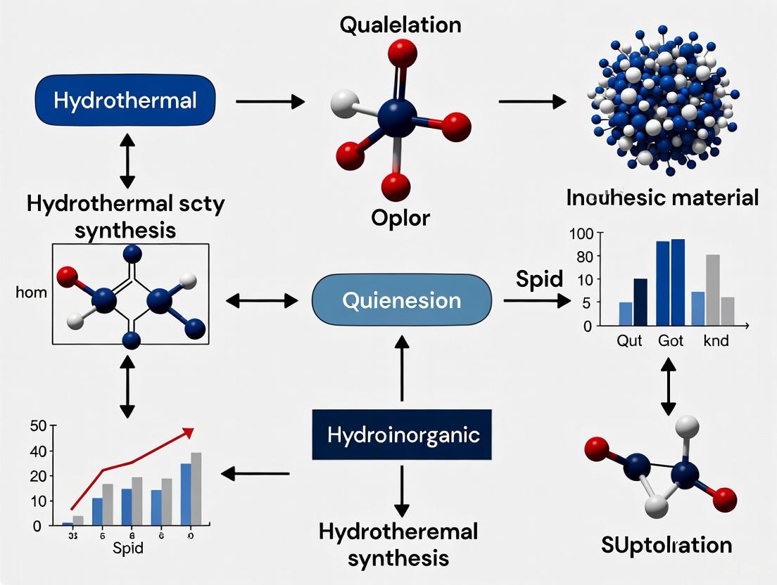
Hydrothermal synthesis is a cornerstone bottom-up methodology for preparing inorganic functional materials and novel compounds through chemical reactions in aqueous media under elevated temperature and pressure. This process occurs in a confined system, typically using high-temperature (200–600 °C) and high-pressure (5–40 MPa) conditions to facilitate reactions in a liquid or supercritical water environment. The defining characteristic of this method is the avoidance of a phase change to steam, which circumvents large enthalpic energy penalties associated with water vaporization, leading to more energetically efficient processing [1]. This technique leverages the unique properties of water near its critical point (374°C, 22.1 MPa), where its dielectric constant, density, and ion dissociation constant change dramatically, creating a superior medium for chemical reactions and crystallization processes that are difficult or impossible under standard conditions [2].
The methodology represents a powerful approach for constructing complex inorganic architectures from molecular precursors, enabling precise control over crystal phase, particle morphology, size distribution, and surface characteristics. The fundamental principle involves dissolving precursor materials in water and subjecting them to controlled temperature and pressure profiles within specially designed reactors (autoclaves). Under these hydrothermal conditions, the precursors undergo nucleation and growth processes, ultimately forming the desired crystalline products. The ability to manipulate reaction parameters and chemical environment makes hydrothermal synthesis exceptionally versatile for producing diverse inorganic materials with tailored properties for advanced applications [3].
Key Applications in Materials Science
Hydrothermal and solvothermal synthesis methods have been extensively applied to create a diverse spectrum of inorganic materials, as summarized in Table 1 below.
Table 1: Applications of Hydrothermal Synthesis in Materials Preparation
| Material Category | Specific Examples | Key Characteristics/Applications |
|---|---|---|
| Metal Oxides | CeOâ‚‚, AlO(OH) (Boehmite) | Nanoparticle formation at supercritical conditions [2] |
| Fluoride Materials | β-NaYF₄ | Host for lanthanide upconversion; laser refrigeration [4] |
| Perovskite-type Compounds | Not specified (multiple) | Functional electronic and magnetic properties [3] |
| Metallic/Non-metallic Compounds | Various | Tailored compositions and structures [3] |
| Advanced Ceramics | Multiple systems | Structural and functional applications [3] |
A particularly sophisticated application involves the synthesis of hexagonal β-phase sodium yttrium fluoride (β-NaYF₄), a leading host material for lanthanide upconversion and anti-Stokes fluorescence laser refrigeration due to its low phonon energies and high upconversion efficiency [4]. Recent research has focused on developing ultra-high aspect ratio β-NaYF₄ disks for specialized applications including optically-levitated sensors for high-frequency gravitational wave detection. Through advanced hydrothermal approaches using methyliminodiacetic acid (MIDA) as a ligand, researchers have achieved hexagonal β-NaYF₄ prisms with corner-to-corner diameters up to 44 µm while maintaining heights around 1 µm, resulting in aspect ratios of approximately 44 [4]. These materials demonstrate exceptional potential for integrated optoelectronic devices, with measured laser refrigeration of up to -4.9±1.0 K in ytterbium-doped disks [4].
Quantitative Data and Process Parameters
The effectiveness of hydrothermal synthesis depends critically on precise control of reaction parameters, which govern the resulting material characteristics. Table 2 summarizes key quantitative data from representative processes.
Table 2: Quantitative Data for Hydrothermal Synthesis Processes
| Process Parameter | Typical Range | Specific Example/Impact |
|---|---|---|
| Temperature | 200–600°C [1] | 523–673 K for CeO₂ and AlO(OH) synthesis [2] |
| Pressure | 5–40 MPa [1] | 30 MPa for metal oxide nanoparticle synthesis [2] |
| Particle Size | Nanoscale to microns | β-NaYF₄ disks: 44 µm diameter [4] |
| Aspect Ratio | Varies with morphology | β-NaYF₄ disks: ~44 [4] |
| Cooling Performance | Material-dependent | Yb-doped β-NaYF₄: -4.9±1.0 K [4] |
| Reaction Rate Change | Supercritical conditions | Increases above critical point [2] |
The synthesis of metal oxide nanoparticles at supercritical conditions demonstrates distinctive kinetic and thermodynamic behavior. Research shows that the Arrhenius plot of the first-order rate constant for Ce(NO₃)₃ and Al(NO₃)₃ hydrolysis follows a straight line in the subcritical region but deviates to higher values above the critical point, indicating enhanced reaction rates under supercritical conditions [2]. Simultaneously, the solubility of metal oxides like Ce(OH)₃ and AlO(OH) gradually decreases with increasing temperature in acidic conditions, then drastically drops above the critical point due to changes in water's dielectric constant. This combination of fast reaction kinetics and low solubility at supercritical conditions creates an ideal environment for rapid nucleation and suppressed crystal growth, facilitating the formation of nanoparticles with controlled size distributions [2].
Experimental Protocols
Protocol: Hydrothermal Synthesis of Ultra-High Aspect Ratio β-NaYF₄ Disks
This protocol describes the synthesis of hexagonal β-NaYF₄ disks using methyliminodiacetic acid (MIDA) as a morphology-directing agent, adapted from recent research [4].
Research Reagent Solutions:
- Yttrium Precursor: Yttrium chloride hexahydrate (YCl₃·6H₂O), ≥99.9%
- Fluoride Source: Sodium fluoride (NaF), analytical grade
- Structure-Directing Agent: Methyliminodiacetic acid (MIDA), 98%
- pH Modulator: Sodium hydroxide (NaOH) solution, 1M and concentrated
- Dopant Source: Ytterbium chloride hexahydrate (YbCl₃·6H₂O) for upconversion properties
- Solvent: Deionized water, 18.2 MΩ·cm resistivity
Procedure:
- Precursor Preparation: Dissolve YCl₃·6H₂O (1.0 mmol) and YbCl₃·6H₂O (0.1 mmol for 10% doping) in 15 mL deionized water in a beaker under magnetic stirring.
- Ligand Addition: Add MIDA (2.0 mmol) to the solution and stir until completely dissolved.
- pH Adjustment: Slowly add NaOH solution (2.00 equivalents relative to MIDA) to adjust the pH to the optimal range for hexagonal disk formation (approximately pH >6.12, above the MIDA inflection point).
- Fluoride Addition: Prepare a separate solution of NaF (4.0 mmol) in 5 mL deionized water, then add dropwise to the reaction mixture with vigorous stirring.
- Reactor Loading: Transfer the final mixture to a Teflon-lined stainless steel autoclave, filling to 70-80% of capacity.
- Hydrothermal Treatment: Seal the autoclave and heat at 180-200°C for 12-24 hours in a laboratory oven.
- Product Recovery: After natural cooling to room temperature, collect the precipitate by centrifugation.
- Purification: Wash the product sequentially with deionized water and ethanol 3 times each to remove impurities.
- Separation: Separate the desired β-NaYF₄ microparticles from any α-NaYF₄ nanoparticle byproducts via supernatant removal from ethanol dispersions, leveraging the mass difference.
- Characterization: Analyze the product using scanning electron microscopy (SEM), X-ray diffraction (XRD), and atomic force microscopy (AFM) to confirm morphology, crystal structure, and surface quality.
Critical Parameters:
- The NaOH:MIDA ratio is crucial for phase purity, with 2.00 equivalents consistently yielding pure β-phase [4].
- Maintaining pH above the MIDA inflection point (pH 6.12) is essential for hexagonal disk morphology rather than rods or semicircular disks.
- Reaction temperature and time control the final particle size and crystallinity.
The Scientist's Toolkit: Essential Research Reagents
Table 3: Essential Research Reagents for Hydrothermal Synthesis
| Reagent/Solution | Function/Purpose | Application Example |
|---|---|---|
| Metal Salts (e.g., Ce(NO₃)₃, Al(NO₃)₃, YCl₃) | Provide metal cation precursors for oxide/compound formation | CeO₂ and AlO(OH) nanoparticle synthesis [2] |
| Structure-Directing Agents (e.g., MIDA, EDTA, Citrate) | Control particle morphology, size, and crystal phase through chelation and surface adsorption | β-NaYF₄ disk morphology control [4] |
| Mineralizers (e.g., NaOH, KOH, HF) | Adjust pH, enhance precursor solubility, and modify reaction kinetics | pH control in MIDA-assisted synthesis [4] |
| Supercritical Water | Reaction medium with tunable properties (dielectric constant, density) | Metal oxide nanoparticle formation [2] |
| 2-Bromo-1,4-dioxane | 2-Bromo-1,4-dioxane, CAS:179690-41-6, MF:C4H7BrO2, MW:167.00 g/mol | Chemical Reagent |
| 4-Propyl-1-indanone | 4-Propyl-1-indanone|C12H14O|Research Chemical | Buy 4-Propyl-1-indanone (C12H14O), a key synthetic intermediate for medicinal chemistry research. This product is For Research Use Only. Not for human or veterinary use. |
Mechanisms and Scientific Principles
The formation of nanoparticles under hydrothermal conditions follows distinct mechanisms that vary between subcritical and supercritical regimes, as illustrated in the diagram below.
The mechanistic pathway illustrates two distinct regimes in hydrothermal synthesis. In subcritical conditions, higher solubility of metal oxides facilitates both nucleation and growth processes, with Ostwald ripening (the dissolution of smaller particles and redeposition on larger particles) leading to larger crystal formations [2]. In contrast, under supercritical conditions (>374°C, >22.1 MPa), a drastic drop in solubility combined with an increased reaction rate creates an environment conducive to rapid nucleation but suppressed crystal growth, resulting in nanoparticle formation [2]. This phenomenon is largely driven by the sharp decrease in water's dielectric constant around the critical point, which reduces the solubility of ionic species and accelerates reaction kinetics. The combination of low solubility preventing Ostwald ripening and fast nucleation rates enables the consistent production of nanoscale materials with controlled size distributions.
The role of organic additives like MIDA and EDTA further enhances morphological control through two complementary functions: as chelating agents that regulate the availability of metal cations in solution, and as surface adsorbents that alter nucleation and growth thermodynamics and kinetics by preferentially binding to specific crystal facets [4]. In the case of β-NaYF₄ disk formation, the fundamental innovation involves increasing surface coverage of ligands during growth by substituting one EDTA molecule with two MIDA molecules, creating higher ligand density on growing crystal surfaces and preferentially inhibiting growth along specific axes to promote high aspect ratio morphologies [4].
Within the broader context of a thesis on the hydrothermal synthesis of inorganic materials, mastering the key process parameters is fundamental to designing materials with tailored properties. Hydrothermal synthesis is a versatile liquid-phase preparation method that involves the use of aqueous solutions under elevated temperatures and pressures in a sealed reaction vessel to crystallize materials directly from solution [5] [6]. This method is prized for its ability to produce high-quality, crystalline powders, including complex oxides, nanostructures, and hybrid materials, often with phases and morphologies unattainable through conventional solid-state routes [7]. The significant advantages of hydrothermal synthesis include high reactivity of reactants, formation of metastable phases, lower air pollution, and low energy consumption [7]. The precise control of temperature, pressure, reaction time, and pH is critical because these parameters collectively govern the reaction kinetics, solubility of precursors, nucleation rates, and ultimate crystal growth, thereby dictating the phase purity, morphology, size, and functional properties of the final product [8] [5].
Systematic Analysis of Key Process Parameters
The following sections provide a detailed analysis of each critical parameter, supported by quantitative data and specific examples from recent research.
Temperature
Temperature is one of the most influential parameters in hydrothermal synthesis. It directly affects the reaction rate, the solubility of precursors, and the crystallinity of the final product.
- Reaction Kinetics and Crystallinity: According to the Arrhenius equation, the reaction rate constant has an exponential relationship with temperature [5]. Higher temperatures generally lead to faster reaction rates and improved product crystallinity. For instance, in the synthesis of
Gd(OH)3:Eunanowires, no nanowire formation occurs below 120 °C. Nanorods are obtained between 120–160 °C, while well-developed nanowires longer than 10 micrometers require temperatures above 160 °C [9]. - Morphology Control: Temperature can also induce dramatic changes in particle morphology. During the synthesis of nanostructured hydroxyapatite (HA), treatment at 180 °C resulted in needle-like particles with dimensions similar to human bone (10-20 nm in diameter, below 100 nm in length), whereas lower temperatures yielded different morphologies [10].
- Solvent Properties: It is crucial to note that temperature simultaneously alters the properties of the solvent, water. As temperature increases, the ionic product of water increases, promoting hydrolysis and ion reaction rates. Conversely, the viscosity and dielectric constant of water decrease, enhancing the mobility of ions and molecules in the solution [5].
Table 1: Effect of Temperature on Hydrothermal Synthesis Outcomes
| Material | Temperature Range | Observed Effect | Reference |
|---|---|---|---|
Gd(OH)3:Eu Nanowires |
< 120 °C | No nanowire formation | [9] |
| 120 - 160 °C | Nanorods with high aspect ratios | [9] | |
| > 160 °C | Long nanowires (> 10 µm) | [9] | |
| Nanostructured Hydroxyapatite (HA) | 180 °C | Needle-like shape (10-20 nm diameter) | [10] |
LiFePO4 Cathode Material |
170 °C | Successful crystallization | [11] |
Pressure
Pressure, often an intrinsic function of temperature and solvent fill in a sealed autoclave, influences the density of the solvent and reaction kinetics, thereby affecting product morphology and reaction rate [8].
- Reaction Kinetics and Solvent Density: Higher pressures can enhance the reaction rate and alter the product morphology. Pressure increases the density of water, which in turn affects properties like viscosity, dielectric constant, and solubility. An increase in density generally leads to increased solubility and a decreased diffusion coefficient [5].
- Complex Interaction with Temperature: In practice, pressure and temperature are intrinsically linked in a closed hydrothermal system. The pressure developed is a consequence of the temperature and the degree of filling of the autoclave. This makes it challenging to deconvolute their individual effects, and they are often studied in tandem.
Reaction Time
Reaction time determines the extent of the reaction and the degree of crystal growth, influencing particle size, crystallinity, and sometimes phase composition.
- Crystallization and Particle Growth: Longer reaction times generally lead to more complete reactions, increased crystallinity, and larger particle sizes due to Ostwald ripening [8]. A landmark study on the hydrothermal synthesis of
VS2demonstrated that phase-pure hierarchical nanosheets could be achieved in a reaction time of just 5 hours, a significant reduction from the conventional 20 hours, while maintaining structural integrity [12]. - Optimization is Crucial: The optimal reaction time is material-dependent and must be determined empirically. Insufficient time may lead to incomplete crystallization, while excessive time may promote particle agglomeration or phase transformations.
Table 2: Effect of Reaction Time and pH on Hydrothermal Synthesis Outcomes
| Parameter & Material | Range/Value | Observed Effect | Reference |
|---|---|---|---|
| Reaction Time | |||
VS2 Nanosheets |
5 hours | Phase purity achieved, reduced from 20 h | [12] |
ZrO2 Crystallization |
24 hours | Formation of tetragonal/monoclinic phases | [5] |
| pH | |||
Gd(OH)3:Eu |
~6 | Plate-type morphology | [9] |
| ~11 | Nanorods to nanowires; strong (110) orientation | [9] | |
| ~14 | Nanospheres and nanotubes | [9] | |
ZrO2 with Hâ‚‚O |
Neutral | 15 nm tetragonal, 17 nm monoclinic | [5] |
pH
The pH of the reaction medium is a powerful tool for exerting morphological control over the growing crystals, as it directly affects the charge state of precursors, complex formation, and the supersaturation ratio.
- Morphological Control: A classic example is the shape-selective synthesis of
Gd(OH)3:Eunanoparticles. By varying the initial pH from about 6 to 14, the morphology can be tuned from plate-type structures and nanorods with different aspect ratios to nanowires, nanospheres, and nanotubes [9]. At a pH of approximately 11, a strong preference for growth along the (110) planes is observed, leading to nanowire formation [9]. - Crystal Phase and Size: pH can also influence the crystalline phase and particle size. As shown in Table 2, the crystallization of
ZrO2in pure water (neutral pH) yields a mixture of tetragonal and monoclinic phases with specific particle sizes, whereas the use of acidic or basic mineralizers can lead to different outcomes [5].
The Scientist's Toolkit: Research Reagent Solutions
The following table lists key reagents and their functions in a typical hydrothermal synthesis protocol.
Table 3: Essential Research Reagents and Materials for Hydrothermal Synthesis
| Reagent/Material | Function | Example |
|---|---|---|
| Precursor Salts | Source of metal cations for the target material. | Vanadyl sulfate for Vâ´âº in VS2 [12]; Cadmium chloride for Cd²⺠in CdS [11]. |
| Mineralizer | Increases solubility of precursors, complexes with ions, accelerates nucleation, influences morphology. | KF, NaOH, LiCl for ZrO2 [5]; KOH for Gd(OH)3:Eu [9]. |
| pH Modulator | Adjusts the pH of the reaction medium to control precursor charge and crystal morphology. | HNO₃, KOH [9]. |
| Structure-Directing Agent (SDA) | Guides the formation of specific porous structures or morphologies. | Surfactants for zeolite synthesis [6]. |
| Solvent | Medium for dissolution, transport, and reaction of precursors; pressure transmission medium. | Deionized water [5]. |
| Reducing Agent | Reduces metal precursors to a lower oxidation state. | Not specified in results. |
| Dopant Precursor | Introduces a specific element into the host lattice to modify properties. | Eu2O3 for doping Gd2O3 [9]. |
| DBtPF | DBtPF, MF:C26H54FeP2, MW:484.5 g/mol | Chemical Reagent |
| Stilben-4-ol | Stilben-4-ol|trans-4-Hydroxystilbene|6554-98-9 | High-purity Stilben-4-ol (trans-4-Hydroxystilbene), a key stilbene derivative for pharmaceutical and biochemical research. For Research Use Only. Not for human use. |
Experimental Protocols
This protocol outlines the optimized synthesis of metallic VS2 nanosheets on a 3D substrate, significantly reducing the conventional reaction time.
- Objective: To synthesize phase-pure hierarchical
VS2nanosheets with controlled morphology for energy storage applications. - Materials:
- Precursors: Ammonium metavanadate (
NH4VO3) and thioacetamide (TAA). - Mineralizer/pH Modulator: Ammonia solution.
- Substrate: Three-dimensional conductive substrate (e.g., carbon cloth).
- Precursors: Ammonium metavanadate (
- Procedure:
- Precursor Preparation: Dissolve stoichiometric molar ratios of
NH4VO3and TAA in an aqueous ammonia solution. The concentration of ammonia is a critical variable. - Reaction Setup: Transfer the solution to a Teflon-lined stainless-steel autoclave. Immerse the 3D substrate in the solution.
- Hydrothermal Reaction: Seal the autoclave and heat it to the optimized reaction temperature (specific value not provided in abstract, but temperature is a key variable). Maintain this temperature for 5 hours.
- Product Recovery: After the reaction, allow the autoclave to cool to room temperature naturally. Remove the substrate, now coated with
VS2nanosheets. Rinse thoroughly with deionized water and ethanol, then dry in a vacuum oven.
- Precursor Preparation: Dissolve stoichiometric molar ratios of
- Key Parameter Insights:
- Molar Ratios: The
NH4VO3: TAA ratio is critical for phase purity. - Temperature & Time: The combination of optimized temperature and a 5-hour reaction time is sufficient for crystallization, replacing longer traditional syntheses.
- Ammonia Concentration: Acts as both a mineralizer and a pH controller, complexing with vanadium and influencing growth.
- Molar Ratios: The
This protocol demonstrates precise morphological control of a nanophosphor by varying a single parameter, the pH.
- Objective: To synthesize
Gd2O3:Eunanophosphors with various morphologies (nanorods, nanowires, nanospheres) for photoluminescence applications. - Materials:
- Precursors:
Gd2O3,Eu2O3. - Solvent/Mineralizer: Nitric acid (
HNO3), Potassium hydroxide (KOH).
- Precursors:
- Procedure:
- Precursor Solution: Dissolve stoichiometric amounts of
Gd2O3andEu2O3in a diluteHNO3solution under stirring until a clear solution is formed. - pH Adjustment: Slowly add an aqueous
KOHsolution to the clear nitrate solution under vigorous stirring until the pH reaches a target value between 6 and 14. This will form a colloidal precipitate.- For nanorods/nanowires: Adjust pH to ~11.
- For nanospheres/nanotubes: Adjust pH to ~14.
- Hydrothermal Reaction: Transfer the colloidal mixture to a Teflon-lined autoclave. Seal and maintain at a temperature between 120 °C and 180 °C for several hours (e.g., 2-24 h). Stirring during the reaction is optional but can improve uniformity.
- Product Recovery: After cooling, collect the solid product
Gd(OH)3:Euby filtration, wash with copious amounts of deionized water, and dry. - Calcination: To convert the hydroxide to the oxide (
Gd2O3:Eu), heat the powder in a furnace at 500 °C for several hours using a slow heating rate (< 3 °C/min) to preserve the nanostructured morphology.
- Precursor Solution: Dissolve stoichiometric amounts of
- Key Parameter Insights:
- pH: This is the primary variable for shape control, directing the crystal growth along specific facets.
- Temperature: Higher temperatures (>160 °C) are required to achieve high-aspect-ratio nanowires at pH ~11.
- Precursor Concentration: Higher concentrations favor the growth of uniform, long nanowires.
Visualization of Parameter Interactions and Mechanisms
The following diagrams illustrate the interconnected nature of hydrothermal parameters and the role of mineralizers.
Diagram 1: Interplay of key hydrothermal parameters and their collective impact on final material properties. Parameters like Temperature and Pressure directly influence fundamental reaction drivers (Kinetics, Solubility), which in turn determine critical outcomes such as Crystallinity and Morphology.
Diagram 2: The functional mechanism of a mineralizer. The mineralizer enhances the dissolution of the poorly soluble metal source, potentially forms complexes with the dissolved ions, and facilitates their transport, leading to controlled nucleation and growth of the final crystalline powder.
In the hydrothermal synthesis of inorganic materials, the transformation from dissolved precursors into functional crystalline solids is governed by a sequence of critical stages: dissolution, nucleation, and crystal growth. Understanding these fundamental reaction mechanisms is paramount for researchers and scientists aiming to design nanomaterials with precise control over their phase, morphology, and size distribution. This process, which occurs in aqueous media under elevated temperature and pressure, leverages the enhanced solubility, reactivity, and mass transfer conditions to facilitate the crystallization of materials that may be unstable through conventional synthetic routes [13] [14]. Within the sealed environment of an autoclave, the dissolution of precursor materials creates a supersaturated solution, providing the thermodynamic driving force for the subsequent nucleation of crystalline embryos and their development into mature crystals [15]. This application note delineates the core mechanisms underpinning hydrothermal crystallization and provides detailed protocols for the kinetic study and synthesis of representative inorganic nanomaterials, specifically TiOâ‚‚ nanoparticles.
Core Mechanisms and Theoretical Framework
The hydrothermal crystallization pathway can be conceptually divided into three interconnected stages. The specific conditions of each stage—including temperature, pressure, precursor concentration, and the presence of mineralizers—exert profound influence on the characteristics of the final crystalline product.
The Three-Stage Reaction Mechanism
1. Dissolution The initial stage involves the dissolution of solid precursor materials in the hydrothermal solvent, typically water. Under high-temperature and high-pressure conditions, the physical and chemical properties of water are markedly altered; its dielectric constant is significantly reduced, which generally enhances the solubility of ionic and polar mineral precursors [15] [13]. This creates a homogeneous reaction medium where metal cations and ligand anions become available for reaction.
2. Nucleation Following dissolution and the achievement of a supersaturated state, nucleation occurs. This is the process where solute molecules in the supersaturated solution organize into tiny, stable clusters (nuclei) that are capable of further growth. A critical concept in hydrothermal synthesis is the role of temperature gradients within the autoclave. As outlined in the response to Jalouli et al., the solute typically dissolves in the hotter region of the vessel and is then transported to the cooler region, where supersaturation is highest, triggering nucleation [15]. It is crucial to distinguish between the low-temperature region (a spatial concept within the reactor) and low temperature in an absolute sense. Furthermore, the viscosity of the solution decreases under hydrothermal conditions, intensifying ion migration and convection, which offsets the solubility reduction from the lowered dielectric constant and enhances nucleation rates [15].
3. Crystal Growth In the final stage, the newly formed nuclei grow into larger crystals via the addition of atoms, ions, or molecules from the solution. The growth is influenced by diffusion processes and the intrinsic crystal structure of the nucleating phase. Kinetic studies of hydrothermal TiOâ‚‚ synthesis have indicated that a diffusion-controlled process is a primary mechanism for crystal growth [16]. The enhanced thermal diffusion coefficient of the solution under these conditions provides a greater convective driving force, which is highly beneficial for uniform crystal growth [15].
Quantitative Kinetic Analysis
The kinetics of hydrothermal crystallization can be quantitatively analyzed using models such as the Avrami-Erofe'ev model. This approach is powerful for probing the mechanism and rate of crystallization. An in situ energy-dispersive X-ray diffraction (EDXRD) study on the hydrothermal crystallization of TiOâ‚‚ from nitric acid-peptized sol-gels provided exemplary kinetic data across different temperatures [16].
Table 1: Kinetic Parameters for Hydrothermal Crystallization of TiOâ‚‚ (Rutile Phase)
| Reaction Temperature (°C) | Induction Time (min) | Maximum Pressure (bar) | Avrami Exponent (n) | Interpreted Mechanism |
|---|---|---|---|---|
| 210 | 34 | 20 | ~0.5 - 1 | Diffusion-controlled process |
| 230 | 33 | 34 | ~0.5 - 1 | Diffusion-controlled process |
| 250 | 31 | 40 | ~0.5 - 1 | Diffusion-controlled process |
| 270 | 30 | 42 | ~0.5 - 1 | Diffusion-controlled process |
The data in Table 1 reveals that the induction time for the emergence of the first crystalline diffraction peaks decreases slightly with increasing temperature, while the autogenous pressure inside the reactor increases. The calculated Avrami exponent values consistently fell between 0.5 and 1 across all temperatures studied, which is a strong indicator of a diffusion-controlled reaction mechanism for the crystallization process [16].
Experimental Protocol: Hydrothermal Synthesis andIn SituKinetic Study of TiOâ‚‚ Nanoparticles
This protocol details the synthesis of TiOâ‚‚ nanoparticles via hydrothermal treatment of a nitric acid-peptized sol-gel and the methodology for an in situ EDXRD kinetic study, as adapted from Rehan et al. [16].
Materials and Equipment
Table 2: Research Reagent Solutions and Essential Materials
| Item Name | Specification / Purity | Function / Role in Experiment |
|---|---|---|
| Titanium Butoxide (Ti(OBu)₄) | Precursor, e.g., ≥97% | Metal precursor serving as the source of titanium. |
| 2-Propanol ((CH₃)₂CHOH) | Anhydrous, 99.5% | Solvent for the titanium precursor. |
| Nitric Acid (HNO₃) | 70%, ACS reagent | Peptizing agent for the formation of a stable sol-gel. |
| Hydrothermal Autoclave | PTFE-lined, thin-walled (e.g., Inconel) | Reaction vessel capable of withstanding high temperature and pressure; thin walls facilitate X-ray penetration for in situ study. |
| Synchrotron Radiation Source | EDXRD setup (e.g., Station 16.4, Daresbury Laboratory SRS) | In situ monitoring tool for collecting time-resolved diffraction data during the reaction. |
Step-by-Step Procedure
Part A: Preparation of HNO₃-Peptized TiO₂ Sol-Gel
- Precursor Solution: Prepare a 0.5 M solution of titanium butoxide in 2-propanol.
- Hydrolysis: Under continuous stirring, add the titanium butoxide solution dropwise to distilled water at a volume ratio of 1:4 (precursor solution to water). Stir the mixture for approximately 1 hour. A white precipitate of hydrolyzed TiOâ‚‚ will form.
- Filtration: Filter the suspension to collect the white precipitate.
- Peptization: To a mixture of 31 g of the collected TiOâ‚‚ precipitate and 90 g of distilled water, add 2.7 mL of 70% nitric acid. Stir the resulting mixture for about 45 minutes until a clear, pale yellow sol-gel is obtained.
Part B: Hydrothermal Synthesis and In Situ EDXRD Data Collection
- Reactor Loading: Transfer 20 mL of the prepared sol-gel into a 30 mL, thin-walled PTFE-lined pressure vessel suitable for in situ EDXRD. Place a PTFE-coated magnetic stirrer inside.
- Experimental Setup: Secure the vessel in the synchrotron EDXRD setup, ensuring the X-ray beam path is aligned and the stirring mechanism is functional.
- Initiation of Reaction: Heat the vessel to the desired reaction temperature (e.g., 210, 230, 250, or 270 °C) at a controlled heating rate of approximately 10 °C minâ»Â¹.
- Data Acquisition: Begin collecting EDXRD spectra simultaneously using solid-state detectors at fixed angles (e.g., 2θ = 7.375°, 4.51°, and 1.61°). Collect spectra at regular intervals (e.g., every 60 seconds) for the duration of the reaction.
- Reaction Completion: After the desired reaction time, allow the autoclave to cool to room temperature.
- Product Recovery: Collect the solid product via centrifugation, wash several times with distilled water and/or ethanol, and dry the resulting TiOâ‚‚ nanoparticles.
Data Analysis and Kinetic Modeling
- Phase Identification: Convert the raw EDXRD data from energy scale to d-spacing using the modified Bragg's law:
E = 6.199 / (d sinθ). Identify the crystalline phases present by matching the observed d-spacings to reference patterns (e.g., JCPDS cards). - Kinetic Profiling: Select a characteristic diffraction peak (e.g., the (110) peak for rutile TiO₂) and monitor the change in its integrated intensity or area over time.
- Model Fitting: Fit the normalized integrated intensity data to the Avrami-Erofe'ev equation to determine the rate constant and the Avrami exponent, which provides insight into the nucleation and growth mechanism.
Application Notes for Drug Development Professionals
For professionals in drug development, controlling the physical form of active pharmaceutical ingredients (APIs) and excipients is critical. While this protocol focuses on inorganic TiOâ‚‚, the principles of hydrothermal synthesis are also applicable to the formation of organic crystals and composite materials.
- Particle Size and Morphology Control: The parameters detailed in Table 1 directly influence the size and shape of the resulting crystals. By fine-tuning temperature, pressure, and heating rate, it is possible to target specific particle size distributions suitable for drug formulation, such as those optimizing dissolution rates or enabling nebulization.
- Enhanced Bioavailability: The capability to produce materials with high crystallinity and controlled polymorphism is invaluable for ensuring the stability and reproducible bioavailability of APIs.
- Green Synthesis: Hydrothermal synthesis utilizes water as the primary solvent, aligning with green chemistry principles by reducing the need for hazardous organic solvents in pharmaceutical manufacturing [17] [13]. This method offers a more environmentally friendly alternative to traditional solvothermal routes.
Within the context of inorganic materials research, the autoclave is a cornerstone piece of equipment for conducting hydrothermal synthesis. This process involves crystallizing substances from hot aqueous solutions at high vapor pressure, enabling the production of advanced materials from transition-metal compounds like oxides, hydroxides, and sulphides [18]. The fundamental principle involves containing a reaction within a sealed vessel that is heated, thereby generating high pressure from the water and steam, which facilitates reactions above the normal boiling point of water [19]. This application note details the designs specific to research-scale hydrothermal synthesis and outlines the critical safety protocols for their operation, providing a framework for researchers and scientists engaged in drug development and materials engineering.
Autoclave Design Specifications for Hydrothermal Synthesis
Hydrothermal synthesis reactors, often called "hydrothermal bombs," are specialized autoclaves designed for crystallizing substances and synthesizing nanomaterials [20] [18]. Unlike large steam-jacketed sterilizing autoclaves, these are typically bench-top vessels designed for small-scale synthesis.
Core Components and Materials
A standard hydrothermal autoclave consists of two primary parts:
- Outer Shell: A robust, pressure-resistant jacket typically machined from stainless steel (grades 304 or 316) for structural integrity and corrosion resistance [20] [21] [22].
- Inner Liner: A removable chamber made from polytetrafluoroethylene (PTFE or Teflon) or, for higher temperature applications, PPL (a polypropylene material) [20] [18]. This liner provides excellent chemical resistance to both highly acidic and alkaline solutions, protecting the steel shell from corrosion [20] [21].
Standard Design Configurations and Specifications
Commercially available hydrothermal reactors come in a range of capacities and share key operational parameters. The following table summarizes the standard specifications for PTFE-lined hydrothermal autoclave reactors.
Table 1: Standard Specifications for PTFE-Lined Hydrothermal Autoclave Reactors
| Specification | Typical Range / Value | Citations |
|---|---|---|
| Available Capacities | 5 mL to 2000 mL | [20] [22] |
| Maximum Operating Temperature | ≤ 240°C | [20] [18] [21] |
| Safe Operating Temperature | 180°C - 200°C | [18] [21] |
| Maximum Working Pressure | ≤ 3 MPa (≈ 30 bar) | [20] [18] [22] |
| Heating/Cooling Rate | ≤ 5°C per minute | [20] [18] [22] |
| Sealing Types | Screw sealing (for capacities up to 500 mL), Flange sealing (for larger capacities) | [20] |
Advanced and Specialized Designs
Research demands have led to more sophisticated autoclave designs. A notable example is a microgravity-compatible autoclave engineered for NASA's SUBSA furnace on the International Space Station. This design addresses unique challenges such as preventing air bubble formation in microgravity and incorporating a "leak-before-burst" failsafe mechanism to mitigate over-pressure risks. The internal surfaces were coated with fluorinated ethylene propylene (FEP) and PTFE to create a chemically inert environment for the synthesis of materials like graphene hydrogel [19].
Safety Considerations and Protocols
Operating vessels at high temperatures and pressures inherently involves risks. Adherence to strict safety protocols is non-negotiable. The hazards can be broadly categorized into those associated with standard sterilization autoclaves and those specific to hydrothermal synthesis reactors.
Universal Autoclave Safety Hazards and Controls
The following table outlines common hazards found in autoclave operations and the necessary controls to mitigate them.
Table 2: Universal Autoclave Safety Hazards and Control Measures
| Hazard Category | Specific Risks | Required Control Measures & Personal Protective Equipment (PPE) |
|---|---|---|
| Heat and Steam Burns | - Steam from opening the door- Hot chamber walls and door- Hot fluids and spillage | - Wear heat-resistant gloves, lab coat, and a rubber apron for liquids [23] [24] [25].- Use face shields with safety glasses for liquids and a splash hazard [23] [24].- Stand behind the door when opening; open slowly to release residual steam [23] [25].- Allow contents to cool inside the chamber for at least 10 minutes before unloading [23] [26]. |
| Explosion and Impact | - Pressure release from seal failure- Boil-over of superheated liquids- Shattering of sealed containers | - Never autoclave liquids in sealed containers; always loosen caps [23] [24] [25].- Use only borosilicate glass (e.g., Pyrex) for liquids [23] [24].- Do not open the door until the cycle is complete and pressure has returned to zero [23] [25].- Inspect door gaskets and seals regularly for wear [26]. |
| Material Incompatibility | - Release of toxic fumes- Corrosion of the autoclave chamber | - Never autoclave flammable, reactive, corrosive, toxic, or radioactive materials [23] [24].- Avoid chlorine, bleach, solvents, and volatile liquids [23] [26].- Use secondary containment made of polypropylene or stainless steel to catch spills [23] [25].- Ensure plastic materials are autoclave-compatible (e.g., Polypropylene #5); do not use polyethylene or polystyrene [23] [24]. |
Specific Safety Protocols for Hydrothermal Synthesis Reactors
In addition to the universal controls above, hydrothermal reactors require specific operational procedures due to their design and application.
- Tightening and Sealing: Ensure the primary and secondary caps are properly tightened according to the manufacturer's instructions to avoid pressure leakage. A torque rod is often provided for this purpose [20] [18].
- Controlled Heating and Cooling: Strictly adhere to the maximum recommended heating and cooling rate of 5°C per minute [20] [18] [22]. Rapid temperature changes can cause thermal shock, potentially damaging the liner or creating unsafe pressure conditions.
- Liner Fill Level: Never operate the autoclave without solvent or overfill the Teflon liner. Sufficient headspace is required to accommodate the expansion of liquids upon heating [18].
- Post-Use Cleaning: Thoroughly clean the PTFE liner after every use to prevent contamination and maintain its chemical resistance [18].
Experimental Protocol: Hydrothermal Synthesis of Inorganic Materials
The following workflow details a standard protocol for the hydrothermal synthesis of inorganic materials, such as nanoparticles or crystals, using a Teflon-lined autoclave.
Detailed Methodology
Materials:
- Hydrothermal synthesis autoclave (e.g., 50 mL capacity, PTFE-lined) [21] [22].
- Precursor chemicals (e.g., metal salts, structure-directing agents).
- Solvent (typically deionized water or other aqueous solutions).
- Oven or furnace capable of precise temperature control.
- Standard laboratory glassware for preparation.
- Personal Protective Equipment (PPE): Heat-resistant gloves, lab coat, closed-toe shoes, and eye protection [23] [24].
Procedure:
- Precursor Preparation: Dissolve the precursor reagents in the solvent to form a homogeneous solution. For example, in the synthesis of LiBaF3 phosphor, stoichiometric ratios of LiF and BaF2 can be used as starting materials [27].
- Loading the Reactor: a. Twist the primary stainless-steel cap counterclockwise to open the autoclave [20] [18]. b. Remove the top SS gasket and carefully lift out the milky white PTFE liner. c. Transfer the precursor solution into the PTFE liner, ensuring it does not exceed the recommended capacity (typically no more than 80% full to allow for expansion). d. Seal the PTFE liner with its cap, ensuring it is airtight to prevent pressure leakage [20].
- Assembling the Autoclave: a. Place the sealed PTFE liner back into the stainless-steel outer jacket. b. Replace the top SS gasket and screw on the primary SS cap, tightening it clockwise until it stops. c. For reactors with a secondary cap, tighten it as well. Use the provided locking rod for final, secure tightening [20] [18].
- Heating Cycle: Place the assembled autoclave in an oven or furnace. Program the oven to heat at a controlled rate of 5°C per minute until the desired process temperature (e.g., 200°C) is reached [20] [18]. Maintain this temperature for the required reaction time (e.g., 3 to 24 hours, depending on the material) [27].
- Cooling Cycle: After the reaction time has elapsed, allow the autoclave to cool naturally inside the switched-off oven. The cooling rate should also be controlled, ideally at 5°C per minute, until it reaches room temperature [20] [18]. Never attempt to force-cool the autoclave by quenching in water.
- Unloading: Once the autoclave is completely cool to the touch, carefully unscrew the caps. Remove the PTFE liner and open it to collect the synthesized product, which may be a suspension or a solid hydrogel [19].
- Product Work-up: Separate the product from the mother liquor via centrifugation or filtration. Wash the product with deionized water and/or ethanol to remove impurities, and dry it in an oven at an appropriate temperature for further characterization [27].
The Scientist's Toolkit: Essential Research Reagent Solutions
The following table lists key reagents and materials commonly used in the hydrothermal synthesis of inorganic materials, along with their primary functions.
Table 3: Essential Reagents and Materials for Hydrothermal Synthesis
| Item | Function/Application | Citation |
|---|---|---|
| Metal Salt Precursors (e.g., LiF, BaFâ‚‚) | Serve as the source of metal cations for constructing the inorganic framework or crystal structure of the target material. | [27] |
| Deionized Water | The most common solvent for creating the high-temperature, high-pressure aqueous environment essential for hydrothermal reactions. | [19] |
| Structure-Directing Agents (Templates) | Organic or inorganic molecules that guide the formation of specific porous structures (e.g., in zeolites or MOFs). | [19] |
| PTFE (Teflon) Liner | The inner vessel of the autoclave; provides excellent chemical resistance to acids and alkalis, preventing corrosion of the steel shell. | [20] [21] |
| Mineralizers (e.g., acids, bases) | Agents that increase the solubility of the precursor materials in the hydrothermal medium, facilitating crystal growth. | [18] |
| Graphene Oxide (GO) Dispersion | A common precursor for the hydrothermal synthesis of graphene-based hydrogels and aerogels. | [19] |
| BOC-ALA-PRO-OH | BOC-ALA-PRO-OH, MF:C13H22N2O5, MW:286.32 g/mol | Chemical Reagent |
| Alpen | Alpen, MF:C16H19N3O4S, MW:349.4 g/mol | Chemical Reagent |
Validation and Efficacy Monitoring
Ensuring that the autoclave process has achieved its intended purpose is critical, particularly for sterilization or consistent material synthesis.
For Sterilization Autoclaves:
- Chemical Indicators: Autoclave tape or integrated indicator strips change color when exposed to sterilization temperatures (e.g., 121°C), providing a basic visual check that the load has been processed [23].
- Biological Indicators: Monthly validation using vials containing spores of Geobacillus stearothermophilus is recommended. The inactivation of these spores after a standard cycle confirms sterilization efficacy [23].
For Hydrothermal Synthesis Reactors:
Process validation is often achieved through material characterization. For instance, a comparative study of LiBaF3 phosphor synthesized with and without an autoclave used X-ray diffraction (XRD) for structural analysis and UV-Vis-NIR spectroscopy and scanning electron microscopy (SEM) to compare optical properties and morphology, thereby validating the impact of the synthesis method [27].
The following diagram summarizes the key safety considerations and their logical relationships when operating an autoclave.
Nanoparticles, defined as particles with at least one dimension between 1 and 100 nanometers, exhibit unique physical, chemical, and optical properties that differ significantly from their bulk counterparts [28] [29]. These distinctive characteristics arise primarily from two fundamental principles: the high surface area-to-volume ratio and quantum confinement effects [30]. As particle size decreases to the nanoscale, a substantial increase in the proportion of surface atoms relative to the total number of atoms occurs, dramatically altering material behavior [29]. This size-dependent behavior is particularly relevant in hydrothermal synthesis of inorganic materials, where precise control over nucleation and growth processes enables tailoring of nanoparticle properties for specific applications in drug delivery, bioimaging, and therapeutic interventions [30] [13].
Fundamental Size-Dependent Properties
Physical Properties
The physical properties of nanoparticles undergo significant transformation as size decreases to nanoscale dimensions. Table 1 summarizes key size-dependent physical properties and their applications.
Table 1: Size-Dependent Physical Properties of Nanoparticles
| Property | Bulk Behavior | Nanoscale Behavior | Critical Size Threshold | Applications |
|---|---|---|---|---|
| Melting Point | High, constant | Dramatically lowered | <50 nm (gold melts at ~300°C vs. 1064°C bulk) [29] | Low-temperature processing, thermal sensors |
| Mechanical Hardness | Malleable/ductile | Superhard materials | <50 nm (copper loses malleability) [29] | Reinforced composites, protective coatings |
| Diffusion & Sintering | High temperature required | Enhanced diffusion at lower temperatures | Size-dependent, increases with surface area [29] | Energy-efficient manufacturing, ceramics |
| Magnetic Behavior | Ferromagnetic | Superparamagnetic | <128 nm (ferrite nanoparticles) [29] | Magnetic data storage, clinical imaging, drug targeting |
The increased surface area to volume ratio in nanoparticles significantly enhances their diffusivity and reduces thermal stability. This property enables sintering processes to occur at substantially lower temperatures compared to bulk materials [29]. In magnetic nanoparticles, size reduction below critical thresholds (e.g., 128 nm for ferrite nanoparticles) induces superparamagnetic behavior, preventing self-agglomeration and enabling applications in clinical imaging and data storage [29].
Chemical Properties
Surface chemistry and reactivity undergo profound changes at the nanoscale. The increased surface energy drives enhanced chemical reactivity, making nanoparticles superior catalysts compared to bulk materials [31]. Zeta potential, a key indicator of colloidal stability, becomes a critical parameter influenced by solution conditions including pH and ionic strength [32] [31]. Nanoparticles with zeta potential values greater than +30 mV or more negative than -30 mV exhibit excellent colloidal stability through electrostatic repulsion [31]. Surface functionalization through ligands, polymers, or surfactants further modulates chemical interactions and stability [32] [29].
Optical Properties
Nanoparticles exhibit unique optical phenomena that are heavily size-dependent. Plasmonic nanoparticles (gold and silver) demonstrate pronounced surface plasmon resonance that varies with size, shape, and local environment [32]. Semiconductor quantum dots exhibit quantum confinement effects, where band gap energy increases with decreasing particle size, enabling precise tuning of fluorescence emission [31]. Table 2 outlines key size-dependent optical properties and their characterization techniques.
Table 2: Size-Dependent Optical Properties and Characterization
| Optical Phenomenon | Size Dependency | Characterization Technique | Applications |
|---|---|---|---|
| Surface Plasmon Resonance | Peak position and intensity sensitive to size, shape, and agglomeration state [32] | UV-Visible Spectroscopy (200-1100 nm) [32] | Biosensing, diagnostic assays, photothermal therapy |
| Quantum Confinement | Bandgap increases with decreasing size (2-10 nm range) [29] | Photoluminescence Spectroscopy, UV-Vis [31] | Bioimaging, LED technology, solar cells |
| Extinction Coefficient | Increases with size; affects light absorption and scattering [32] | UV-Visible Spectroscopy [32] | Photovoltaic cells, therapeutic applications |
| Color Emission | Varies with size and aspect ratio [29] | Dark Field Microscopy, Spectrophotometry [32] | Cellular labeling, multiplexed detection |
Gold and silver nanoparticles exhibit intense colors in solution that depend on their size, aspect ratio, and nanostructure morphology. For instance, 20 nm gold nanoparticles produce a wine-red solution, while 20 nm platinum nanoparticles yield a yellowish-gray solution [29]. These tunable optical properties enable applications in bioimaging, where nanoparticles can be engineered to produce varying color intensities by manipulating nanoshell thickness and aspect ratio [29].
Experimental Protocols for Hydrothermal Synthesis and Characterization
Hydrothermal Synthesis of Metal Oxide Nanoparticles
Hydrothermal synthesis represents a powerful method for producing high-quality inorganic nanoparticles with controlled size and morphology through reactions in aqueous solutions at elevated temperature and pressure [13]. The following protocol details the synthesis of ZnO nanorods as a representative example:
Materials:
- Zinc acetate dehydrate (Zn(CH₃COO)₂·2H₂O)
- Sodium hydroxide (NaOH) or potassium hydroxide (KOH)
- Distilled water and methanol (solvents)
- Teflon-lined stainless steel autoclave (100-200 mL capacity)
- Laboratory oven or water bath
- Centrifuge and centrifugation tubes
Procedure:
- Precursor Preparation: Prepare two separate solutions by dissolving zinc acetate dehydrate (0.1 M) in 20 mL of distilled water:methanol (1:1 v/v) mixture and NaOH/KOH (0.4 M) in 20 mL of the same solvent mixture using ultrasonic agitation for 15 minutes [13].
Reaction Mixture: Combine the two solutions under continuous stirring, which will result in a milky suspension indicating the formation of zinc hydroxide complexes.
Hydrothermal Treatment: Transfer the resultant solution to a Teflon-lined stainless steel autoclave, filling 70-80% of its capacity to maintain appropriate pressure conditions. Seal the autoclave securely and place it in a preheated oven or water bath at 60°C for 21 hours [13].
Product Recovery: After the reaction period, carefully open the autoclave after it cools to room temperature. Collect the white precipitate by centrifugation at 10,000 rpm for 10 minutes.
Purification: Wash the precipitate three times with distilled water and twice with ethanol to remove impurities and unreacted precursors. Dry the final product in an oven at 60°C for 12 hours to obtain ZnO nanorods [13].
Critical Parameters:
- Temperature variations of ±5°C significantly impact nucleation rates and final particle size
- Reaction time controls the aspect ratio of nanorods
- Alkali concentration (mineralizer) influences crystallization kinetics
- Solvent composition affects particle morphology and size distribution
Advanced Hydrothermal Synthesis Workflow
The following diagram illustrates the complete hydrothermal synthesis workflow from precursor preparation to final characterization:
Protocol for Size and Surface Charge Characterization
Accurate characterization of nanoparticle size and surface properties is essential for understanding structure-property relationships. The following integrated protocol employs multiple complementary techniques:
Dynamic Light Scattering (DLS) for Hydrodynamic Size:
- Sample Preparation: Dilute the nanoparticle sample in appropriate buffer (typically 1:100 v/v) to achieve optimal scattering intensity. For biologically relevant data, use PBS (pH 7.4) or cell culture medium matching intended application conditions [31].
- Dispersion: Sonicate the sample using a bath sonicator for 5-10 minutes to ensure complete dispersion and break weak agglomerates.
- Measurement: Transfer the sample to a disposable sizing cuvette and place in the DLS instrument (e.g., Malvern Zetasizer Nano ZS). Set measurement parameters to 25°C, equilibration time of 60 seconds, and minimum 10 runs per measurement [32].
- Data Analysis: Record the Z-average hydrodynamic diameter and polydispersity index (PDI). PDI values <0.2 indicate monodisperse populations, while values >0.5 suggest broad size distributions [31].
Transmission Electron Microscopy (TEM) for Core Size and Morphology:
- Sample Grid Preparation: Deposit 5-10 μL of appropriately diluted nanoparticle suspension onto a carbon-coated copper TEM grid and allow to adhere for 1 minute [32].
- Staining: Carefully wick away excess solution using filter paper. For biological samples, negative staining with 1% uranyl acetate may be required.
- Imaging: Insert the grid into the TEM instrument (e.g., JEOL 1010 at 100 keV accelerating voltage) and acquire images at multiple magnifications (typically 50,000x-150,000x) [32].
- Size Analysis: Use image analysis software (e.g., ImageJ) to measure core diameters of at least 200 particles from multiple images to generate statistically relevant size distribution histograms.
Zeta Potential Measurement:
- Sample Preparation: Prepare nanoparticle suspension in 1 mM KCl or appropriate buffer to maintain consistent ionic strength. Adjust pH as needed to study charge dependence.
- Measurement: Load sample into a clear disposable zeta cell, ensuring no air bubbles are present. Apply voltage (typically 150 V) and measure electrophoretic mobility using laser Doppler electrophoresis [32].
- Data Interpretation: Convert electrophoretic mobility to zeta potential using the Smoluchowski approximation. Report mean zeta potential from at least 3 measurements with standard deviation [31].
Characterization Techniques and Data Interpretation
Integrated Characterization Workflow
The comprehensive characterization of nanoparticle properties requires an integrated approach combining multiple analytical techniques as illustrated below:
Comparative Analysis of Characterization Techniques
Table 3 provides a comprehensive comparison of major nanoparticle characterization techniques, their applications, and limitations:
Table 3: Nanoparticle Characterization Techniques: Capabilities and Limitations
| Technique | Size Range | Information Obtained | Limitations | Sample Requirements |
|---|---|---|---|---|
| Dynamic Light Scattering (DLS) | 1 nm - 5 μm [33] | Hydrodynamic diameter, size distribution, polydispersity [32] [31] | Poor resolution for multimodal samples; assumes spherical shape [31] | Dilute suspensions (0.1-1 mg/mL); transparent solutions [31] |
| Transmission Electron Microscopy (TEM) | 1 nm - 1 μm [32] | Core size, morphology, size distribution, crystallinity [32] | Sample drying artifacts; limited statistics; expensive equipment [33] [31] | Dry samples on grids; high vacuum compatible; conductive coating may be needed [32] |
| UV-Visible Spectroscopy | 2-100 nm (plasmonic) [32] | Optical properties, concentration, aggregation state, size estimation [32] | Indirect size measurement; requires reference standards | Clear solutions; appropriate concentration for absorbance range 0.1-1 [32] |
| Zeta Potential Analysis | 3 nm - 10 μm | Surface charge, colloidal stability [32] [31] | Sensitive to pH and ionic strength; difficult with polydisperse samples [31] | Dilute suspensions in specific buffers; known conductivity [32] |
| Nanoparticle Tracking Analysis (NTA) | 10-1000 nm [31] | Particle size distribution, concentration, aggregation state [31] | Requires extreme dilution; lower throughput than DLS | Highly diluted samples (10â·-10â¹ particles/mL) [31] |
| Scanning Electron Microscopy (SEM) | 10 nm - 1 μm | Surface topography, size, morphology, elemental composition [33] | Sample charging; requires conductive coatings; vacuum conditions | Solid, dry samples; conductive coating required for non-conductive materials [33] |
Advanced Characterization Techniques
For comprehensive nanoparticle analysis, several advanced techniques provide additional insights:
Small-Angle X-Ray Scattering (SAXS): Probes particle size, shape, and internal structure in the range of several nanometers to hundreds of nanometers without requiring sample dilution or drying [33]. SAXS is particularly valuable for analyzing the long-period structure of nanomaterials and studying spatial correlations in solution.
Inductively Coupled Plasma Mass Spectrometry (ICP-MS): Provides extremely sensitive elemental analysis with detection limits in the parts-per-trillion range [32]. Single-particle ICP-MS mode enables particle size distribution characterization by measuring the intensity of ionic plumes generated from individual nanoparticles [32].
X-Ray Diffraction (XRD): Determines crystal structure, phase composition, and crystallite size through analysis of diffraction patterns. The technique is indispensable for characterizing crystallographic properties of inorganic nanoparticles synthesized via hydrothermal methods [34].
The Scientist's Toolkit: Essential Research Reagents and Materials
Table 4 outlines critical reagents, materials, and equipment required for hydrothermal synthesis and characterization of inorganic nanoparticles:
Table 4: Essential Research Reagents and Materials for Nanoparticle Research
| Category | Item | Specification/Function | Application Notes |
|---|---|---|---|
| Precursor Materials | Metal salts (acetates, chlorides, nitrates) | High purity (>99.9%) sources of target elements | Determine solubility and reactivity in hydrothermal conditions [13] |
| Mineralizers | NaOH, KOH, NHâ‚„OH | pH regulation, hydrolysis control, and solubility enhancement | Critical for controlling nucleation rates and final morphology [13] |
| Solvents | Deionized water, ethanol, methanol | Reaction medium, dispersion solvent | High purity essential to avoid contamination; affects reaction kinetics [13] |
| Surface Modifiers | Silicon derivatives, phosphoric acid, surfactants | Surface stabilization, prevent agglomeration [29] | Improve compatibility with biological systems; enhance colloidal stability |
| Characterization Consumables | TEM grids (carbon-coated) | Sample support for electron microscopy | Ensure proper adhesion of nanoparticles without aggregation [32] |
| Buffer Systems | KCl, PBS, specific pH buffers | Control ionic strength and pH for DLS and zeta potential | Critical for obtaining biologically relevant characterization data [31] |
| Hydrothermal Equipment | Teflon-lined stainless steel autoclaves | Withstand high temperature/pressure conditions | Various sizes (100-2000 mL) for different scale syntheses [13] |
| Characterization Instruments | DLS/Zeta potential analyzer, UV-Vis spectrometer, TEM/SEM | Size, charge, optical properties, and morphology analysis | Multimodal approach essential for comprehensive characterization [32] [31] |
| Diacetylbiopterin | Diacetylbiopterin, CAS:62933-57-7, MF:C13H15N5O5, MW:321.29 g/mol | Chemical Reagent | Bench Chemicals |
| Benocyclidine-d10 | Benocyclidine-d10, MF:C19H25NS, MW:309.5 g/mol | Chemical Reagent | Bench Chemicals |
The size-dependent physical, chemical, and optical properties of nanoparticles represent a fundamental aspect of nanotechnology with profound implications for materials science and biomedical applications. Through precise control of hydrothermal synthesis parameters including temperature, reaction time, precursor concentration, and mineralizer content, researchers can tailor nanoparticle characteristics for specific functions. The integration of multiple characterization techniques—including DLS, TEM, UV-Vis spectroscopy, and zeta potential analysis—provides complementary insights into nanoparticle properties and behavior in relevant environments. As nanotechnology continues to advance, understanding and exploiting these size-dependent relationships will enable the development of increasingly sophisticated materials for drug delivery, diagnostic imaging, and therapeutic interventions with enhanced efficacy and reduced side effects.
Within the broader context of research on the hydrothermal synthesis of inorganic materials, the selection of a synthetic pathway is paramount, fundamentally directing the structural characteristics and ensuing functionality of the resulting material. The paradigm is broadly divided into top-down methods, which involve the physical or chemical breakdown of bulk materials into nanostructures, and bottom-up approaches, which construct materials atom-by-atom or molecule-by-molecule from precursor solutions [35]. Hydrothermal synthesis is a quintessential bottom-up technique, defined as a chemical process for crystallizing substances directly from high-temperature aqueous solutions at high pressures, typically within an autoclave [13] [36]. This method offers distinct and significant advantages over top-down routes, primarily through its unparalleled ability to generate products with superior crystallinity and precise morphological control—features that are often unattainable through comminution or etching-based top-down processes [37] [38]. These advantages are critical for applications ranging from electrocatalysis and energy storage to drug development, where specific crystal phases and nanostructured morphologies directly dictate performance metrics such as catalytic activity, charge storage capacity, and ion diffusion rates [37].
Table 1: Fundamental Comparison of Bottom-Up Hydrothermal and Top-Down Approaches.
| Feature | Bottom-Up Hydrothermal Synthesis | Typical Top-Down Methods |
|---|---|---|
| Fundamental Principle | Constructs materials from molecular precursors in a solution phase [35] | Breaks down bulk materials into nanostructures [35] |
| Typical Crystallinity | High, often single-crystal quality [38] [36] | Often lower, with introduced defects and strain [35] |
| Morphological Control | High; enables complex shapes (nanosheets, rods, spheres) [37] [13] | Limited by the starting bulk material and process [35] |
| Primary Advantages | High crystallinity, complex morphology control, pure phases [36] | Scalability, applicability to a wide range of materials [35] |
| Primary Disadvantages | Requires high-pressure equipment, often batch processing [8] [36] | Surface defects, contamination, limited shape diversity [35] |
The Hydrothermal Advantage: Mechanisms and Manifestations
Achieving Enhanced Crystallinity
The pursuit of high crystallinity is central to materials science, as it minimizes defect sites that can impede electron transport, act as recombination centers in photocatalytic applications, or reduce overall chemical stability. Hydrothermal synthesis excels in this regard by replicating the natural geological conditions under which minerals form over millennia, but on a drastically reduced timescale. The mechanism hinges on the properties of water at elevated temperatures and pressures. As temperature increases towards the critical point (374°C, 22.1 MPa), the dielectric constant of water decreases significantly, reducing the solubility of ionic species and creating a state of high supersaturation that triggers rapid nucleation [38]. Furthermore, the enhanced mobility of molecules and ions in the hydrothermal fluid facilitates their correct integration into the growing crystal lattice, leading to highly ordered structures with minimal imperfections [38] [36]. This environment allows for the direct crystallization of thermodynamically stable phases, often eliminating the need for post-synthesis high-temperature calcination, which can induce particle agglomeration or phase transitions [38]. A key advantage is the method's ability to grow crystalline phases that are not stable at the material's melting point, a significant limitation for many melt-based synthesis routes [36].
Precise Morphological Control
Beyond crystallinity, the ability to dictate a material's morphology—its shape, size, and architecture—is where hydrothermal synthesis truly distinguishes itself from top-down methods. Top-down approaches, such as milling or lithography, are often limited in the nanostructures they can produce and can introduce surface contaminants or damage [35]. In contrast, the bottom-up hydrothermal process offers a powerful and versatile toolkit for morphological engineering. By carefully modulating synthesis parameters, researchers can guide self-assembly processes to yield a diverse array of nanostructures.
The following diagram illustrates the logical relationship between key hydrothermal synthesis parameters and the resulting material properties, demonstrating how precise morphological control is achieved.
For instance, in energy storage applications, vertically aligned NiCo-LDH nanosheet arrays can be grown directly on conductive substrates, providing short ion diffusion paths and high electrical conductivity, enabling excellent cycling stability [37]. In catalysis, flower-like LDHs that incorporate macro- and mesopores can be engineered to facilitate enhanced diffusion of reactants and products, thereby boosting catalytic efficiency [37]. Similarly, the synthesis of complex hierarchical structures like hollow spheres or core-shell nanoparticles is readily achievable through hydrothermal methods, offering functionalities such as large surface areas and the ability to buffer volume changes in battery electrodes [37].
Table 2: Influence of Hydrothermal Synthesis Parameters on Material Properties.
| Synthesis Parameter | Influence on Crystallinity | Influence on Morphology | Exemplary Outcome |
|---|---|---|---|
| Temperature | Higher temperatures generally improve crystallinity and phase stability [8] | Governs reaction kinetics and determines the predominant crystal facet growth [37] | Formation of large, well-faceted single crystals at high T [36] |
| Reaction Duration | Longer times allow for Ostwald ripening, increasing crystal size and perfection [37] | Extended times can lead to overgrowth and transformation between morphologies [37] | Transformation from nanosheets to more thermodynamically stable 3D structures [37] |
| Precursor Concentration | Lower concentrations can favor uniform nucleation and monodisperse crystals [39] | High supersaturation drives nucleation of nanoparticles; lower concentration favors growth [38] | Low precursor concentration suppresses Ostwald ripening for uniform ~10 nm TiOâ‚‚ [39] |
| Solvent & Additives | Minimal direct effect, but organic solvents can modify reaction kinetics | Structure-directing agents (templates) can dictate final nano-architecture (rods, spheres) [37] | Butanol solvent reduces surface tension for smaller, ~7.7 nm TiOâ‚‚ [39] |
Experimental Protocols for Hydrothermal Synthesis
Protocol: Hydrothermal Synthesis of Yttrium-Doped Titanium Dioxide Nanoparticles
This protocol outlines the synthesis of Y-doped TiOâ‚‚ nanoparticles, a material with enhanced photocatalytic activity due to the formation of energy levels within the band gap that reduce charge carrier recombination [40].
3.1.1. Research Reagent Solutions
Table 3: Essential Reagents for Y-Doped TiOâ‚‚ Synthesis.
| Reagent/Solution | Function in Synthesis | Specific Example / Note |
|---|---|---|
| Titanium Precursor | Provides the source of Ti ions for the formation of TiOâ‚‚. | Tetrabutyl titanate or titanium isopropoxide [40] [39] |
| Yttrium Dopant Source | Introduces Y³⺠ions into the TiO₂ lattice to modify electronic structure. | Yttrium nitrate (Y(NO₃)₃) or yttrium chloride (YCl₃) [40] |
| Mineralizer / pH Modifier | Controls the alkalinity or acidity of the solution, influencing hydrolysis and condensation rates. | Sodium hydroxide (NaOH) or potassium hydroxide (KOH) [13] |
| Solvent | The reaction medium for hydrothermal synthesis. | Deionized water, ethanol, butanol, or water-alcohol mixtures [39] |
3.1.2. Step-by-Step Procedure
- Precursor Solution Preparation: Dissolve the titanium precursor (e.g., tetrabutyl titanate) in a suitable solvent, such as ethanol or butanol, under vigorous stirring to form a homogeneous solution. In a separate container, dissolve the yttrium salt in deionized water. The molar ratio of Y:Ti should be calculated based on the desired doping level (e.g., 1-5 at%) [40].
- Mixing and Aging: Slowly add the yttrium salt solution to the titanium precursor solution under continuous stirring. A white precipitate may form. Continue stirring for 30-60 minutes to ensure complete hydrolysis and homogeneous mixing of the precursors.
- Hydrothermal Reaction: Transfer the resulting suspension into a Teflon (PTFE)-lined stainless-steel autoclave, filling it to 70-80% of its total capacity to maintain an appropriate pressure regime. Seal the autoclave securely. Place the autoclave in a preheated oven and maintain a temperature between 150-200°C for a period of 6-24 hours [40].
- Product Recovery and Washing: After the reaction is complete and the autoclave has cooled naturally to room temperature, open it carefully. Collect the resulting precipitate via centrifugation. Wash the precipitate multiple times with deionized water and ethanol to remove any residual ions or organic impurities.
- Post-Synthesis Treatment (Optional): Dry the washed powder in an oven at 60-80°C overnight. To further improve crystallinity and remove residual organics, the powder may be calcined in a muffle furnace at 400-500°C for 1-2 hours in an air atmosphere [40].
The experimental workflow for this protocol is summarized in the following diagram:
Protocol: Supercritical Hydrothermal Synthesis of Nano-Titanium Dioxide
This advanced protocol utilizes water above its critical point (374°C, 22.1 MPa) to achieve extremely rapid nucleation and produce small, highly crystalline, and dispersible nanoparticles [38] [39].
3.2.1. Key Procedure Steps:
- System Setup: Utilize a continuous-flow reactor system designed to withstand supercritical conditions, comprising high-pressure pumps, a preheater, a reactor coil, and a cooling and pressure let-down system.
- Precursor Preparation: Prepare an aqueous solution of the titanium precursor (e.g., tetrabutyl titanate). The concentration is critical; a low concentration around 0.01 mol/L is recommended to suppress Ostwald ripening and optimize particle uniformity [39].
- Supercritical Reaction: Pump the precursor solution through the preheater to rapidly raise its temperature to supercritical conditions (e.g., >374°C, >22.1 MPa). The extremely low dielectric constant of supercritical water causes instantaneous hydrolysis and nucleation, leading to the formation of nanocrystalline particles.
- Quenching and Collection: The fluid is immediately cooled after the reaction zone to quench particle growth. The suspension is then passed through a back-pressure regulator to depressurize, and the nanoparticles are collected.
- Key Insight - Solvent Engineering: Replacing ethanol with butanol as a co-solvent in a 3:7 alcohol-to-water ratio can significantly reduce particle surface tension due to an interfacial dispersion effect, yielding smaller TiOâ‚‚ particles with an average size of 7.70 nm [39].
Advanced Characterization for Validation
The advantages of crystallinity and morphological control conferred by hydrothermal synthesis must be validated through a suite of advanced characterization techniques.
- X-ray Diffraction (XRD): This is the primary tool for assessing crystallinity. It provides information on phase identification, crystal structure, lattice parameters, and crystallite size. The sharpness and intensity of diffraction peaks are direct indicators of high crystallinity [37].
- Electron Microscopy (SEM/TEM): Scanning and Transmission Electron Microscopy offer direct visualization of morphology, particle size, size distribution, and structural details like hollow or core-shell architectures. TEM can further provide insights into crystal structure through selected area electron diffraction (SAED) [37].
- Specific Surface Area and Porosity Analysis (BET): Techniques based on gas adsorption (e.g., Nâ‚‚) are used to determine the specific surface area, pore volume, and pore size distribution of the synthesized materials, which are critical parameters for applications in catalysis and adsorption [37].
- Spectroscopic Techniques: Fourier Transform Infrared (FTIR) Spectroscopy can identify functional groups and surface modifications. Diffuse Reflectance Spectroscopy (DRS) is used to determine the band gap energy of semiconductors, which is crucial for photocatalytic applications [40].
Advanced Hydrothermal Techniques and Their Biomedical Applications
Hydrothermal synthesis is a versatile and powerful wet-chemical technique for producing a wide range of inorganic materials with precise control over their structure and properties. This method utilizes elevated temperatures and pressures in aqueous or solvent-based solutions to facilitate the crystallization of materials that are difficult to obtain under standard conditions. The hydrothermal environment promotes enhanced reactant solubility, increased diffusion rates, and unique reaction pathways, enabling the formation of complex oxides, nanocrystals, and composite materials with tailored characteristics. Within materials research, this technique has become indispensable for synthesizing advanced metal oxides for energy applications, biocompatible hydroxyapatites for medical use, versatile perovskites for optoelectronics, and fluorescent carbon quantum dots for sensing and drug delivery.
The significance of hydrothermal synthesis lies in its ability to produce highly crystalline materials with controlled morphology, size, and composition while often being more environmentally friendly than high-temperature solid-state methods. Researchers can manipulate critical parameters including temperature, pressure, reaction time, pH, and precursor chemistry to engineer materials with specific functionalities. The following sections provide detailed application notes and experimental protocols for synthesizing and characterizing four key material classes, supported by quantitative data comparisons and visual workflow representations to guide research implementation.
Application Notes: Material Properties and Performance
Table 1: Performance Characteristics of Hydrothermally Synthesized Materials
| Material Category | Specific Composition | Key Properties | Performance Metrics | Potential Applications |
|---|---|---|---|---|
| Double Perovskites | Rb₂SnBr₆ [41] | Bandgap: 2.97 eV; Semiconductor; Light-yellow crystals | Large crystal growth (mm- to cm-scale); High stability; Low defect levels | Photocatalysis; Photovoltaics; Optoelectronics |
| Carbon Quantum Dots | N-CQDs from wood [42] | Green fluorescence; Particle size: ~5.02 nm; Water-soluble | High yield: 42% (5.04 g); Quantum yield: ~54%; Stable for >2 months | Fe(III) detection in water (0.1-1000 μmol/L); Composite materials |
| Halide Perovskites | CsPbBr₃/Sr [43] | Cubic phase; Lattice spacing: 0.59 nm (100 plane); Enhanced water stability | Stable in aqueous solution for 264 hours; Blue-shifted photoluminescence | Optoelectronic materials; Devices; Sensing |
| Hydroxyapatite | Caâ‚â‚€(POâ‚„)₆(OH)â‚‚ [44] | Needle-shaped nanoparticles; High crystallinity; High bioactivity | Particle size: 8-39 nm (tunable); Forms apatite layer in SBF | Bone defect repair; Dental/orthopedic implants; Drug delivery |
| Titanate Perovskites | Naâ‚€.â‚…Yâ‚€.₃₉Ybâ‚€.â‚Erâ‚€.â‚€â‚TiO₃ [45] | Orthorhombic phase (Pnma); Uniform faceted particles | Size: 200 nm - 2.0 μm (viscosity-controlled); Dispersity: <10% | Optical temperature sensing; Anti-counterfeiting; Fingerprint recognition |
| Metal Oxide Composites | NaFe₂O₃-GO [46] | Composite structure with graphene oxide | High discharge capacity: ~720 mAh/g | Lithium-ion battery cathodes; Energy storage |
Experimental Protocols
Protocol 1: Hydrothermal Growth of Large Rb₂SnBr₆ Double Perovskite Crystals
Objective: To synthesize millimeter- to centimeter-scale Rb₂SnBr₆ double perovskite crystals for photocatalysis and optoelectronic applications [41].
Materials:
- Rubidium bromide (RbBr, 99%)
- Tin(IV) bromide (SnBrâ‚„, 99%)
- Hydrobromic acid (HBr, 48%)
- Deionized water
Equipment:
- Teflon-lined stainless steel autoclave (25 mL capacity)
- Programmable furnace
- Glove box (for handling hygroscopic SnBrâ‚„)
Procedure:
- Preparative Steps: Conduct all precursor handling in a glove box due to the hygroscopic nature of SnBrâ‚„.
- Precursor Loading: Weigh stoichiometric quantities of RbBr and SnBrâ‚„ into a 25 mL Teflon liner.
- Solution Preparation: Add 2.0 mL of HBr:Hâ‚‚O (1:3 ratio) solution to the mixture in the Teflon liner. The acidic environment is crucial for maintaining reaction stability.
- Reactor Assembly: Seal the Teflon liner within the stainless steel autoclave to prevent pressure loss.
- Hydrothermal Reaction: Place the autoclave in a furnace and program the temperature protocol:
- Ramp to 220°C
- Maintain at 220°C for 24 hours
- Implement a slow cooling rate of 3°C per hour to promote crystal growth
- Product Recovery: After the system cools to room temperature, open the autoclave and collect the light-yellow crystals.
- Characterization: Analyze crystal structure by powder X-ray diffraction (PXRD) and single-crystal X-ray diffraction (SCXRD). Determine band gap (approximately 2.97 eV) using UV-Vis diffuse reflectance spectroscopy with Tauc plot transformation [41].
Protocol 2: Gram-Scale Synthesis of Nitrogen-Doped Carbon Quantum Dots from Wood
Objective: To produce gram quantities of green-emissive nitrogen-doped carbon quantum dots (N-CQDs) from poplar wood for Fe(III) detection in aqueous environments [42].
Materials:
- Poplar wood blocks (20 × 20 × 40 mm)
- Aqueous ammonia solution (25%)
- Hydrogen peroxide (30%)
- Ferric chloride hexahydrate (FeCl₃·6H₂O) for sensing tests
- Deionized water
Equipment:
- Teflon-lined stainless steel autoclave (100 mL capacity)
- Blast drying oven
- Ultrasonic bath
- Vacuum oven
- Fluorescence spectrophotometer
Procedure:
- Wood Pretreatment: Soak poplar wood blocks in deionized water until saturated and submerged (approximately one week).
- Hydrothermal Carbonization:
- Place saturated wood blocks in a 100 mL Teflon-lined autoclave
- Heat in a blast drying oven at 200°C for 10 hours with a ramp rate of 5°C/min
- Allow natural cooling to room temperature
- Carbon Collection: Crush the carbonized wood block and wash with water via agitation and vacuum filtration (repeat three times).
- Nitrogen Doping and Oxidation:
- Mix the filtered solid biocarbon with a solution of deionized water (40%), Hâ‚‚Oâ‚‚ (40%), and ammonia solution (20%) by volume
- Stir under ultraviolet light exposure for 4 hours
- Product Recovery: Dry the N-CQDs solution in a vacuum oven at 95°C for 12 hours to obtain the final powder product.
- Quality Control: Characterize using TEM for morphology (average size ~5.02 nm), XRD for crystallinity, and fluorescence spectroscopy for emission properties (green fluorescence under UV light).
- Fe(III) Sensing Application:
- Prepare N-CQDs solution (0.25 mg/mL) in deionized water with 10 min sonication
- Add Fe(III) solutions across concentration range (0.1-1000 μmol/L)
- Measure fluorescence quenching (Fâ‚€/F) which shows linear correlation with Fe(III) concentration [42]
Protocol 3: Viscosity-Controlled Hydrothermal Synthesis of Multinary Titanate Perovskites
Objective: To synthesize uniform faceted particles of multinary titanate perovskites through viscosity control for optical applications [45].
Materials:
- Titanium chloride (TiClâ‚„, as Tiâ´+ source)
- Rare earth chlorides (YCl₃, YbCl₃, ErCl₃, etc.)
- Sodium hydroxide (NaOH)
- Sodium chloride (NaCl)
- Sodium acetate (NaAc)
- Deionized water
Equipment:
- High-pressure hydrothermal reactors
- Viscometer
- Ice bath with magnetic stirrer
- High-temperature oven
Procedure:
- Precursor Preparation: Prepare aqueous solutions containing 1 mmol Tiâ´+ and 0.5 mmol total rare earth ions (RE³âº) in 10 mL total volume.
- Viscosity Modulation:
- Add NaOH (20-200 mmol) to the precursor solution while stirring in an ice bath
- Alternatively, use combinations of NaOH with NaCl or NaAc to achieve specific viscosity thresholds
- Measure final viscosity (target >100 cP for uniform particles)
- Hydrothermal Reaction:
- Transfer the gelled solution to a hydrothermal reactor
- Heat at 240°C for 1-6 hours (depending on target particle size)
- Product Collection: After cooling, filter the resulting particles and wash with deionized water and ethanol.
- Characterization:
- Analyze crystal structure by XRD (orthorhombic Pnma phase confirmed by Rietveld refinement)
- Examine particle size and morphology by SEM
- Confirm narrow size distribution (dispersity index <10% when viscosity >100 cP) [45]
Table 2: Viscosity Control Parameters for Titanate Perovskite Synthesis
| Additive System | Concentration Range | Resulting Viscosity | Particle Morphology | Size Range |
|---|---|---|---|---|
| NaOH only | 20-200 mmol | 47.9-232.8 cP | Cubes to corner-truncated cubes | 0.78-2.07 μm |
| 6 M NaOH + 5 M NaCl | Fixed ratio | >100 cP | Uniform faceted particles | Tunable with time |
| 4 M NaOH + 5 M NaAc | Fixed ratio | >100 cP | Uniform faceted particles | Tunable with time |
| Critical Threshold | Varies by system | ~100 cP | Transition from polydisperse to monodisperse | 200 nm - 2.0 μm |
Protocol 4: Hydrothermal Synthesis of Hydroxyapatite Nanoparticles with Parameter Optimization
Objective: To synthesize hydroxyapatite (HAp) nanoparticles with controlled size and high crystallinity for biomedical applications using Response Surface Methodology (RSM) optimization [44].
Materials:
- Calcium nitrate tetrahydrate (Ca(NO₃)₂·4H₂O)
- Diammonium hydrogen phosphate ((NHâ‚„)â‚‚HPOâ‚„)
- Ammonia solution (NH₃, for pH adjustment)
- Nitric acid (HNO₃, for pH adjustment)
- Deionized water
- Ethanol
Equipment:
- Teflon vessel (150 mL capacity)
- pH meter
- Centrifuge
- Drying oven
Procedure:
- Solution Preparation:
- Prepare 1 M calcium nitrate tetrahydrate solution
- Prepare 0.67 M diammonium hydrogen phosphate solution
- Precipitation:
- Add the phosphate solution dropwise to the calcium solution under stirring
- Maintain stoichiometric Ca/P ratio of 1.67
- Adjust pH to target value (1-13) using ammonia or nitric acid
- Hydrothermal Treatment:
- Transfer suspension to Teflon vessel
- Treat at varied temperatures (70-190°C) and times (1-13 hours) based on experimental design
- Product Isolation:
- Filter the white suspension after cooling
- Wash three times with deionized water and ethanol (1:1 volume ratio)
- Dry at 60°C for 10 hours
- Bioactivity Assessment:
- Immerse HAp pellets in Simulated Body Fluid (SBF) at 37°C
- Monitor apatite layer formation on surface using SEM/EDX after 7-14 days
- Characterization and Optimization:
- Use RSM with Central Composite Design to model effects of pH, temperature, and time
- Characterize products using FTIR, XRD, TEM, and SEM [44]
Workflow Visualization
Experimental Workflow: This diagram outlines the comprehensive research workflow for hydrothermal synthesis projects, from initial planning to final application testing.
Viscosity Control: This specialized workflow illustrates the critical viscosity control process for synthesizing uniform multinary titanate perovskite particles.
The Scientist's Toolkit: Essential Research Reagents and Materials
Table 3: Key Research Reagents for Hydrothermal Synthesis Experiments
| Reagent/Material | Typical Purity | Primary Function | Example Applications | Handling Considerations |
|---|---|---|---|---|
| Rubidium Bromide (RbBr) | ≥99% | Alkali metal source for perovskite formation | Rb₂SnBr₆ double perovskites [41] | Standard handling; hygroscopic |
| Tin(IV) Bromide (SnBr₄) | ≥99% | Metal cation source for crystal structure | Rb₂SnBr₆ double perovskites [41] | Hygroscopic; handle in glove box |
| Hydrobromic Acid (HBr) | 48% | Provides acidic reaction environment | Maintaining solution acidity [41] | Corrosive; use in fume hood |
| Calcium Nitrate Tetrahydrate | Reagent grade | Calcium source for HAp synthesis | Hydroxyapatite nanoparticles [44] | Standard handling |
| Diammonium Hydrogen Phosphate | Reagent grade | Phosphate source for HAp synthesis | Hydroxyapatite nanoparticles [44] | Standard handling |
| Titanium Chloride (TiCl₄) | Reagent grade | Titanium source for titanate perovskites | Na₀.₅RE₀.₅TiO₃ multinary oxides [45] | Moisture sensitive; corrosive |
| Rare Earth Chlorides | ≥99.9% | Provide rare earth cations for doping | Optical titanate perovskites [45] | Standard handling |
| Sodium Hydroxide (NaOH) | Reagent grade | Mineralizer and viscosity modifier | pH control; gel formation [45] | Corrosive; exothermic dissolution |
| N,N-Dimethylacetamide (DMA) | ≥99.9% | Solvent for aqueous-phase synthesis | CsPbBr₃ perovskite preparation [43] | Standard solvent handling |
| Cesium Trifluoroacetate (Cs-TFA) | ≥98% | Cesium source for perovskite synthesis | CsPbBr₃ quantum dots [43] | Moisture sensitive |
| Oleylamine (OLA) | 80-90% | Surface ligand for nanocrystal control | Quantum dot surface passivation [43] | Air sensitive; store under inert gas |
| 4-Bromobutyric Acid (BBA) | ≥98% | Provides bromine-rich environment | CsPbBr₃ synthesis [43] | Standard handling |
| Wood Biomass | Natural | Carbon source for CQD synthesis | N-CQDs from poplar wood [42] | Grind to appropriate size |
| Hydrogen Peroxide (Hâ‚‚Oâ‚‚) | 30% | Oxidizing agent for carbonization | Wood-derived CQD synthesis [42] | Oxidizer; store properly |
| Aqueous Ammonia | 25% | Nitrogen source for doping | N-CQD synthesis [42] | Volatile; use in well-ventilated area |
| Mpc-meca | Mpc-meca | Immune Signaling Probe | For Research Use | Mpc-meca is a potent research chemical for immunology studies. For Research Use Only. Not for human or veterinary diagnostic or therapeutic use. | Bench Chemicals | |
| MDP-rhodamine | MDP-Rhodamine | Mitochondrial Probe | For Research | MDP-rhodamine is a cell-permeant mitochondrial dye for live-cell imaging & apoptosis research. For Research Use Only. Not for human or veterinary use. | Bench Chemicals |
The hydrothermal synthesis of inorganic materials represents a powerful and versatile approach in advanced materials research. This method enables precise control over the morphology of nanomaterials—such as nanorods, nanotubes, and hierarchical structures—by manipulating synthesis parameters like temperature, pressure, pH, and chemical composition [14]. Such morphological control is paramount because the physical and chemical properties of nanomaterials are intrinsically linked to their shape and architecture [9]. Consequently, achieving precise morphological control unlocks applications across diverse fields, including drug delivery, energy storage, sensing, and catalysis [47] [48] [49].
This protocol provides detailed methodologies for the hydrothermal synthesis of key inorganic nanomaterials with controlled morphologies, specifically focusing on tungsten oxide hierarchical hollow spheres, gadolinium oxide-based nanostructures, and bismuth telluride hierarchical assemblies. The document is structured to offer researchers a practical guide, complete with quantitative data tables, standardized experimental procedures, and visual workflows to ensure reproducibility and deepen the understanding of synthesis mechanisms.
Application Notes: Morphology-Property Relationships
The controlled synthesis of specific nanostructures directly translates to enhanced performance in various applications. The following examples illustrate this critical structure-function relationship.
Tungsten Oxide Hierarchical Hollow Spheres for Gas Sensing: Hierarchical WO₃ hollow spheres, synthesized via a template-free hydrothermal method, exhibit superior gas sensing performance, particularly for NO₂ [48]. The enhancement is attributed to their high surface area and porous structure, which facilitates gas diffusion and surface reactions. The hierarchical organization of nanosheets into a hollow sphere morphology provides abundant active sites while maintaining structural stability.
Gadolinium Oxide-Based Nanophosphors for Photoluminescence: Gadolinium oxide doped with europium (Gd₂O₃:Eu) demonstrates how morphology influences photoluminescence efficiency [9]. Hydrothermally synthesized Gd₂O₃:Eu nanostructures can be morphologically tuned from nanorods to nanowires and nanotubes by precisely controlling the pH of the precursor solution. The aspect ratio and crystallinity of these one-dimensional nanostructures directly affect their luminescence intensity and concentration quenching behavior, making them suitable for applications in displays and biological labeling.
Bismuth Telluride Hierarchical Structures for Thermoelectrics: Biomolecule-assisted hydrothermal synthesis can produce Bi₂Te₃ with a nanostring-cluster hierarchical structure [50]. This unique morphology, composed of aligned, platelet-like crystals, contributes to a favorable combination of a high Seebeck coefficient, moderate electrical resistivity, and low thermal conductivity. This structural engineering is a key strategy for enhancing the thermoelectric figure of merit (ZT) in these materials.
Experimental Protocols
Protocol 1: Synthesis of Self-Assembled WO₃ Hierarchical Hollow Spheres
This protocol describes the template-free hydrothermal synthesis of tungsten oxide (WO₃) hierarchical hollow spheres (HS) for enhanced gas sensing applications [48].
- Objective: To fabricate self-assembled WO₃ hierarchical hollow spheres and investigate their NO₂ gas sensing properties.
- Principal Synthesis Mechanism: The formation involves a chelation-assisted nucleation and growth process, where oxalic acid acts as a chelating agent to control the morphology. The subsequent self-assembly of WO₃ nanosheets into hollow spheres is driven by Ostwald ripening and the minimization of surface energy.
### Workflow: WO3 Hollow Sphere Synthesis
Step-by-Step Procedure:
- Precursor Preparation: Dissolve 2.5 mmol of sodium tungstate (Naâ‚‚WOâ‚„) in 30 mL of deionized water under magnetic stirring.
- Chelation and pH Adjustment: Slowly add a predetermined amount of oxalic acid (Hâ‚‚Câ‚‚Oâ‚„) to the solution. The amount of Hâ‚‚Câ‚‚Oâ‚„ is critical for morphology evolution. Stir for 30 minutes to form a clear solution.
- Hydrothermal Reaction: Transfer the final solution into a 50 mL Teflon-lined stainless-steel autoclave. Seal the autoclave and maintain it at a temperature of 180–220 °C for 12–24 hours in a laboratory oven.
- Product Recovery: After the reaction, allow the autoclave to cool naturally to room temperature. Collect the resulting greenish precipitate by centrifugation at 8,000 rpm for 5 minutes.
- Washing and Drying: Wash the precipitate sequentially with deionized water and absolute ethanol three times each to remove ionic residues. Dry the final product in an oven at 60 °C for 12 hours.
- Post-treatment: For gas sensing applications, calcine the powder in air at 300–500 °C for 2 hours to obtain crystalline WO₃.
### Research Reagent Solutions
| Reagent | Function | Specific Role in Synthesis |
|---|---|---|
| Sodium Tungstate (Na₂WO₄) | Tungsten Source | Primary precursor providing W ions for WO₃ formation. |
| Oxalic Acid (Hâ‚‚Câ‚‚Oâ‚„) | Chelating Agent / Morphology Director | Controls nucleation and growth rate by forming complexes with W ions, guiding self-assembly into hierarchical hollow spheres. |
| Deionized Water | Solvent / Reaction Medium | Medium for dissolution, hydrolysis, and crystallization. |
| Ethanol | Washing Solvent | Removes water and organic residues during purification. |
Table 1: Key Reaction Parameters and Their Impact on WO₃ Morphology [48]
| Parameter | Investigated Range | Optimal Value for Hollow Spheres | Impact on Morphology |
|---|---|---|---|
| Oxalic Acid Concentration | Variable | Specific threshold value | Determines the degree of chelation; low concentration leads to irregular particles, while an optimal amount promotes nanosheet self-assembly into hollow spheres. |
| Reaction Temperature | 180 - 220 °C | ~200 °C | Higher temperatures accelerate crystallization and Ostwald ripening, which is essential for forming the hollow structure. |
| Reaction Time | 12 - 24 hours | >18 hours | Longer durations allow for complete self-assembly and crystal growth of the hierarchical structure. |
Protocol 2: pH-Dependent Hydrothermal Synthesis of Gd₂O₃:Eu Nanostructures
This protocol outlines the shape-selective synthesis of Gd(OH)₃:Eu precursors and their subsequent conversion to Gd₂O₃:Eu nanophosphors with morphologies including nanorods, nanowires, nanospheres, and nanotubes [9].
- Objective: To selectively synthesize various morphologies of Gd₂O₃:Eu nanostructures by controlling the pH during hydrothermal synthesis.
- Principal Synthesis Mechanism: The pH of the precursor solution directly influences the crystallization kinetics and growth direction of the lanthanide hydroxide. Gadolinium and europium ions co-precipitate as a hydroxide, which grows anisotropically under hydrothermal conditions. The specific crystallographic plane favored for growth is highly pH-dependent, leading to different aspect ratios and shapes.
### Workflow: Gd₂O₃:Eu Nanostructure Synthesis
Step-by-Step Procedure:
- Precursor Dissolution: Dissolve stoichiometric amounts of Gd₂O₃ and Eu₂O₃ (e.g., 95% Gd, 5% Eu) in a dilute nitric acid (HNO₃) solution under stirring until a clear solution is obtained.
- Precipitation and pH Adjustment: Slowly add an aqueous potassium hydroxide (KOH) solution to the above mixture under vigorous stirring until the pH reaches a predefined value between 6 and 14. A colloidal precipitate of Gd(OH)₃:Eu will form.
- Hydrothermal Treatment: Transfer the colloidal mixture into a Teflon-lined autoclave. Seal and heat the autoclave to a temperature between 120 °C and 180 °C for several hours. For nanowire growth, temperatures above 160 °C are recommended.
- Collection of Hydroxide Precursor: After natural cooling, collect the solid Gd(OH)₃:Eu product by filtration or centrifugation. Wash thoroughly with deionized water and dry.
- Formation of Oxide Nanophosphor: Place the dried Gd(OH)₃:Eu powder in a furnace and heat to 500 °C for several hours using a slow heating rate (< 3 °C/min) to facilitate controlled dehydration and conversion to Gd₂O₃:Eu without morphological damage.
### Research Reagent Solutions
| Reagent | Function | Specific Role in Synthesis |
|---|---|---|
| Gadolinium Oxide (Gd₂O₃) | Host Matrix Source | Forms the primary lattice of the nanophosphor. |
| Europium Oxide (Eu₂O₃) | Activator / Dopant | Provides luminescent centers within the Gd₂O₃ host. |
| Nitric Acid (HNO₃) | Solvent / Dissolution Agent | Dissolves lanthanide oxides to form soluble nitrate precursors. |
| Potassium Hydroxide (KOH) | Precipitating Agent / pH Controller | Causes hydroxide precipitation and is the primary parameter for morphological control. |
Table 2: Optimization of Synthesis Parameters for Gd₂O₃:Eu Morphological Control [9]
| Parameter | Effect on Nanostructure Morphology |
|---|---|
| pH of Precursor Solution | pH ~7: Leads to the formation of nanorods with lower aspect ratios.pH ~11: Results in high-aspect-ratio nanowires due to preferred growth along (110) planes.pH >12: Yields isotropic nanospheres or nanotubes. |
| Reaction Temperature | Temperatures below 120 °C inhibit nanowire formation. Optimal nanowire growth occurs above 160 °C. |
| Precursor Concentration | Higher precursor concentrations favor the growth of longer, more uniform nanowires. |
| Heating Rate during Dehydration | A slow heating rate (< 3 °C/min) is crucial to preserve the nanoscale morphology during the conversion from hydroxide to oxide. |
Protocol 3: Biomolecule-Assisted Synthesis of Bi₂Te₃ Hierarchical Nanostructures
This protocol utilizes a biomolecule-assisted hydrothermal method to fabricate hierarchical Bi₂Te₃ nanostructures with potential thermoelectric applications [50].
- Objective: To fabricate Bi₂Te₃ thermoelectric nanomaterials with a nanostring-cluster hierarchical structure using alginic acid as a biomolecular template.
- Principal Synthesis Mechanism: The synthesis is a two-step self-assembly process. First, alginic acid acts as a reductant and template for the formation of Te nanorods. Subsequently, Bi³⺠ions react with the Te nanorods in a solution-phase diffusion process, leading to the directed growth of Bi₂Te₃ nanoplatelets on the Te surface. These nanostrings further self-assemble side-by-side to form the final ordered hierarchical clusters.
Step-by-Step Procedure:
- Reaction Mixture Preparation: Dissolve sodium alginate (alginic acid salt) and bismuth salt (e.g., Bi(NO₃)₃) in deionized water. In a separate container, dissolve tellurium dioxide (TeO₂) or a similar Te source.
- NaOH Concentration Adjustment: Combine the solutions and add sodium hydroxide (NaOH) to achieve the desired concentration. The NaOH concentration is a critical parameter controlling the final particle size and morphology.
- Hydrothermal Reaction: Transfer the mixture into an autoclave and react at 220 °C for 24 hours.
- Product Isolation: After cooling, collect the solid product by centrifugation, wash with water and ethanol, and dry under vacuum.
Table 3: Thermoelectric Properties of Bi₂Te₃ Hierarchical Nanostructures [50]
| Property | Value | Measurement Conditions |
|---|---|---|
| Seebeck Coefficient | -172 µV Kâ»Â¹ | Room Temperature |
| Electrical Resistivity | 1.97 x 10â»Â³ Ω·m | Room Temperature |
| Thermal Conductivity | 0.29 W mâ»Â¹ Kâ»Â¹ | Room Temperature |
Critical Control Parameters and Troubleshooting
Successful morphological control hinges on precise management of several interdependent parameters. Below is a guide to key variables and common issues.
### Key Parameters for Morphological Control
| Parameter | Influence on Morphology | Recommendation |
|---|---|---|
| pH | Dictates the surface charge of growing nuclei, influencing growth kinetics and crystallographic direction. Arguably the most critical parameter for shape selection. | Systematically explore a wide pH range (e.g., 6-14) for a new material system. Use buffers for precise control. |
| Precursor Concentration & Chemistry | Affects supersaturation, nucleation rate, and growth. Chelating agents (e.g., Hâ‚‚Câ‚‚Oâ‚„) can selectively bind to crystal facets. | Optimize concentration to balance nucleation and growth. Use chelating agents to direct specific morphologies. |
| Temperature & Time | Higher temperatures increase reaction kinetics and crystallinity. Time determines the extent of growth and Ostwald ripening. | Use higher temperatures (>160°C) for 1D structures. Longer times are needed for hierarchical self-assembly. |
| Template Use | Soft templates (biomolecules, surfactants) or hard templates (porous membranes) can directly confine growth to a desired shape. | Alginic acid can template Te nanorods [50]. Template removal (calcination, etching) is a necessary additional step. |
### Common Issues and Troubleshooting
| Problem | Possible Cause | Suggested Remedy |
|---|---|---|
| Agglomeration of particles | High surface energy of nanoparticles. Rapid nucleation. | Use surfactants or capping agents. Ensure sufficient washing and consider solvent exchange before drying. |
| Irregular morphology | Inconsistent temperature or pH. Impure precursors. Insufficient reaction time. | Calibrate equipment. Use high-purity reagents (ACS grade). Extend reaction duration. |
| Low yield or no product | Incorrect precursor concentration. Temperature too low. Leaking autoclave. | Verify calculations and procedure. Ensure autoclave is properly sealed and maintained. |
| Poor crystallinity | Temperature too low. Reaction time too short. | Increase reaction temperature or time. Consider a post-synthesis annealing step. |
Hydrothermal synthesis is a well-established method for preparing inorganic materials, utilizing aqueous solutions under elevated temperature and pressure to crystallize substances that are otherwise insoluble under normal conditions [51]. This technique has been foundational in developing advanced functional materials for applications in piezoelectrics, ferroelectrics, and ceramic powders [51]. A significant advancement in this field is the integration of microwave irradiation, creating Microwave-Assisted Hydrothermal (MAH) synthesis. This hybrid approach leverages microwave dielectric heating to directly energize reaction mixtures, resulting in dramatically accelerated reaction kinetics, improved product purity, and unique morphological control compared to conventional hydrothermal methods [51] [52]. This protocol details the application of MAH synthesis for the rapid production of inorganic functional materials, providing researchers with a framework to exploit its advantages for accelerated materials development.
Principles and Mechanisms of MAH Synthesis
Fundamental Mechanisms of Microwave Heating
In MAH synthesis, heating occurs through the interaction of microwave electromagnetic radiation (typically at 2.45 GHz) with the reaction mixture. This interaction primarily generates heat through:
- Dipole Polarization: Molecules with a dipole moment (like water) continuously reorient themselves to align with the rapidly oscillating electric field, resulting in molecular friction and volumetric heating [53].
- Conduction Loss: In systems containing ions or conductive materials, charge carriers move back and forth under the electric field, generating current and resulting in resistive heating [53].
The efficiency of these mechanisms is quantified by the dielectric loss tangent (tanδ), which compares a material's ability to dissipate electrical energy as heat (dielectric loss factor, ε″) versus its ability to store energy (dielectric constant, ε′) [53]. This direct energy transfer leads to near-instantaneous heating throughout the reaction volume, bypassing the slow thermal conduction gradients of conventional ovens and enabling superheating of solvents [52].
Kinetic Enhancement in MAH Synthesis
The primary advantage of MAH synthesis is the significant acceleration of reaction kinetics. This is achieved through:
- Rapid Heating and Cooling: Microwave systems can achieve target temperatures in seconds to minutes, minimizing non-isothermal periods and allowing for precise kinetic studies [54].
- Enhanced Reaction Rates: The direct coupling of microwave energy with molecular dipoles and ions lowers the activation energy for nucleation, leading to higher nucleation rates and faster crystallization [51].
- Reduced Processing Time: Reactions that require hours or days under conventional hydrothermal conditions can often be completed in minutes or a few hours with microwave assistance [55] [52].
Table 1: Comparative Analysis of Conventional vs. Microwave-Hydrothermal Synthesis
| Parameter | Conventional Hydrothermal | Microwave-Hydrothermal |
|---|---|---|
| Heating Mechanism | Conductive, convection-based heat transfer | Direct, volumetric dielectric heating |
| Heating Rate | Slow (minutes to hours) | Rapid (seconds to minutes) |
| Reaction Duration | Several hours to days [51] | Minutes to a few hours [55] [52] |
| Energy Efficiency | Lower (heats entire vessel) | Higher (directly heats reaction mixture) |
| Temperature Uniformity | Gradient-dependent | More uniform [53] |
| Product Crystallinity | High | High, with potential for improved purity [55] |
| Morphology Control | Good | Enhanced, enables novel nanostructures [55] |
The following diagram illustrates the fundamental mechanisms through which microwaves interact with reaction mixtures to enhance synthesis kinetics.
Experimental Protocols
Protocol 1: General MAH Synthesis of Metal Oxide Nanoparticles
This protocol outlines the general procedure for synthesizing metal oxide nanoparticles, such as MnFeâ‚‚Oâ‚„, using the MAH method [55].
Reagent Preparation
- Metal Precursors: Dissolve stoichiometric amounts of metal salts (e.g., MnCl₂·4H₂O and FeCl₃·6H₂O for MnFe₂O₄) in deionized water under vigorous stirring.
- Mineralizer/Precipitating Agent: Prepare an aqueous solution of a base (e.g., NaOH) or other mineralizer. The type and concentration of the mineralizer significantly influence the product's morphology and crystalline size [51].
- Surfactant (Optional): For controlled morphology and to prevent agglomeration, add a surfactant like citric acid (CA) directly to the precursor solution for in-situ functionalization [55].
Reaction and Crystallization
- Loading: Transfer the mixed precursor solution into a Teflon-lined microwave hydrothermal autoclave, filling it to 50-80% of its total capacity to maintain an appropriate pressure upon heating.
- Sealing: Secure the autoclave according to the manufacturer's instructions to ensure it is properly sealed and can withstand the generated autogenous pressure.
- MAH Reaction:
- Place the sealed vessel into the microwave reactor cavity.
- Program the reactor with the desired parameters. For MnFe₂O₄ nanorods, typical conditions are a temperature of 180-200 °C for a duration of 3-6 hours [55].
- Initiate the microwave program. The system will rapidly heat the mixture to the set temperature and maintain it isothermally.
- Cooling: After the reaction time elapses, the system should be programmed for rapid cooling, often using forced air or a compressed gas jet, to quench the reaction and prevent further crystal growth or phase changes [54].
Product Recovery
- Collection: Once the vessel has cooled to room temperature, carefully open it and collect the resulting suspension.
- Washing: Separate the solid product via centrifugation. Wash the precipitate multiple times with deionized water and/or ethanol to remove impurities, unreacted precursors, and mineralizers.
- Drying: Dry the purified powder in an oven at 60-80 °C for several hours.
- Calcination (Optional): If necessary, calcine the powder at a specified temperature (e.g., 300-500 °C) to achieve the desired crystallinity or remove residual organics.
Protocol 2: Ultrafast Synthesis of Colloidal Magnetic Nanoparticles
This specialized protocol demonstrates the extreme kinetic acceleration possible with MAH, producing colloidal MnFeâ‚‚Oâ‚„ nanoparticles in only 30 minutes [55].
Key Modifications for Ultrafast Synthesis
- Surfactant Integration: Citric acid (CA) is used as a chelating surfactant. It coordinates with metal ions (Fe³âº, Mn²âº) in the precursor solution, which directly influences the crystallization pathway during the MAH process [55].
- Optimized MAH Parameters:
- Temperature: 175 °C
- Time: 30 minutes
- The short duration is critical to prevent degradation of the CA, which is essential for forming stable, pure-phase colloidal nanoparticles [55].
Outcome
This optimized rapid protocol yields a stable colloid comprising 100% pure spinel MnFe₂O₄ nanoparticles with a size of ≤32 ± 10 nm and exhibiting very soft magnetic properties, directly suitable for applications in biomedicine and catalysis [55].
Table 2: Quantitative Kinetic Data from MAH Synthesis Studies
| Material Synthesized | Synthesis Method | Optimal Temperature | Optimal Time | Key Kinetic Outcome |
|---|---|---|---|---|
| MnFe₂O₄ Nanorods | MAH | 200 °C | 6 h | Phase purity up to 97% [55] |
| MnFe₂O₄ Colloidal NPs | MAH (with CA) | 175 °C | 30 min | 100% phase purity; 43.4 emu/g saturation magnetization [55] |
| Wheat Straw Hydrolysate | Pressurized Microwave | 180-220 °C | Minutes | Primary solubilisation E₠= 148 kJ/mol [54] |
| Various Electroceramics | Microwave-Hydrothermal | Varies | Significantly reduced | Synthesis demonstrated for BaTiO₃, PbTiO₃, PZT, etc. [56] |
The Scientist's Toolkit: Key Reagents and Equipment
Successful MAH synthesis relies on a specific set of reagents and specialized equipment. The table below details these essential components.
Table 3: Essential Research Reagent Solutions and Equipment for MAH Synthesis
| Item Name | Function / Role in MAH Synthesis |
|---|---|
| Teflon-lined Microwave Autoclave | A sealed reaction vessel that withstands high temperature and pressure, is microwave-transparent, and is chemically inert. |
| Metal Salt Precursors | Starting materials (e.g., chlorides, nitrates, sulfates) that dissolve to provide metal ions for the formation of the target oxide or ceramic material. |
| Mineralizer (e.g., NaOH, KOH) | Increases the solubility of the precursor materials in the hydrothermal medium, controls the pH, and can direct crystal growth and morphology [51]. |
| Surfactant (e.g., Citric Acid) | Used for in-situ functionalization to control particle size, prevent agglomeration, stabilize colloids, and influence the magnetic properties of the final product [55]. |
| Microwave Reactor | A scientific instrument capable of delivering controlled microwave power with temperature and pressure feedback control, ensuring reproducible and safe reactions. |
| syn-Norelgestromin | syn-Norelgestromin | High Purity | For Research Use |
| Normeperidine-D4.HCl | Normeperidine-D4.HCl | Deuterated Internal Standard |
The workflow for planning and executing an MAH experiment, from precursor preparation to final product characterization, is summarized in the following diagram.
Microwave-assisted hydrothermal synthesis represents a powerful advancement in the toolkit of inorganic materials research. By enabling rapid, volumetric heating, it directly addresses the kinetic limitations of conventional hydrothermal methods, slashing reaction times from days to minutes while offering superior control over product crystallinity, phase purity, and morphology. The detailed protocols and mechanistic insights provided in this application note equip researchers to harness MAH synthesis for accelerating the development of functional materials, from soft magnetic colloids for biomedicine to complex electroceramics for electronics. As microwave reactor technology continues to evolve, particularly toward scalable continuous-flow systems, the role of MAH synthesis in both academic and industrial applications is poised for significant growth [53].
Zinc oxide nanorods (ZnO NRs) represent a significant advancement in the field of nano-antibiotics, offering a potent weapon against the growing threat of antimicrobial resistance. These one-dimensional nanostructures leverage the intrinsic properties of zinc oxide—a wide bandgap (3.3-3.4 eV) semiconductor with high exciton binding energy (60 meV)—while exploiting the enhanced physicochemical characteristics afforded by their rod-like morphology [57] [58]. The unique combination of high aspect ratio, increased surface-to-volume ratio, and tunable surface chemistry enables ZnO NRs to interact with pathogenic microorganisms through multiple mechanisms simultaneously, thereby reducing the likelihood of resistance development [59] [60]. Within the broader context of hydrothermal synthesis research for inorganic materials, ZnO NRs stand out for their versatility, cost-effectiveness, and biocompatibility, making them particularly suitable for biomedical applications including wound healing, medical device coatings, and antibacterial therapeutics [61] [60].
The hydrothermal synthesis approach, characterized by its simplicity, scalability, and precise morphological control, enables researchers to tailor ZnO NR dimensions and surface properties to optimize antibacterial efficacy while maintaining biocompatibility with human cells [62] [57]. This application note provides a comprehensive technical resource for researchers and drug development professionals seeking to harness the potential of ZnO NRs as effective nano-antibiotics, with particular emphasis on synthesis protocols, structure-activity relationships, and mechanistic insights.
Synthesis and Characterization of ZnO Nanorods
Hydrothermal Synthesis Protocol
The hydrothermal method represents the most widely utilized approach for synthesizing well-aligned ZnO NRs with controlled dimensions and orientations. The following optimized protocol yields ZnO NRs with enhanced antibacterial properties:
Materials and Equipment:
- Substrate: Silicon wafer (n-type, 100) or glass slides
- Zinc precursor: Zinc nitrate hexahydrate (Zn(NO₃)₂•6H₂O)
- Base solution: Hexamethylenetetramine (HMT, C₆Hâ‚â‚‚Nâ‚„)
- Seed layer solution: Zinc acetate dihydrate (Zn(CH₃COO)₂•2H₂O) in ethanol
- pH modifiers: Hydrochloric acid (HCl) and sodium hydroxide (NaOH)
- Equipment: RF magnetron sputtering system, autoclave, oven, centrifugation equipment
Step-by-Step Procedure:
Substrate Preparation: Clean substrates using standard RCA cleaning protocol. For silicon wafers, sequential ultrasonic cleaning in acetone, isopropanol, and deionized water is recommended [62] [58].
Seed Layer Deposition:
- Prepare 0.005 M zinc acetate solution in ethanol with 0.5 M ethanolamine as stabilizer
- Deposit seed layer via spin coating at 3000 rpm for 30 seconds (repeat 3 times)
- Anneal at 400°C for 10 minutes between coatings for optimal seed formation [62]
- Alternative: RF sputtering of ZnO target (200 nm thickness) followed by annealing at 400°C [58]
Hydrothermal Growth Solution Preparation:
Hydrothermal Reaction:
Post-Synthesis Processing:
- Remove substrates, rinse thoroughly with deionized water
- Dry at 60-80°C for 2 hours
- Characterize using SEM, XRD, and UV-Vis spectroscopy
Critical Parameters for Morphological Control:
- Seed annealing temperature: 400°C produces uniform, nanosized seeds optimal for NR growth [62]
- Precursor concentration: 0.1 M produces NRs approximately 770 nm long with 80 nm top diameter [62]
- pH: Neutral to slightly basic conditions (pH 7-10) promote anisotropic growth [57]
- Growth duration: 4 hours sufficient for most applications; extended time increases NR length [62]
Surface Functionalization for Enhanced Antimicrobial Efficacy
Surface modification of ZnO NRs can significantly improve their antimicrobial performance and stability:
PEGylation Protocol [61]:
- Prepare 4 mM polyethylene glycol (PEG 6000) solution in distilled water
- Add PEG solution to ZnO NRs (0.1g/10mL ratio)
- Stir mixture for 48 hours at room temperature
- Centrifuge at 2000 rpm to collect PEG-coated ZnO NRs
- Dry under vacuum and store for further applications
Drug Loading Protocol [61]:
- Prepare 0.010 M ciprofloxacin solution in suitable solvent
- Add drug solution to PEG-coated ZnO NRs
- Stir for 24 hours at room temperature
- Centrifuge to remove unbound drug and PEG
- Determine encapsulation efficiency spectrophotometrically at 295 nm
Table 1: Optimization Parameters for Hydrothermal Synthesis of ZnO Nanorods
| Parameter | Optimal Range | Effect on Morphology | Impact on Antibacterial Activity |
|---|---|---|---|
| Precursor Concentration | 0.05-0.1 M | Higher concentration increases diameter | Moderate effect; controls surface area |
| Growth Temperature | 80-90°C | Higher temperature accelerates growth | Indirect effect through morphology control |
| Solution pH | 7-10 | Acidic pH inhibits growth; basic pH promotes anisotropy | Significant; affects surface charge and ROS generation |
| Reaction Time | 4-24 hours | Longer time increases length and aspect ratio | High; aspect ratio correlates with penetration efficiency |
| Seed Annealing Temperature | 400°C | Higher temperature produces smaller, uniform seeds | Critical for aligned growth and increased active sites |
Antibacterial Efficacy and Structure-Activity Relationships
Quantitative Assessment of Antibacterial Performance
ZnO NRs exhibit broad-spectrum antibacterial activity against both Gram-positive and Gram-negative pathogens. The table below summarizes key efficacy data from multiple studies:
Table 2: Antibacterial Efficacy of ZnO Nanorods Against Common Pathogens
| Bacterial Strain | Gram Character | Inhibition Zone (mm) | MIC (μg/mL) | MBC (μg/mL) | Key Observations |
|---|---|---|---|---|---|
| Staphylococcus aureus | Positive | 11-15 [61] | 5-25 [59] | 10-50 [59] | More sensitive than E. coli due to cell wall structure |
| Escherichia coli | Negative | 9-13 [61] | 25-50 [59] | 50-100 [59] | Higher resistance due to outer membrane |
| Pseudomonas aeruginosa | Negative | 10-12 [60] | 25-100 [60] | 50-200 [60] | Moderate sensitivity; biofilm formation affects efficacy |
| Bacillus subtilis | Positive | 12-16 [60] | 5-20 [60] | 10-40 [60] | High sensitivity due to peptidoglycan structure |
The antibacterial efficacy of ZnO NRs is strongly influenced by their specific physicochemical properties. Key structure-activity relationships include:
Aspect Ratio Enhancement: NRs with higher aspect ratios (length:width) demonstrate superior antibacterial activity due to increased membrane penetration capability [59] [60]. Optimal dimensions observed at 770 nm length and 80 nm diameter [62].
Surface Area Considerations: The specific surface area of ZnO NRs typically ranges between 20-90 m²/g, with higher values correlating with enhanced antibacterial activity due to increased contact area with bacterial cells [59] [60].
Concentration Dependency: Antibacterial effects follow dose-response patterns, with MIC values typically between 5-50 μg/mL depending on bacterial strain and NR morphology [59].
Crystallographic Orientation: NRs with strong (002) peak in XRD patterns, indicating c-axis preferred orientation, demonstrate enhanced bioactivity due to increased polar facets and surface reactivity [62] [57].
Comparison with Other ZnO Morphologies
The nanorod morphology offers distinct advantages over other ZnO nanostructures for antibacterial applications:
Table 3: Comparative Antibacterial Efficacy of Different ZnO Morphologies
| Morphology | Specific Surface Area (m²/g) | Antibacterial Efficacy | Advantages | Limitations |
|---|---|---|---|---|
| Nanorods | 20-90 [59] [60] | High [59] | Enhanced penetration, directional growth, high aspect ratio | Potential cytotoxicity at high concentrations |
| Nanoparticles | 40-100 [59] | Moderate to High [59] | High surface area, ease of synthesis | Aggregation tendency, random orientation |
| Hierarchical Structures | 4.5-30 [59] | Moderate [59] | Complex architecture, multiple interaction sites | Reduced accessible surface area |
| Tetrapods | ~29 [59] | Moderate [59] | 3D structure, interlocking capability | Complex synthesis, lower surface area |
| Thin Films | N/A | Low to Moderate [62] | Surface coating applications | Limited active sites, fixed orientation |
Mechanisms of Antimicrobial Action
The antibacterial activity of ZnO NRs involves multiple concurrent mechanisms that collectively contribute to their efficacy against diverse pathogens. The primary modes of action include membrane disruption, reactive oxygen species generation, and ion release, with the relative contribution of each mechanism dependent on NR properties and environmental conditions.
Membrane Disruption and Physical Damage
The high aspect ratio and sharp tips of ZnO NRs facilitate direct physical interaction with bacterial membranes:
Penetration Mechanism: The nanoscale dimensions and rod-like morphology enable penetration through bacterial cell walls, creating physical pores that compromise membrane integrity [59] [60]. This effect is particularly pronounced in Gram-positive bacteria due to their thicker peptidoglycan layer.
Membrane Lipid Peroxidation: ZnO NRs catalyze the oxidation of membrane phospholipids, leading to increased fluidity and permeability. This process is enhanced by the generation of reactive oxygen species at the NR surface [60] [63].
Proton Motive Force Disruption: Direct contact with ZnO NRs dissipates the proton gradient across bacterial membranes, impairing energy production and active transport mechanisms essential for bacterial survival [63].
Reactive Oxygen Species (ROS) Generation
The semiconductor properties of ZnO NRs facilitate photocatalytic generation of reactive oxygen species under ambient light conditions:
Superoxide Radicals (Oâ‚‚â»): Photoexcited electrons reduce molecular oxygen to superoxide anions, initiating a cascade of oxidative reactions [60] [63].
Hydroxyl Radicals (OHâ»): The hydroxyl radicals generated through photocatalytic processes represent the most damaging ROS species, capable of oxidizing virtually all cellular components including DNA, proteins, and lipids [63].
Hydrogen Peroxide (Hâ‚‚Oâ‚‚): The two-electron reduction pathway produces hydrogen peroxide, which readily diffuses into cells and induces oxidative damage to intracellular components [60].
The ROS generation capacity of ZnO NRs is influenced by multiple factors including surface defects, crystallinity, and aspect ratio. NRs with optimal surface oxygen vacancies demonstrate enhanced ROS production due to improved charge separation and reduced electron-hole recombination [57] [63].
Ion Release and Intracellular Damage
The gradual dissolution of ZnO NRs in aqueous environments releases antimicrobial Zn²⺠ions:
Zn²⺠Toxicity: Released zinc ions disrupt cellular metabolism by binding to sulfhydryl groups in enzymes and competing with essential metal cofactors in catalytic sites [60] [63].
Intracellular Accumulation: Zn²⺠ions transported into the bacterial cell interior disrupt zinc homeostasis and interfere with various enzymatic processes including glycolysis and electron transport [63].
Synergistic Effects: The combination of physical membrane disruption, ROS-mediated oxidation, and Zn²⺠toxicity creates a multi-mechanistic antibacterial approach that minimizes the development of bacterial resistance [60].
Experimental Protocols for Antibacterial Assessment
Standardized Antibacterial Susceptibility Testing
Disc Diffusion Method [61] [60]:
- Prepare bacterial suspensions in nutrient broth adjusted to 0.5 McFarland standard (approximately 1.5 × 10⸠CFU/mL)
- Swab bacterial cultures uniformly on nutrient agar plates
- Impregnate sterile filter paper discs (6 mm diameter) with ZnO NR suspensions (5 μg/μL in DMSO)
- Apply discs to inoculated agar plates and incubate at 37°C for 24 hours
- Measure zones of inhibition (including disc diameter); ≥11 mm considered effective [61]
Microbroth Dilution for MIC/MBC Determination [61] [60]:
- Prepare two-fold serial dilutions of ZnO NRs in nutrient broth (concentration range: 0.5-256 μg/mL)
- Inoculate with standardized bacterial suspension (final density: 5 × 10ⴠCFU/mL)
- Incubate at 37°C for 24 hours with shaking
- Determine MIC as the lowest concentration showing no visible growth
- Subculture aliquots from clear wells on nutrient agar to determine MBC (lowest concentration killing ≥99.9% of inoculum)
Time-Kill Kinetics Assay:
- Expose bacterial suspensions (10ⶠCFU/mL) to sub-MIC and MIC concentrations of ZnO NRs
- Sample at predetermined intervals (0, 2, 4, 8, 12, 24 hours)
- Perform serial dilution and plate counting on nutrient agar
- Plot log CFU/mL versus time to determine bactericidal (≥3-log reduction) versus bacteriostatic activity
Characterization of Antibacterial Mechanisms
ROS Detection Protocol [63]:
- Prepare bacterial suspension (10â· CFU/mL) in PBS
- Add 10 μM 2',7'-dichlorodihydrofluorescein diacetate (H₂DCFDA) probe
- Expose to ZnO NRs at MIC concentration
- Incubate in dark for 30 minutes at 37°C
- Measure fluorescence (excitation 485 nm, emission 535 nm) at timed intervals
- Compare with negative (bacteria only) and positive (Hâ‚‚Oâ‚‚-treated) controls
Membrane Integrity Assessment:
- Harvest bacterial cells after ZnO NR treatment by centrifugation
- Resuspend in PBS with propidium iodide (PI, 10 μg/mL)
- Incubate for 15 minutes in dark at room temperature
- Analyze by fluorescence microscopy or flow cytometry
- Calculate percentage of PI-positive cells (indicating membrane compromise)
Zn²⺠Release Quantification:
- Incubate ZnO NRs in relevant biological buffer at 37°C with shaking
- Collect supernatant at predetermined time points by centrifugation
- Filter through 3 kDa molecular weight cut-off filters
- Analyze zinc concentration by atomic absorption spectroscopy or ICP-MS
- Correlate release kinetics with antibacterial activity time course
The Scientist's Toolkit: Essential Research Reagents and Materials
Table 4: Essential Research Reagents for ZnO Nanorod Synthesis and Antibacterial Evaluation
| Reagent/Material | Specifications | Function/Purpose | Storage/Handling |
|---|---|---|---|
| Zinc Nitrate Hexahydrate | ≥99% purity, molecular weight 297.49 g/mol | Primary zinc source for hydrothermal growth | Desiccator, room temperature |
| Hexamethylenetetramine (HMT) | ≥99% purity, molecular weight 140.19 g/mol | Alkaline source and structure-directing agent | Sealed container, room temperature |
| Zinc Acetate Dihydrate | ≥99% purity, molecular weight 219.50 g/mol | Seed layer formation | Desiccator, moisture protection |
| Polyethylene Glycol (PEG) | MW 6000, pharmaceutical grade | Surface modification for enhanced stability and drug loading | Sealed container, room temperature |
| Ciprofloxacin | ≥98% purity, pharmaceutical standard | Model antibiotic for combination therapy | 4°C, protected from light |
| Nutrient Agar/Broth | Microbiology grade, standardized | Bacterial culture for efficacy testing | According to manufacturer instructions |
| DMSO | Anhydrous, ≥99.9% purity | Solvent for hydrophobic compounds and stock solutions | Sealed, anhydrous conditions |
| Propidium Iodide | Molecular biology grade, 1 mg/mL solution | Membrane integrity assessment | -20°C, protected from light |
| H₂DCFDA | ≥95% purity, cell biology grade | ROS detection probe | -20°C, desiccated, protected from light |
| MDMB-3en-BUTINACA | MDMB-3en-BUTINACA|Cannabinoid Research Standard | High-purity MDMB-3en-BUTINACA for forensic and clinical research. Study SCRAs and their effects. For Research Use Only. Not for human consumption. | Bench Chemicals |
| Dithiaden | Dithiaden | Dithiaden (Bisulepin) is a potent H1-antagonist and anti-allergic research compound. It is for research use only (RUO). Not for human consumption. | Bench Chemicals |
Application Protocols and Technical Implementation
Wound Dressing Functionalization Protocol
ZnO NRs demonstrate significant potential for advanced wound care applications, particularly in managing infected wounds:
Materials Preparation:
- Synthesize ZnO NRs according to Section 2.1 protocol
- Prepare 2% (w/v) sodium alginate solution in deionized water
- Sterilize materials by gamma irradiation (25 kGy) or ethylene oxide treatment
Composite Fabrication:
- Disperse ZnO NRs in alginate solution to achieve 0.5-2% (w/w) loading
- Mix thoroughly using high-shear homogenizer (10,000 rpm for 5 minutes)
- Cast solution onto leveled surface to achieve 0.5 mm thickness
- Crosslink by immersion in 2% (w/v) calcium chloride solution for 30 minutes
- Rinse with sterile water and dry at 37°C for 12 hours
- Cut into appropriate sizes and package sterilely
Quality Control Parameters:
- Uniform NR distribution confirmed by SEM imaging
- NR concentration verification by TGA analysis
- Mechanical properties: Tensile strength ≥2 MPa, elongation ≥150%
- Antibacterial efficacy: Zone of inhibition ≥12 mm against S. aureus
Medical Device Coating Application
Substrate Preparation:
- Clean implant surfaces (titanium, stainless steel, polymers) with sequential solvent washing
- Apply oxygen plasma treatment (100 W, 5 minutes) to enhance adhesion
- Deposit ZnO seed layer by RF sputtering (50 nm thickness)
Hydrothermal Coating:
- Prepare growth solution according to Section 2.1
- Suspend substrates in autoclave ensuring complete immersion
- Hydrothermal treatment at 85°C for 6-8 hours
- Rinse gently with deionized water to remove loosely adhered particles
- Sterilize by autoclaving (121°C, 15 psi, 20 minutes)
Coating Characterization:
- Coating uniformity by SEM at multiple magnification levels
- Adhesion strength by tape test per ASTM D3359
- ZnO NR alignment and density: ≥80% vertical orientation, 20-40 NRs/μm²
- Antibacterial efficacy: ≥3-log reduction in bacterial adhesion compared to uncoated surfaces
ZnO nanorods represent a promising platform for next-generation antimicrobial applications, combining potent antibacterial activity with favorable biocompatibility and synthetic versatility. Their multi-mechanistic action, encompassing physical membrane disruption, ROS generation, and ion-mediated toxicity, provides a comprehensive approach to combating pathogenic microorganisms while minimizing resistance development.
The hydrothermal synthesis method offers exceptional control over NR dimensions, orientation, and surface properties, enabling researchers to tailor these nanomaterials for specific biomedical applications. Current research directions focus on enhancing target specificity through surface functionalization, optimizing synergistic combinations with conventional antibiotics, and developing advanced delivery systems for controlled antimicrobial release.
As research progresses, key challenges including long-term toxicity profiles, in vivo performance validation, and scalable manufacturing require continued investigation. The integration of ZnO NRs into medical devices, wound dressings, and antimicrobial surfaces holds significant promise for addressing the growing threat of antibiotic-resistant infections across clinical and community settings.
Carbon quantum dots (CQDs) represent a class of zero-dimensional fluorescent carbon nanomaterials that have garnered significant attention in biomedical imaging due to their superior biocompatibility, tunable photoluminescence, and simple synthesis routes [64]. These nanoparticles, typically less than 10 nm in size, were first discovered accidentally in 2004 during the purification of single-walled carbon nanotubes [65]. The intrinsic fluorescence property of CQDs, combined with their chemical inertness and low toxicity, makes them particularly advantageous over traditional semiconductor quantum dots for biological applications [64]. Within the broader context of hydrothermal synthesis research for inorganic materials, CQDs stand out for their adaptability to green chemistry principles and their compatibility with aqueous biological environments [66]. This application note details the synthesis, characterization, and implementation of CQDs specifically for fluorescence-based bioimaging applications, providing researchers with standardized protocols and analytical frameworks.
The fundamental mechanisms responsible for the fluorescence capability of CQDs are still debated, with evidence supporting both size-dependent quantum confinement effects and surface state recombination [65]. This unique combination of properties enables precise tuning of their optical characteristics through controlled synthesis parameters and surface engineering [64]. Recent advancements have demonstrated CQDs with quantum yields exceeding 86%, making them competitive with traditional fluorophores while offering substantially improved biocompatibility [67]. Their application spans from basic cellular imaging to advanced multimodal diagnostic platforms, positioning CQDs at the forefront of nanomaterial-based bioimaging research [68].
Synthesis and Optimization Protocols
Hydrothermal Synthesis of Near-Infrared Emitting CQDs
The hydrothermal method represents one of the most widely employed bottom-up approaches for CQD synthesis, offering control over size, surface functionality, and optical properties through modulation of temperature, pressure, and precursor selection [64]. This protocol describes the synthesis of near-infrared emission CQDs optimized for deep-tissue imaging applications.
Materials:
- Reduced glutathione (≥98% purity)
- Formamide (ACS reagent grade)
- Deionized water (18.2 MΩ·cm resistivity)
- Teflon-lined stainless steel autoclave (100 mL capacity)
- Dialysis membrane (500 Da and 2000 Da molecular weight cutoff)
- Centrifuge and ultracentrifugation equipment
Procedure:
- Precursor Preparation: Dissolve 1.5 g of reduced glutathione in 60 mL of formamide under continuous stirring at 400 rpm for 30 minutes to ensure complete dissolution and homogeneous mixing.
- Hydrothermal Reaction: Transfer the precursor solution to a 100 mL Teflon-lined autoclave, ensuring the vessel is filled to 70-80% capacity to maintain appropriate pressure conditions. Seal the autoclave and place it in a preheated oven at 180°C for 8 hours [69].
- Cooling and Recovery: After the reaction time, carefully remove the autoclave from the oven and allow it to cool naturally to room temperature (approximately 3-4 hours). The color change from pale yellow to dark brown indicates successful CQD formation.
- Purification: Centrifuge the crude product at 12,000 rpm for 20 minutes to remove large aggregates. Collect the supernatant and subject it to dialysis against deionized water using a 500 Da molecular weight cutoff membrane for 24 hours, with water changes every 6 hours.
- Storage: Lyophilize the purified CQDs for long-term storage or maintain as an aqueous solution at 4°C for immediate use. The synthesized CQDs exhibit stable fluorescence for several weeks without precipitation or degradation [70].
Optimization Notes:
- Temperature variation between 150-200°C directly influences particle size and emission wavelength, with higher temperatures generally producing red-shifted emission.
- Reaction time between 4-12 hours affects crystallinity and quantum yield, with optimal results typically observed at 8 hours.
- Precursor molar ratios can be adjusted to modify surface functional groups and subsequent biocompatibility.
Heteroatom Doping for Enhanced Fluorescence
Doping CQDs with heteroatoms such as nitrogen, sulfur, or metal ions significantly improves their quantum yield and tuning of optical properties [68]. This protocol describes the synthesis of magneto-fluorescent CQDs doped with nitrogen and manganese for multimodal imaging.
Materials:
- D-glucose (≥99.5% purity)
- m-phenylenediamine (mPDA, reagent grade)
- Manganese sulfate monohydrate (MnSO₄·H₂O, ACS reagent)
- Teflon-lined stainless steel autoclave (50 mL capacity)
- Isopropanol (HPLC grade)
- Freeze-drying apparatus
Procedure:
- Precursor Solution: Prepare an aqueous solution containing 0.1 M D-glucose, 0.1 M mPDA, and 0.05 M MnSO₄·H₂O in 20 mL of deionized water. Mix thoroughly until complete dissolution.
- Hydrothermal Treatment: Transfer the solution to a 50 mL autoclave and maintain at 150°C for 3 hours [68].
- Purification: After cooling, centrifuge the reaction mixture at 6,000 rpm for 15 minutes to remove large particles. Purify the CQDs through antisolvent precipitation using isopropanol (1:3 volume ratio) followed by centrifugation at 6,000 rpm for 30 minutes.
- Characterization: The resulting Mn-, N-, S-doped CQDs exhibit multicolor emission with quantum yields of 17.7% at 460 nm, 16.5% at 490 nm, and 53.9% at 515 nm, along with magnetic properties suitable for MR imaging [68].
Characterization and Analytical Data
Comprehensive characterization of CQDs is essential for correlating their structural properties with imaging performance. The following tables summarize key parameters and their measurement standards.
Table 1: Structural and Compositional Characterization of CQDs
| Parameter | Analytical Technique | Typical Results | Significance |
|---|---|---|---|
| Size distribution | HR-TEM | 2-6 nm diameter [68] | Determines renal clearance and cellular uptake |
| Crystallinity | XRD, Raman spectroscopy | ID/IG ratio: 0.69-0.94 [70] | Indicates graphitization level and structural defects |
| Surface functional groups | FTIR | -OH (3440 cmâ»Â¹), C=C (1610 cmâ»Â¹), N-H (1510 cmâ»Â¹) [70] | Influences solubility and bioconjugation potential |
| Elemental composition | EDS | C, O, N, S, and doping elements [68] | Confirms successful doping and chemical makeup |
| Quantum yield | Fluorescence spectroscopy with reference standard | 8.9% (folic acid-derived) to 53.9% (doped CDs) [70] [68] | Determines fluorescence efficiency |
Table 2: Optical Properties of Representative CQDs
| CQD Type | Excitation (nm) | Emission (nm) | Quantum Yield (%) | Application |
|---|---|---|---|---|
| NIR-CQDs (glutathione/formamide) | 325 | 470 (blue) | Not specified | Cortisol sensing and imaging [69] |
| Folic acid-derived CQDs | 325 | 470 | 8.9 [70] | Cancer cell targeting |
| Mn-, N-, S-doped CQDs | 460 | 515 | 53.9 [68] | Multimodal MR/fluorescence imaging |
| Carbohydrate-derived CQDs | Variable | Tunable 400-600 | Up to 83 [66] | Broad analytical applications |
Table 3: Stability Assessment of CQDs Under Different Conditions
| Condition | Test Parameters | Performance | Reference |
|---|---|---|---|
| Photostability | UV irradiation (365 nm) for 264 hours | >70% intensity retention (salt-embedded) [71] | |
| pH stability | pH 2-12 | Maximum intensity at neutral pH [70] | |
| Thermal stability | 50°C exposure | 90% intensity retention [70] | |
| Salt tolerance | 0.1-2 mM NaCl | No significant intensity change [70] | |
| Long-term storage | 4°C, aqueous solution | Several weeks without precipitation [70] |
Bioimaging Applications and Protocols
In Vitro Fluorescence Imaging
CQDs demonstrate exceptional performance in cellular imaging due to their tunable emission, biocompatibility, and facile cellular uptake [64]. The following protocol describes the implementation of CQDs for standard cell imaging applications.
Materials:
- Synthesized CQDs (aqueous solution, 1 mg/mL)
- Cell culture (e.g., Human Fibroblast (HFB) or Umbilical Vein Endothelial Cells (HUVEC))
- Cell culture medium (RPMI-1640 or DMEM with 10% FBS)
- Phosphate buffered saline (PBS, pH 7.4)
- 35 mm glass-bottom culture dishes
- Fluorescence microscope with appropriate filter sets
Procedure:
- Cell Culture: Seed cells at a density of 10â´ cells per well in 35 mm glass-bottom dishes and culture for 24 hours until 70-80% confluence is achieved.
- CQD Treatment: Replace medium with fresh medium containing 100 μg/mL of CQDs and incubate for 3 hours at 37°C in a 5% CO₂ atmosphere [68].
- Washing: Remove CQD-containing medium and gently wash cells three times with PBS to remove uninternalized CQDs.
- Imaging: Observe cells under fluorescence microscopy using appropriate excitation/emission filters matched to the CQDs' optical properties. The synthesized Mn-, N-, S-doped CQDs show rapid uptake and high-quality fluorescence imaging in both HFB and HUVEC cells without significant toxicity [68].
Technical Notes:
- CQD concentration between 10-200 μg/mL maintains cell viability above 80% for most cell types.
- Incubation time can be adjusted from 1-6 hours depending on desired labeling intensity.
- CQDs typically localize in the cytoplasm with minimal nuclear accumulation.
Aptamer-Functionalized CQDs for Targeted Sensing
Surface functionalization of CQDs with biomolecules enables targeted imaging and specific analyte detection [69]. This protocol describes the development of an aptamer-based cortisol sensor for stress monitoring applications.
Materials:
- Synthesized NIR-CQDs (aqueonous solution, 1 mg/mL)
- Cortisol-specific aptamer sequence
- Graphene oxide (GO) suspension (0.1 mg/mL)
- Cross-linking reagents (EDC/NHS chemistry)
- Dialysis membrane (10 kDa MWCO)
- Buffer solutions (10 mM PBS, pH 7.4)
Procedure:
- Aptamer Conjugation: Activate carboxyl groups on CQDs using EDC/NHS chemistry in PBS buffer (pH 7.4) for 30 minutes. Add cortisol aptamer (1:5 molar ratio) and incubate for 12 hours at 4°C with gentle stirring.
- Purification: Remove unreacted aptamers by dialysis against PBS using a 10 kDa MWCO membrane for 24 hours.
- Sensor Assembly: Mix NIR-CQDs-Apt with graphene oxide (1:2 volume ratio) and incubate for 1 hour to allow π-π stacking, resulting in fluorescence quenching ("off" state).
- Cortisol Detection: Introduce samples containing cortisol (0.4-500 nM range). Cortisol binding to the aptamer disrupts the CQD-GO interaction, restoring fluorescence ("on" state) with a detection limit of 0.13 nM [69].
In Vivo Multimodal Imaging
The combination of fluorescence and magnetic properties in doped CQDs enables multimodal imaging approaches for enhanced diagnostic capability [68].
Materials:
- Magnet-fluorescent CQDs (Mn-, N-, S-doped, 5 mg/mL in saline)
- Animal model (e.g., mouse tumor model)
- MRI scanner (minimum 1.5 Tesla)
- Fluorescence imaging system
- Injection equipment (sterile syringes, needles)
Procedure:
- Contrast Agent Administration: Intravenously inject CQD solution (100 μL containing 5 mg/kg) via the tail vein.
- Multimodal Imaging: Perform MR imaging at predetermined time points (e.g., 0, 1, 2, 4 hours post-injection). The doped CQDs exhibit high r1 relaxivity of 32.3 mMâ»Â¹sâ»Â¹, enabling high-contrast tumor visualization.
- Fluorescence Imaging: Subsequently, perform fluorescence imaging using appropriate excitation/emission settings to correlate MR findings with fluorescence signals.
- Ex Vivo Validation: Sacrifice animals and harvest tissues for histological analysis and fluorescence microscopy to confirm specific accumulation.
The Scientist's Toolkit
Table 4: Essential Research Reagent Solutions for CQD Bioimaging
| Reagent/Chemical | Function | Application Notes |
|---|---|---|
| Reduced glutathione | Sulfur-containing precursor | Enhances fluorescence and provides surface functionality for bioconjugation [69] |
| D-glucose | Carbon source | Economical, biocompatible precursor for green synthesis of CDs [68] |
| m-phenylenediamine (mPDA) | Nitrogen dopant | Significantly improves quantum yield (up to 53.9%) when used as co-dopant [68] |
| Metal sulfates (MnSOâ‚„, FeSOâ‚„, etc.) | Metal dopants | Imparts magnetic properties for multimodal imaging; enhances optical properties [68] |
| Graphene oxide (GO) | Fluorescence quencher | Enables "off-on" sensing platforms through π-π stacking with CQDs [69] |
| Aptamers | Targeting ligands | Provide specific molecular recognition for targeted imaging and sensing [69] |
| Dialysis membranes | Purification tools | Molecular weight-based separation of CQDs from precursors and reaction byproducts [70] |
| Polyethylene glycol (PEG) | Surface passivator | Enhances biocompatibility, water solubility, and quantum yield [65] |
| H-D-Ser(SOH)-OH | H-D-Ser(SOH)-OH|D-Serine Sulfenic Acid|RUO | |
| MK-421 (D5 maleate) | MK-421 (D5 maleate), MF:C24H32N2O9, MW:497.5 g/mol | Chemical Reagent |
Workflow and Signaling Pathways
The following diagrams illustrate key experimental workflows and mechanistic pathways for CQD synthesis and application in bioimaging.
CQD Synthesis and Application Workflow
Aptamer-CQD Cortisol Sensing Mechanism
Silicon-Substituted Hydroxyapatite for Bone Regeneration
Silicon-substituted hydroxyapatite (Si-HA) represents a significant advancement in biomaterials for bone tissue engineering. Synthetic hydroxyapatite (HA) has long been valued for its similarity to the mineral component of natural bone tissue, but its clinical application is limited by a low degradation rate [72]. The incorporation of silicon into the HA structure addresses this limitation by enhancing bioactivity and degradation kinetics. Silicon plays a crucial biological role in bone formation and calcification, with studies demonstrating that silicate ions actively stimulate osteoblast differentiation and collagen production [73]. Silicon substitution in the HA crystal lattice occurs primarily through the replacement of phosphate groups (PO₄³â») with silicate ions (SiOâ‚„â´â»), creating a charge imbalance that leads to increased solubility and subsequent release of bioactive ions [72] [73]. This ionic release profile enhances cellular responses and accelerates bone regeneration, making Si-HA a superior material for bone graft substitutes, scaffolds, and coatings in orthopedic and dental applications [72] [74] [75].
The hydrothermal synthesis method offers distinct advantages for producing Si-HA with controlled morphology and crystallinity. This technique facilitates the formation of complex crystalline structures under elevated temperature and pressure conditions, enabling precise control over particle size, shape, and substitution efficiency [10] [76] [14]. Recent research has demonstrated that Si-HA scaffolds produced through hydrothermal methods and manufactured via robocasting exhibit enhanced bone regeneration capabilities, even in challenging pathological conditions such as osteoporosis [74] [75].
Key Properties and Performance Data
The incorporation of silicon into the hydroxyapatite structure significantly alters its physical, chemical, and biological properties. The following table summarizes the key characteristics of Si-HA compared to conventional HA:
Table 1: Comparative Properties of Hydroxyapatite (HA) and Silicon-Substituted Hydroxyapatite (Si-HA)
| Property | Conventional HA | Si-Substituted HA | Biological Significance |
|---|---|---|---|
| Crystal Structure | Hexagonal P6₃/m [75] | Hexagonal P6₃/m with lattice distortions [75] | Enhanced solubility and bioactivity |
| Degradation Rate | Low [72] | Higher due to silicate substitution [72] | Increased ionic release promoting bone formation |
| Osteoconductivity | Good [72] | Enhanced [72] [74] | Improved bone growth and scaffold integration |
| Osteoinductivity | Limited | Demonstrated [74] [76] | Stimulates stem cell differentiation into osteoblasts |
| In Vivo Bone Regeneration | Moderate | Significantly enhanced, even in osteoporotic conditions [74] | Higher ossification degree and thicker trabeculae |
| Protein Adsorption | Moderate | Increased [76] | Enhanced cell-material interactions |
The biological performance of Si-HA scaffolds has been quantitatively evaluated in both in vitro and in vivo settings. In critical-sized bone defects in osteoporotic sheep models, Si-HA scaffolds decorated with vascular endothelial growth factor (VEGF) demonstrated significantly higher bone regeneration compared to controls, with enhanced ossification degree, thicker trabeculae, and greater presence of osteoblasts and blood vessels [74] [75]. The table below presents experimental data on the cellular response to Si-HA materials:
Table 2: Biological Performance of Silicon-Substituted Hydroxyapatite
| Evaluation Model | Experimental Findings | Key Metrics |
|---|---|---|
| In vitro - Adipose Stem Cells | Enhanced cellular proliferation and matrix mineralization [72] | High ALP activity, collagen production |
| In vitro - Osteoblasts | Stimulated differentiation and maturation [73] | Increased osteogenic gene expression |
| In vivo - Osteoporotic Sheep | Superior bone regeneration with VEGF decoration [74] | 68% greater bone ingrowth vs. controls |
| In vivo - Subcutaneous Rat | Osteoinductive properties in ectopic sites [77] | de novo bone formation without osteogenic supplements |
| Composite Scaffolds - PEO/Si-HA | Mimics extracellular matrix of bone [73] | Fiber diameters 100-500 nm, non-cytotoxic |
Hydrothermal Synthesis Protocol for Si-HA Nanoparticles
Principle
The hydrothermal synthesis method enables the production of crystalline Si-HA nanoparticles through a reaction between calcium and phosphorus precursors in the presence of a silicon source under elevated temperature and pressure. This method facilitates the formation of highly crystalline materials with controlled morphology and efficient silicon incorporation into the apatite structure [76] [14].
Materials and Reagents
Table 3: Required Reagents for Si-HA Hydrothermal Synthesis
| Reagent | Specification | Function |
|---|---|---|
| Calcium nitrate tetrahydrate (Ca(NO₃)₂·4H₂O) | ≥99% purity | Calcium source |
| Ammonium phosphate dibasic ((NH₄)₂HPO₄) | ≥98% purity | Phosphorus source |
| Tetraethyl orthosilicate (Si(OC₂H₅)₄) | ≥99% purity | Silicon source |
| Ammonium hydroxide solution (NHâ‚„OH) | 25% ACS grade | pH adjustment |
| Deionized water | HPLC grade | Reaction solvent |
| Ethanol | Absolute, ≥99.8% | Washing solvent |
Equipment
- High-pressure stainless steel autoclave with PTFE liner
- Programmable muffle furnace
- Precision analytical balance (±0.0001 g)
- pH meter with temperature compensation
- Magnetic stirrer with hotplate
- Centrifuge (capable of 10,000 rpm)
- Vacuum drying oven
- Ultrasonic bath
Step-by-Step Procedure
Solution Preparation
- Dissolve 0.5 mol Ca(NO₃)₂·4H₂O in 200 mL deionized water.
- Dissolve 0.287 mol (NHâ‚„)â‚‚HPOâ‚„ in 200 mL deionized water (maintaining Ca/(P+Si) ratio of 1.67).
- Add the appropriate amount of tetraethyl orthosilicate (typically 0.013 mol for 0.4 wt% Si substitution) to the phosphate solution.
- Stir both solutions separately for 30 minutes at 60°C until completely clear.
Precipitation Reaction
- Add the calcium solution dropwise to the phosphate-silicate solution at a rate of 2 mL/min under vigorous stirring.
- Maintain the reaction temperature at 60°C throughout the addition.
- Adjust the pH to 10 using 25% ammonium hydroxide solution.
- Continue stirring for 1 hour after complete addition until a milky suspension forms.
Hydrothermal Treatment
- Transfer the suspension to a PTFE-lined stainless steel autoclave, filling 70% of its capacity.
- Seal the autoclave and place it in a preheated oven at 180°C for 12 hours [76].
- Allow the autoclave to cool naturally to room temperature.
Product Recovery
- Collect the precipitate by centrifugation at 10,000 rpm for 10 minutes.
- Wash the product three times with deionized water and twice with ethanol.
- Dry the resulting Si-HA nanoparticles at 120°C for 24 hours in a vacuum oven.
Post-Synthesis Treatment (Optional)
- For enhanced crystallinity, calcine the powder at 700°C for 2 hours in air atmosphere [75].
- Characterize the final product using XRD, FTIR, and SEM to verify phase purity and morphology.
Critical Parameters for Success
- pH Control: Maintain strict pH at 10 throughout precipitation to ensure proper silicate incorporation.
- Temperature Ramp: Use controlled heating rate of 3°C/min to reach hydrothermal temperature.
- Precursor Purity: Use high-purity reagents to prevent impurity incorporation.
- Mixing Efficiency: Ensure vigorous stirring during precipitation for homogeneous particle size.
Scaffold Fabrication and Functionalization Protocols
Robocasting of Macroporous Si-HA Scaffolds
Principle: Robocasting, also known as direct ink writing, is an additive manufacturing technique that enables the fabrication of 3D macroporous scaffolds with controlled architecture and high interconnectivity, essential for bone ingrowth and vascularization [74] [75].
Procedure:
- Prepare a ceramic ink by mixing Si-HA powder (70 wt%) with a 2 wt% methylcellulose solution as a binder.
- Load the ink into a syringe barrel and extrude through a conical nozzle (diameter 400 μm) using pressurized air.
- Deposit the filament in a layer-by-layer pattern with a spacing of 500 μm between parallel filaments.
- Dry the green scaffolds at room temperature for 24 hours.
- Sinter at 1200°C for 2 hours with heating and cooling rates of 5°C/min to achieve mechanical stability.
Electrospinning of PEO/Si-HA Composite Fibers
Principle: Electrospinning creates nanofibrous scaffolds that mimic the native extracellular matrix, providing an optimal environment for cell attachment and proliferation [73].
Procedure:
- Prepare 5% w/v polyethylene oxide (PEO) solution by dissolving PEO (600,000 MW) in deionized water/ethanol mixture (volume ratio 1:3).
- Add 0.2-0.6 wt% Si-HA nanoparticles to the PEO solution and stir for 24 hours.
- Load the solution into a syringe with a metallic needle (21 gauge).
- Apply a voltage of 5 kV with a flow rate of 20 μL/min and a working distance of 130 mm between the needle tip and the collector.
- Collect fibers on a rotating mandrel (500 rpm) for 10-20 minutes.
- Characterize fiber morphology using scanning electron microscopy.
VEGF Functionalization of Si-HA Scaffolds
Principle: Decorating scaffolds with vascular endothelial growth factor (VEGF) enhances vascularization, which is crucial for successful bone regeneration, particularly in compromised conditions such as osteoporosis [74] [75].
Procedure:
- Sterilize Si-HA scaffolds by autoclaving at 121°C for 20 minutes.
- Prepare VEGF solution at a concentration of 50 ng/mL in phosphate-buffered saline (PBS).
- Immerse scaffolds in the VEGF solution (100 μL per scaffold) and incubate at 4°C for 24 hours under gentle agitation.
- Remove scaffolds from the solution and air-dry under sterile conditions.
- Validate VEGF adsorption by ELISA measurement of the remaining solution concentration.
Biological Mechanisms and Signaling Pathways
The enhanced osteogenic capacity of Si-HA materials is mediated through multiple biological mechanisms. Silicon incorporation increases the solubility of HA, leading to elevated concentrations of soluble silica species and calcium ions in the local microenvironment [72] [75]. These ions stimulate osteoblasts at various stages of differentiation and promote angiogenesis through several interconnected signaling pathways.
Silicate ions released from Si-HA have been shown to upregulate the expression of osteogenic genes including Runx2, osteocalcin, and collagen type I [73] [76]. Additionally, the incorporation of silicon creates a more negatively charged surface that enhances protein adsorption, particularly fibronectin and vitronectin, which facilitates osteoblast attachment through integrin-mediated signaling [75].
The coupling between angiogenesis and osteogenesis is particularly important for successful bone regeneration. VEGF-functionalized Si-HA scaffolds enhance this coupling by promoting endothelial cell proliferation and vascular network formation, which improves oxygen and nutrient supply to newly forming bone tissue [74] [75].
The Scientist's Toolkit: Essential Research Reagents and Materials
Table 4: Key Research Reagent Solutions for Si-HA Synthesis and Application
| Reagent/Material | Specification | Function | Application Notes |
|---|---|---|---|
| Tetraethyl Orthosilicate | ≥99% purity, molecular weight: 208.33 g/mol | Silicon precursor for substitution | Hydrolyzes in aqueous solutions; use fresh stock |
| Calcium Nitrate Tetrahydrate | ≥99% purity, ACS reagent | Calcium source | Hygroscopic; store in desiccator |
| Ammonium Phosphate Dibasic | ≥98% purity, molecular biology grade | Phosphorus source | Maintain Ca/(P+Si) ratio of 1.67 |
| Polyethylene Oxide | MW 600,000, powder form | Polymer matrix for electrospinning | Forms viscous solutions at 5% w/v concentration |
| VEGF165 | Recombinant human, carrier-free | Angiogenic growth factor | Functionalize at 50 ng/mL concentration |
| Methylcellulose | MW 88,000, viscosity 4000 cP | Binder for robocasting | Provides appropriate rheology for extrusion |
| Rh-BMP-2 Peptide | 73-92 residue sequence | Osteoinductive factor | Covalent grafting enhances activity [77] |
| PLGA | 75:25 LA:GA ratio, MW 80,000 | Biodegradable polymer matrix | Enhances mechanical properties of composites |
| BLI-489 free acid | BLI-489 free acid, CAS:635322-76-8, MF:C13H11N3O4S, MW:305.31 g/mol | Chemical Reagent | Bench Chemicals |
| Sapitinib difumurate | Sapitinib difumurate, CAS:1196531-39-1, MF:C31H33ClFN5O11, MW:706.1 g/mol | Chemical Reagent | Bench Chemicals |
Characterization and Quality Control
Comprehensive characterization of Si-HA materials is essential to verify successful silicon incorporation and appropriate material properties for bone regeneration applications.
Structural Characterization:
- X-ray Diffraction (XRD): Confirm apatite phase purity and hexagonal crystal structure (space group P6₃/m). Silicon incorporation typically causes slight peak shifts due to lattice parameter changes [75].
- Fourier-Transform Infrared Spectroscopy (FTIR): Identify characteristic phosphate bands (560-600 cmâ»Â¹ and 960-1100 cmâ»Â¹) and silicate incorporation (approximately 875 cmâ»Â¹) [73] [76].
- Scanning Electron Microscopy (SEM): Evaluate particle morphology, size distribution, and scaffold microstructure. Hydrothermally synthesized Si-HA typically exhibits nanorod morphology with dimensions of 10-20 nm diameter and below 100 nm length [10] [76].
Biological Validation:
- In Vitro Cell Culture: Assess cytotoxicity using adipose-derived stem cells (ADSCs) or MC3T3-E1 pre-osteoblast cells according to ISO 10993-5 standards [72] [76].
- Osteogenic Differentiation: Evaluate alkaline phosphatase (ALP) activity, calcium deposition, and expression of osteogenic markers (osteocalcin, Runx2, collagen type I) after 7, 14, and 21 days of culture [76].
- In Vivo Evaluation: Implant scaffolds in critical-sized bone defects (e.g., osteoporotic sheep model) for 8-12 weeks followed by histological analysis and micro-CT quantification of bone volume/total volume (BV/TV) ratio [74] [75].
Applications and Future Perspectives
Silicon-substituted hydroxyapatite has demonstrated significant potential across various bone regeneration scenarios. In osteoporosis treatment, Si-HA/VEGF scaffolds have shown remarkable efficacy in promoting bone regeneration despite the challenging pathological environment [74] [75]. For craniofacial and dental applications, the enhanced bioactivity of Si-HA promotes osseointegration of implants and healing of maxillofacial defects [72] [73].
Future research directions include the development of multifunctional Si-HA materials with additional capabilities such as MRI visibility through gadolinium co-doping [76]. The integration of Si-HA with advanced manufacturing techniques, including 4D printing with stimuli-responsive polymers, represents another promising avenue for creating smart bone graft substitutes that dynamically interact with the physiological environment.
The combination of hydrothermal synthesis for material optimization and advanced fabrication techniques for structural control positions Si-HA as a cornerstone material in the next generation of bone regeneration strategies, potentially enabling personalized implants tailored to specific patient needs and defect characteristics.
Application Notes
Enhanced Drug Targeting and Bioavailability
Functionalized porous nanocarriers represent a significant advancement over traditional drug delivery systems by providing improved solubility, stability, and targeted delivery of therapeutic agents [78]. These nanocarriers are engineered to navigate biological barriers, enabling precise drug delivery to specific tissues, cells, or even subcellular compartments. Their high surface area-to-volume ratio and tunable pore structures make them particularly suitable for loading and controlling the release of various bioactive molecules, from small-molecule drugs to peptides and nucleic acids [79] [80]. This targeted approach minimizes systemic side effects and enhances therapeutic efficacy, making them invaluable for treating complex diseases including cancer, cardiovascular disorders, and neurological conditions [81] [82].
The integration of these nanocarriers within hydrothermal synthesis research allows for the precise engineering of inorganic nanocarriers with tailored porosity, crystallinity, and surface functionality. This is crucial for developing next-generation drug delivery systems with optimized loading capacity and release kinetics.
Key Therapeutic Applications
Oncology and Angiogenesis Targeting In cancer therapy, nanocarriers can be designed to exploit the Enhanced Permeability and Retention (EPR) effect for passive tumor targeting [82]. Advanced strategies now leverage active transport mechanisms; for instance, nanocarriers responsive to gamma-glutamyl transferase (GGT) on tumor endothelial cells can convert passive diffusion into active transcytosis, significantly improving drug delivery efficiency and antitumor effects [82]. Ligand-functionalized nanocarriers can also target specific molecular markers on cancer cells for selective drug delivery.
Vascular and Neurological Disorders Targeting vascular endothelial cells (ECs) is a promising strategy for treating atherosclerosis and other inflammatory vascular diseases [82]. By functionalizing nanocarriers with ligands that recognize adhesion molecules or other markers on dysfunctional ECs, drugs can be precisely delivered to sites of vascular inflammation. Furthermore, nanocarriers show exceptional promise for crossing the blood-brain barrier (BBB) by utilizing transport molecules such as transferrin and insulin, opening new avenues for treating stroke, Alzheimer's disease, and brain tumors [81] [82].
Peptide and Biologics Delivery Nanocarriers effectively protect sensitive bioactive peptides from enzymatic degradation in the gastrointestinal tract and bloodstream, thereby enhancing their oral bioavailability and stability [80]. Liposomes, polymeric nanoparticles, and micelles have all been successfully employed to encapsulate peptides like ACE inhibitors and antigenic peptides, improving their absorption and therapeutic performance [80].
Commercial and Clinical Landscape
The translation of nanocarrier technology is evidenced by several FDA-approved nanomedicines and a growing market. The global nanocarrier drug delivery market was valued at US$ 10.7 Bn in 2024 and is projected to reach US$ 49.5 Bn by 2034 [83].
Table 1: Selected FDA-Approved Nanocarrier-Based Therapeutics
| Product Name | Nanocarrier Type | Active Ingredient | Therapeutic Application |
|---|---|---|---|
| Doxil / Caelyx | Liposome | Doxorubicin | Ovarian cancer, multiple myeloma, Kaposi's sarcoma [82] |
| Abraxane | Albumin-bound nanoparticle | Paclitaxel | Breast cancer, non-small cell lung cancer, pancreatic cancer [82] |
| Onpattro | Lipid nanoparticle | siRNAs (Patisiran) | Hereditary transthyretin-mediated amyloidosis [82] |
| mRNA Vaccines | Lipid nanoparticle | mRNA | COVID-19 [83] [82] |
Table 2: Market Segmentation of Nanocarrier Drug Delivery (2024) [83]
| Segmentation Category | Leading Segment | Key Driving Factors |
|---|---|---|
| By Type | Liposomes | Established safety, biocompatibility, ability to encapsulate both hydrophilic/hydrophobic drugs (e.g., Doxil) [83]. |
| By Application | Oncology | Rising global cancer incidence; need for targeted, less toxic therapies [83]. |
| By Region | North America | Strong presence of pharmaceutical companies, substantial R&D investment, supportive government policies [83]. |
Experimental Protocols
Protocol: Hydrothermal Synthesis of Mesoporous Silica Nanoparticles (MSNs)
Objective: To synthesize monodisperse mesoporous silica nanoparticles (MSNs) with controlled pore size and high surface area for drug loading applications.
Principle: This method utilizes a hydrothermal process to facilitate the condensation of silica precursors around a surfactant template, resulting in a highly ordered mesoporous structure. The template is subsequently removed to create the porous network.
Materials and Reagents:
- Tetraethyl orthosilicate (TEOS) as silica source
- Cetyltrimethylammonium bromide (CTAB) as structure-directing agent
- Sodium hydroxide (NaOH) solution
- Deionized water
- Ethanol (absolute)
- Ammonium nitrate or acidic ethanol for template removal
- Optional: 3-Aminopropyltriethoxysilane (APTES) for amine functionalization
Procedure:
- Solution Preparation: Dissolve 0.5 g of CTAB in 240 mL of deionized water. Add 1.75 mL of 2 M NaOH solution under vigorous stirring (500 rpm) at 80°C until the solution becomes homogeneous.
- Silica Precursor Addition: Slowly add 2.5 mL of TEOS dropwise to the surfactant solution. Continue stirring for 2-4 hours to allow for the formation of a white precipitate.
- Hydrothermal Treatment: Transfer the reaction mixture into a Teflon-lined stainless-steel autoclave. Seal the autoclave and maintain it at 100°C for 24-48 hours in a forced-air oven to promote framework condensation and pore ordering.
- Product Recovery: After cooling to room temperature, collect the resulting white precipitate by centrifugation (e.g., 15,000 rpm for 20 minutes).
- Template Removal (Extraction): Wash the precipitate with a mixture of ethanol and ammonium nitrate (or HCl in ethanol) and reflux for 4-6 hours to remove the CTAB template. Repeat the centrifugation and washing steps with ethanol and water.
- Drying: Dry the final product in a vacuum oven at 60°C overnight.
- Functionalization (Optional - Post-synthesis): To functionalize the MSNs with amine groups, disperse the dried MSNs in anhydrous toluene. Add APTES (e.g., 1 mmol per g of MSNs) and reflux under an inert atmosphere for 24 hours. Recover the functionalized MSNs by centrifugation and wash thoroughly with toluene and ethanol.
Protocol: Characterization of Synthesized Nanocarriers
Objective: To comprehensively characterize the physicochemical properties of the synthesized nanocarriers, which are critical for predicting their in-vivo behavior and performance [79].
A. Particle Size, Polydispersity Index (PDI), and Zeta Potential Analysis by Dynamic Light Scattering (DLS)
Principle: DLS determines the hydrodynamic diameter of particles in suspension by measuring the fluctuations in scattered light intensity caused by Brownian motion. Zeta potential is derived from the electrophoretic mobility of particles under an applied electric field, indicating surface charge and colloidal stability [79].
Procedure:
- Sample Preparation: Dilute the nanocarrier suspension in an appropriate aqueous buffer (e.g., 1 mM KCl for zeta potential) to achieve a faintly opalescent solution. Filter the sample through a 0.45 μm or 0.2 μm syringe filter to remove dust.
- Instrument Calibration: Calibrate the DLS instrument using a standard of known size (e.g., latex beads).
- Measurement: Transfer the sample into a disposable sizing cuvette. For size/PDI measurement, set the instrument to the appropriate parameters (temperature: 25°C, equilibration time: 60 s). Perform measurements at a fixed scattering angle (e.g., 173°). Record the intensity-weighted mean diameter (Z-average) and PDI from a minimum of 3 runs.
- Zeta Potential: Transfer the sample into a dedicated folded capillary cell. Measure the zeta potential based on laser Doppler velocimetry. Report the average value from multiple measurements.
B. Morphological Analysis by Transmission Electron Microscopy (TEM)
Principle: TEM provides high-resolution, direct imaging of nanocarriers by transmitting a beam of electrons through an ultra-thin specimen. It is crucial for confirming size, shape, and internal porous structure [79].
Procedure:
- Sample Preparation: Dilute the nanocarrier suspension in ethanol (or water) and sonicate briefly. Deposit a single drop (5-10 μL) onto a carbon-coated copper TEM grid.
- Staining (if necessary): For lipid-based or polymeric nanocarriers, negative staining with uranyl acetate or phosphotungstic acid may be required to enhance contrast.
- Drying: Allow the grid to air-dry completely under a dust-free cover.
- Imaging: Insert the grid into the TEM microscope. Acquire images at various magnifications to assess the overall morphology, size, and uniformity of the nanocarriers.
C. In-vitro Drug Release Kinetics
Objective: To evaluate the release profile of the loaded drug from the nanocarriers under simulated physiological conditions.
Procedure:
- Dialysis Method: Place a known volume of drug-loaded nanocarrier dispersion (with a precise drug concentration) into a dialysis membrane tube (appropriate MWCO).
- Incubation: Immerse the sealed dialysis bag in a large volume of release medium (e.g., Phosphate Buffered Saline, PBS, at pH 7.4 or simulated gastric/intestinal fluid) maintained at 37°C under constant agitation.
- Sampling: At predetermined time intervals, withdraw a fixed aliquot of the external release medium and replace it with an equal volume of fresh pre-warmed medium to maintain sink conditions.
- Analysis: Quantify the drug concentration in the withdrawn samples using a validated analytical method (e.g., HPLC, UV-Vis spectroscopy).
- Data Processing: Calculate the cumulative percentage of drug released and plot it against time to generate the release profile.
Table 3: Key Characterization Techniques for Nanocarriers [79]
| Property | Characterization Technique | Brief Principle | Critical Insights |
|---|---|---|---|
| Size & PDI | Dynamic Light Scattering (DLS) | Measures Brownian motion to derive hydrodynamic diameter [79]. | Biodistribution, cellular uptake, stability; PDI indicates homogeneity [79]. |
| Surface Charge | Zeta Potential Measurement | Analyzes electrophoretic mobility of particles [79]. | Colloidal stability, interaction with cell membranes, targeting potential [79]. |
| Morphology & Size | Transmission Electron Microscopy (TEM) | High-resolution imaging via electron transmission [79]. | Direct visualization of particle shape, core-shell structure, and porosity [79]. |
| Surface Chemistry | X-ray Photoelectron Spectroscopy (XPS) | Analyzes elemental and chemical state composition of surfaces [78]. | Confirmation of successful surface functionalization. |
Visualization: Nanocarrier Synthesis and Characterization Workflow
The following diagram outlines the key stages from synthesis to characterization of porous, functionalized nanocarriers.
The Scientist's Toolkit
Table 4: Essential Reagents and Materials for Nanocarrier Development and Evaluation
| Item | Function/Application | Key Considerations |
|---|---|---|
| Tetraethyl Orthosilicate (TEOS) | A common precursor for the sol-gel synthesis of silica-based nanocarriers [84]. | High purity ensures uniform particle formation. Hydrolysis rate controls reaction kinetics. |
| Cetyltrimethylammonium Bromide (CTAB) | Surfactant template for creating mesoporous structures (e.g., in MCM-41 silica) [84]. | Critical micelle concentration and alkyl chain length determine pore size and geometry. |
| 3-Aminopropyltriethoxysilane (APTES) | A common silane coupling agent for introducing primary amine groups onto silica surfaces [78]. | Enables covalent conjugation of targeting ligands, proteins, and other biomolecules. |
| Polyethylene Glycol (PEG) | Polymer for surface functionalization to impart "stealth" properties, reducing opsonization and extending circulation half-life [82]. | PEG molecular weight and grafting density significantly impact biocompatibility and pharmacokinetics. |
| Dialysis Membranes | Used for purifying nanocarrier suspensions and conducting in-vitro drug release studies. | Molecular Weight Cut-Off (MWCO) must be selected to retain the nanocarrier while allowing free drug diffusion. |
| Caco-2 Cell Line | A human colon adenocarcinoma cell line used as an in-vitro model of the intestinal epithelial barrier for permeability studies [80]. | Passage number and culture conditions (e.g., days post-confluence) are critical for reproducible barrier integrity. |
| Pinealon Acetate | Pinealon Acetate, MF:C17H30N6O10, MW:478.5 g/mol | Chemical Reagent |
Optimizing Hydrothermal Synthesis: Response Methodology and Process Control
Response Surface Methodology for Parameter Optimization
Response Surface Methodology (RSM) is a powerful collection of statistical and mathematical techniques for developing, improving, and optimizing complex processes, particularly when multiple variables influence performance outcomes. In hydrothermal synthesis of inorganic materials, RSM provides a systematic approach to understanding parameter interactions while reducing the number of experimental trials required. This methodology enables researchers to efficiently model relationships between experimental factors and responses, identify optimal conditions, and predict performance within the experimental domain.
Unlike traditional one-factor-at-a-time approaches, RSM captures interaction effects between variables through carefully designed experiments and mathematical modeling. This capability is particularly valuable in hydrothermal synthesis, where parameters such as temperature, reaction time, precursor concentration, and pH often exhibit complex interdependent effects on material characteristics including crystallinity, morphology, phase purity, and functional properties. The application of RSM in inorganic materials research has demonstrated significant advantages in process efficiency, resource optimization, and mechanistic understanding.
Fundamental Principles and Design Considerations
Core Mathematical Framework
RSM employs empirical models to describe the relationship between independent variables (factors) and dependent variables (responses). The methodology typically utilizes second-order polynomial equations to approximate the response surface:
[Y = \beta0 + \sum{i=1}^{k}\betaiXi + \sum{i=1}^{k}\beta{ii}Xi^2 + \sum{i=1}^{k-1}\sum{j=i+1}^{k}\beta{ij}XiXj + \varepsilon]
Where Y represents the predicted response, β₀ is the constant coefficient, βi represents linear coefficients, βii represents quadratic coefficients, βij represents interaction coefficients, Xi and Xj are independent variables, and ε is the random error term.
Experimental Design Selection
The choice of experimental design depends on the research objectives, number of variables, and resource constraints:
Table: Common Experimental Designs in Hydrothermal Synthesis Research
| Design Type | Applications | Advantages | Limitations |
|---|---|---|---|
| Central Composite Design (CCD) | Ideal for sequential experimentation; widely used in process optimization [85] | Efficient and practical; provides high-quality predictions | Requires more runs than Box-Behnken |
| Box-Behnken Design (BBD) | Suitable when extreme conditions should be avoided; requires fewer runs [86] [87] | Requires only 3 levels per factor; no extreme condition tests | Cannot test all factors simultaneously at extremes |
| Three-Factor, Three-Level Design | Optimizing critical parameters with limited resources [86] | Balanced design with center points for curvature detection | Limited to specific factor combinations |
Application in Hydrothermal Synthesis: Case Studies
Optimization of Biogenic Silica Extraction from Rice Husk and Straw Ash
In a comprehensive study on silica extraction from rice husk and straw ash, researchers employed RSM to optimize ash digestion parameters including sodium hydroxide concentration (1-3 M), temperature (60-120°C), and reaction time (1-3 hours) [85]. A quadratic model successfully correlated the interaction effects of these independent variables to maximize silica production.
The experimental results demonstrated that temperature emerged as the most statistically significant parameter (indicated by the largest F-value), followed by NaOH concentration, then digestion time. Through RSM optimization, the researchers achieved silica with purity exceeding 97.35 wt.% from the hybrid RH/RS material, confirming its potential for applications in construction, ceramics, and specialized materials like silica gels [85].
Table: Optimization Parameters for Biogenic Silica Extraction
| Factor | Range | Significance | Influence on Silica Production |
|---|---|---|---|
| Temperature | 60-120°C | Most significant (highest F-value) | Higher temperatures enhance digestion efficiency |
| NaOH Concentration | 1-3 M | Second most significant | Increased concentration improves silica yield |
| Time | 1-3 hours | Least significant | Moderate effect within tested range |
Hydrothermal Synthesis of Pharmacosiderite-Type Titanosilicates
RSM has been successfully applied to optimize the hydrothermal synthesis parameters of pharmacosiderite-type titanosilicates for extracting ^137^Cs and ^90^Sr from high-salinity liquid media [88]. Researchers systematically investigated how hydrothermal synthesis duration influenced sorption capacity, structural phase composition, surface morphology, and textural characteristics.
The optimization process revealed critical interactions between synthesis time and the resulting material properties that would be difficult to identify using conventional approaches. Through careful experimental design and response surface analysis, the research team developed titanosilicates with enhanced performance for radionuclide extraction, demonstrating RSM's value in designing specialized inorganic materials for environmental remediation applications [88].
Development of ZnO/MIP-202(Zr) Hybrid Photocatalyst
In the synthesis of a novel ZnO/MIP-202(Zr) (ZMIP) heterostructure for photocatalytic applications, RSM was employed to optimize operational parameters including irradiation time, pH, initial carbamazepine concentration, and catalyst dosage [89]. The hybrid material was fabricated through a mild hydrothermal process incorporating green-synthesized ZnO nanoparticles derived from water lettuce extract.
The RSM optimization resulted in exceptional photocatalytic performance, with carbamazepine removal reaching 99.37% and total organic carbon mineralization of 84.39% under optimal conditions (90 minutes, pH 6, 15 mg/L carbamazepine, 1.25 g/L catalyst) [89]. The catalyst maintained stable performance over five reuse cycles, demonstrating the practical utility of RSM-optimized materials for environmental applications.
Experimental Protocols for RSM in Hydrothermal Synthesis
Protocol 1: Standard RSM Workflow for Hydrothermal Parameter Optimization
Protocol 2: Hydrothermal Synthesis with Real-Time Parameter Monitoring
This protocol outlines the specific steps for conducting hydrothermal experiments with parameter monitoring and response measurement:
Experimental Design Phase:
- Select appropriate experimental design (CCD or BBD based on research objectives)
- Define factor ranges based on preliminary experiments or literature values
- Determine required number of experimental runs with replication
Hydrothermal Reaction Setup:
- Prepare precursor solutions according to experimental design specifications
- Adjust pH using appropriate acids or bases if required
- Transfer solutions to hydrothermal autoclaves with PTFE liners
- Secure closures and place in preheated ovens or heating mantles
Process Execution:
- Maintain precise temperature control (±1°C) using calibrated equipment
- Record actual temperature profiles throughout reactions
- Control heating and cooling rates as experimental factors
- Monitor pressure development in systems with pressure measurement
Product Recovery and Characterization:
- Allow autoclaves to cool naturally or use controlled cooling
- Collect products by filtration or centrifugation
- Wash with appropriate solvents to remove impurities
- Dry at specified temperatures and times
- Characterize using relevant analytical techniques (XRD, SEM, BET, FTIR)
Data Analysis and Modeling:
- Record all response measurements systematically
- Input data into statistical software (Design Expert, JMP, R)
- Develop regression models and perform ANOVA
- Evaluate model adequacy using diagnostic plots
- Identify significant factors and interaction effects
Protocol 3: Model Validation and Optimization
Model Validation:
- Conduct confirmation experiments at predicted optimal conditions
- Compare experimental results with model predictions
- Calculate prediction error and validate model adequacy
- If discrepancy exceeds acceptable limits, refine model with additional experiments
Optimization and Visualization:
- Generate response surface and contour plots
- Identify regions satisfying multiple response criteria
- Establish design space for robust operation
- Determine optimal factor settings using desirability functions
Implementation:
- Document final optimized parameters
- Establish control strategies for critical process parameters
- Transfer optimized process to production scale if applicable
The Scientist's Toolkit: Essential Research Reagents and Materials
Table: Key Research Reagents for Hydrothermal Synthesis and RSM Optimization
| Reagent/Material | Function | Application Examples | Considerations |
|---|---|---|---|
| Sodium Hydroxide (NaOH) | Mineralizer; enhances solubility of precursors [85] | Silica extraction from rice husk ash [85] | Concentration significantly affects reaction kinetics |
| Hydrochloric Acid (HCl) | pH adjustment; product recovery [87] | Stripping solution in membrane processes [87] | Concentration affects crystallization and particle morphology |
| Choline Chloride | Hydrogen bond acceptor in deep eutectic solvents [86] | Green solvent for extraction processes [86] | Enables environmentally benign synthesis routes |
| Metal Precursors (Chlorides, Nitrates, Sulfates) | Source of metal ions in inorganic frameworks [88] | Titanosilicate synthesis [88] | Purity affects crystallinity and phase composition |
| Structure-Directing Agents | Control pore architecture and morphology | Zeolite synthesis [90] | Significantly influences material characteristics |
| Distilled/Deionized Water | Reaction medium; solvent for precursor solutions | All hydrothermal processes [85] [86] | Purity critical for reproducible results |
Data Analysis and Interpretation
Statistical Evaluation and Model Diagnostics
The analysis of variance (ANOVA) is essential for evaluating the significance and adequacy of RSM models. Key statistical parameters include:
- F-value: Determines the overall significance of the model; larger values indicate greater significance
- P-value: Identifies statistically significant terms (typically p < 0.05)
- R²: Measures the proportion of variance explained by the model
- Adjusted R²: Modifies R² to account for the number of terms in the model
- Adequate Precision: Measures the signal-to-noise ratio; values greater than 4 are desirable
In the silica extraction study, the quadratic model demonstrated high statistical significance with temperature showing the largest F-value, confirming its dominant influence on silica production [85].
Response Surface Visualization and Interpretation
Implementation in Pharmaceutical and Industrial Applications
The application of RSM extends beyond laboratory-scale research to industrial pharmaceutical development and manufacturing. In pharmaceutical wastewater treatment using advanced materials, RSM has optimized critical parameters for degrading refractory pharmaceuticals including carbamazepine, doxorubicin, tetracycline, paracetamol, and ibuprofen [89]. The methodology enabled researchers to simultaneously maximize degradation efficiency while minimizing resource consumption.
For industrial implementation, RSM facilitates:
- Scale-up from laboratory to production scale
- Robust process parameter establishment
- Quality-by-Design (QbD) implementation
- Regulatory compliance through documented design space
- Continuous process verification and improvement
The successful application of RSM in diverse areas including silica extraction [85], pharmaceutical degradation [89], rutin extraction [86], and membrane processes [87] demonstrates its versatility and effectiveness in optimizing complex processes across the materials science and pharmaceutical development spectrum.
In the hydrothermal synthesis of inorganic materials, controlling particle agglomeration is a fundamental challenge that directly impacts the material's physicochemical properties and its performance in applications ranging from drug delivery to energy storage. Agglomeration, the undesirable adhesion of particles, reduces effective surface area, alters dissolution kinetics, and compromises batch-to-batch reproducibility—particularly critical in pharmaceutical development. Within the context of hydrothermal synthesis, where crystallization occurs from high-temperature aqueous solutions under pressure, the strategic use of surfactants and surface modification protocols provides a powerful means to exert control over nucleation, growth, and ultimate particle dispersion. This document details practical application notes and experimental protocols for employing these strategies, framed within a research thesis on advancing hydrothermal methodologies.
Theoretical Foundations: Mechanisms of Action
The Role of Surfactants in Hydrothermal Synthesis
Surfactants function by adsorbing to the surface of nascent particles during synthesis, creating a protective barrier that mitigates uncontrolled agglomeration. Their amphiphilic nature allows them to interact with both the polar aqueous medium and the growing crystal faces. Under hydrothermal conditions—typically involving temperatures above 100°C and pressures above 1 atm in a sealed autoclave—this interaction is enhanced [36] [5]. The high-temperature, high-pressure environment significantly alters the properties of water, reducing its viscosity and surface tension while increasing its ion product, which accelerates reaction kinetics and influences surfactant assembly and adsorption behavior [5].
Surface Modification Strategies
Beyond surfactants, post-synthetic surface modification represents a complementary approach. This can involve the grafting of organic ligands, polymers, or inorganic shells onto particle surfaces after their initial formation. For instance, n-butylamine and caprylic acid have been successfully used to modify the surface of ZnO and TiO2 nanoparticles under mild hydrothermal conditions, altering their surface chemistry and colloidal stability [91]. Similarly, microfluidic technologies offer a route for the continuous surface engineering of inorganic nanoparticles, providing exceptional control over the modification process and resulting in superior uniformity compared to conventional batch methods [92].
Application Notes: Surfactant Selection and Impact
The choice of surfactant is critical and depends on the desired material properties. The table below summarizes the profound impact of different surfactant types on the characteristics of hydrothermally synthesized inorganic materials.
Table 1: Impact of Surfactants on Hydrothermally Synthesized Materials
| Material | Surfactant | Key Findings | Reference |
|---|---|---|---|
| NiCoâ‚‚Oâ‚„ (Spinel) | CTAB (Cationic) | Specific capacitance of 2253 F gâ»Â¹ at 1 A gâ»Â¹; 90% capacitance retention after 10,000 cycles. | [93] |
| NiCoâ‚‚Oâ‚„ (Spinel) | PVP (Non-ionic) | Provided high specific capacitance and excellent cyclic stability. | [93] |
| NiCoâ‚‚Oâ‚„ (Spinel) | PEG-4000 (Non-ionic) | Effective in controlling physical and electrochemical properties. | [93] |
| Pyrite (FeS₂) | CTAB (Cationic) | Achieved a high specific surface area of 35.69 m²/g and 94.39% photodegradation of Rhodamine B. | [94] |
| Pyrite (FeSâ‚‚) | PEG (Non-ionic) | Resulted in a lower specific surface area compared to CTAB-synthesized pyrite. | [94] |
| Cu₃V₂O₇(OH)₂·2H₂O | CTAB (Cationic) | Directed the self-assembly of nanoplates into uniform 3D flower-like microstructures. | [95] |
The following diagram illustrates the logical decision-making process for selecting an appropriate agglomeration control strategy based on the desired particle morphology and application.
Detailed Experimental Protocols
Protocol 1: Surfactant-Assisted Hydrothermal Synthesis of NiCoâ‚‚Oâ‚„ Nanoparticles for Supercapacitors
This protocol is adapted from a study demonstrating high specific capacitance and excellent cyclic stability [93].
4.1.1 Research Reagent Solutions
Table 2: Essential Reagents for NiCoâ‚‚Oâ‚„ Synthesis
| Reagent | Function | Specifications |
|---|---|---|
| Nickel Chloride Hexahydrate (NiCl₂·6H₂O) | Nickel ion source | Analytical grade, ≥98% purity |
| Cobalt Chloride Hexahydrate (CoCl₂·6H₂O) | Cobalt ion source | Analytical grade, ≥98% purity |
| Urea (CHâ‚„Nâ‚‚O) | Precipitating and complexing agent | Analytical grade |
| Selected Surfactant (e.g., CTAB, PVP, PEG-4000) | Structure-directing agent to control agglomeration and morphology | Analytical grade, molecular weight as specified (e.g., PEG-4000) |
| Distilled Water | Reaction solvent | Deionized, resistivity ≥18 MΩ·cm |
4.1.2 Step-by-Step Procedure
- Solution Preparation: Dissolve 2.6 mmol of NiCl₂·6H₂O and 5.2 mmol of CoCl₂·6H₂O in 35 mL of distilled water using magnetic stirring until a homogeneous solution is obtained.
- Additive Incorporation: To this solution, add 41.6 mmol of urea and the selected surfactant (e.g., CTAB, PVP, or PEG-4000 at a predetermined optimal ratio relative to metal salts). Stir vigorously for 30 minutes to ensure complete dissolution and mixing.
- Hydrothermal Reaction: Transfer the final mixture into a 50 mL Teflon-lined stainless-steel autoclave. Seal the autoclave and place it in a preheated oven at 120°C for 6.5 hours.
- Product Recovery: After natural cooling to room temperature, collect the resultant precipitate by filtration.
- Washing and Drying: Wash the precipitate sequentially with distilled water and acetone several times to remove residual ions and solvent. Dry the resulting precursor at ambient temperature.
- Annealing: Based on thermal analysis (TGA/DTA), calcine the dry precursor in a muffle furnace at 350°C for 5 hours in air to crystallize the final spinel NiCo₂O₄ phase.
4.1.3 Critical Parameters and Troubleshooting
- Surfactant Concentration: Systematically vary the surfactant-to-metals ratio, as it is a critical parameter controlling morphology and agglomeration.
- Annealing Temperature: The annealing step is crucial for obtaining the crystalline oxide phase. Always use thermal analysis (TGA/DTA) to determine the appropriate calcination temperature for a specific precursor.
- Mixing Efficiency: Inadequate stirring before the hydrothermal step can lead to inhomogeneous precursor solutions and inconsistent results.
Protocol 2: CTAB-Directed Hydrothermal Synthesis of Flower-like Cu₃V₂O₇(OH)₂·2H₂O Microstructures
This protocol details the synthesis of complex 3D architectures, demonstrating the power of surfactants in morphology control beyond simple dispersion [95].
4.2.1 Step-by-Step Procedure
- Preparation of Copper Solution: Dissolve 0.5 mmol of CTAB in 4 mL of deionized water at room temperature. Then, add 0.1 mmol of Cu₂(OH)₂CO³ under constant stirring.
- Preparation of Vanadium Solution: Separately, dissolve 0.3 mmol of NHâ´VO³ in another 4 mL of deionized water at 80°C.
- Mixing: Slowly add the clear, warm NHâ´VO³ solution to the copper-CTAB solution under strong magnetic stirring. This results in the formation of a reaction mixture.
- Hydrothermal Treatment: Transfer the final mixture to a 15 mL Teflon-lined autoclave. Maintain the autoclave at 80°C for 24 hours.
- Work-up: After natural cooling, collect the yellow precipitate by filtration, wash with distilled water and absolute ethanol, and dry under vacuum at 60°C for 4 hours.
4.2.3 Critical Parameters and Troubleshooting
- Temperature Control: Precise control of the temperature during the dissolution of NHâ´VO³ and the hydrothermal reaction is essential for reproducibility.
- Mixing Rate: The slow addition of the vanadium solution to the copper-surfactant mixture is key to facilitating controlled nucleation and the subsequent self-assembly process.
- Reaction Time: The 24-hour duration is necessary for the complete evolution of the nanoplate building blocks into the final flower-like superstructures.
The workflow for these hydrothermal synthesis protocols, from preparation to final product, can be visualized as follows:
The strategic implementation of surfactants and surface modification protocols is indispensable for controlling particle agglomeration in hydrothermal synthesis. As detailed in these application notes, the choice of surfactant—whether cationic like CTAB or non-ionic like PVP—can dictate the final material's architecture, from discrete nanoparticles to complex 3D hierarchical structures, with direct consequences for performance metrics in energy storage and catalysis. The provided protocols offer a reliable foundation for researchers to replicate and build upon these strategies. Future work in this domain will continue to refine these approaches, particularly through the integration of advanced techniques like microfluidic engineering [92], to achieve unprecedented control over inorganic nanomaterial design for specialized pharmaceutical and technological applications.
Within the broader context of research on the hydrothermal synthesis of inorganic materials, achieving high crystallinity is a paramount objective for optimizing material performance in applications ranging from energy storage to photocatalysis. Crystallinity directly influences essential properties such as electrical conductivity, chemical stability, and catalytic activity. This protocol details a systematic approach to optimizing the two most critical parameters in hydrothermal synthesis—temperature and reaction duration—to enhance the crystallinity of inorganic materials. The guidelines and data presented are synthesized from recent, high-impact research to serve the needs of researchers and scientists developing advanced materials.
Core Principles and the Impact of Synthesis Parameters
Hydrothermal synthesis involves the crystallization of materials from aqueous solutions at elevated temperatures and pressures. The process fundamentally hinges on manipulating solubility and supersaturation to drive nucleation and crystal growth [13]. Under the right conditions, this method can produce materials with superior crystallinity compared to other synthetic routes.
The relationship between key synthesis parameters and the final product's properties can be summarized as follows:
As the diagram illustrates, temperature and reaction time are primary levers for controlling crystallization. Temperature directly accelerates reaction kinetics and influences the solvent's properties, which can dramatically affect phase purity and morphology [96] [38]. Reaction duration determines the extent of crystal growth and Ostwald ripening, directly impacting crystal size and the elimination of defects [12] [97].
Optimized Experimental Protocols
Systematic Optimization Procedure
The following workflow provides a step-by-step methodology for determining the optimal temperature and time for a new material system. This procedure ensures a structured approach to parameter screening.
- Define Baseline Parameters: Based on literature, establish initial precursor concentrations, a solvent fill ratio (typically 70-80% of autoclave volume), and a starting pH. A common baseline temperature is 160-200°C, with a duration of 12-24 hours [96].
- Fix Time, Vary Temperature: Conduct a series of experiments at a fixed, moderate reaction time (e.g., 12 hours) while varying the temperature across a wide range (e.g., 100°C to 250°C) [96].
- Characterize Products: Analyze the resulting powders from step 2 using X-ray diffraction (XRD) to determine phase purity and crystallite size (via Scherrer's equation). Use scanning electron microscopy (SEM) to observe morphology.
- Fix Optimal Temperature, Vary Time: Once the temperature yielding the best crystallinity and phase purity is identified, fix this value and perform a new series of experiments where the reaction time is varied (e.g., 2 hours to 48 hours) [12] [97].
- Final Validation: Synthesize a final batch using the optimized temperature and duration parameters. Perform comprehensive characterization to confirm the material's phase, crystallinity, morphology, and functional performance.
Case Studies and Data Analysis
The following table synthesizes quantitative data from recent studies, demonstrating how temperature and duration directly influence crystallinity and morphology in specific material systems.
Table 1: Optimization of Crystallinity via Temperature and Duration in Hydrothermal Synthesis
| Material | Optimal Temperature (°C) | Optimal Duration (hours) | Impact on Crystallinity & Morphology | Key Finding |
|---|---|---|---|---|
| VS2 Nanosheets [12] | Not Specified | 5 | High phase purity & structural integrity | Reaction time successfully reduced from conventional 20 hours while maintaining quality. |
| BiOCl Photocatalyst [97] | 200 | 4 | Highest crystallinity; well-defined sheet-like morphology | Dominant (001) facet exposure enhanced charge separation and photocatalytic activity. |
| ZnO Microstructures [96] | 160 | 24 (with CA)12 (pH series) | Formation of flake-like roses (160°C); well-formed hexagonal pellets (pH 8.0-8.5) | Temperature and pH were critical for controlling superstructure morphology. |
| LiFePO4 Cathode [98] | 300 | Not Specified | High purity, reduced Fe3O4 impurities, improved crystallinity | A slow heating rate (4°C/min) to a threshold of 130-150°C was crucial before fast ramp to 300°C. |
| VO2 (M/R) Nanorods [99] | 280 | 72 | High crystallinity and thermochromic phase content | Extended duration was necessary to form the desired crystalline phase with optimal properties. |
Protocol for Hydrothermal Synthesis of Crystalline ZnO
This specific protocol, adapted from research on ZnO superstructures, demonstrates the practical application of these optimization principles [96].
- Solution Preparation: Dissolve 4.4 g of zinc acetate dihydrate in 1320 mL of distilled water under vigorous stirring. Add 2.2 g of citric acid monohydrate and stir for 10 minutes.
- Precipitation: Slowly drip 440 mL of 1 M sodium hydroxide solution into the mixture at a controlled rate (e.g., 20 drops per minute).
- Hydrothermal Reaction: Transfer the resultant suspension to a 2 L Teflon-lined stainless-steel autoclave. Seal the autoclave and heat it to 160°C in a preheated oven. Maintain this temperature for 24 hours.
- Product Recovery: After natural cooling to room temperature, collect the white precipitate by filtration. Wash the solid thoroughly with deionized water and ethanol several times to remove residual ions and organics.
- Drying: Dry the purified powder in an oven at 60°C for 24 hours.
Characterization: Analyze the final product by XRD, which will show diffraction peaks corresponding to the wurtzite structure of ZnO. The crystallite size can be estimated using Scherrer's analysis of the (101) peak. SEM will reveal a homogeneous morphology of flake-like roses.
The Scientist's Toolkit: Essential Research Reagent Solutions
The following table lists key reagents and their critical functions in hydrothermal synthesis for crystallinity control.
Table 2: Essential Reagents for Hydrothermal Synthesis Optimization
| Reagent / Material | Function / Rationale | Example from Literature |
|---|---|---|
| Metal Salts (e.g., Acetates, Nitrates) | Precursor for metal cation source. Acetates often preferred for better diffusion and formation of homogeneous particles [96]. | Zinc acetate for ZnO [96]; Iron sulfate for LiFePO4 [98]. |
| Mineralizing Agents (e.g., NaOH, KOH) | Provides OH- ions to drive hydrolysis and precipitation; critical for pH control, which dictates product phase and morphology [13] [96]. | NaOH used to control pH for ZnO pellet formation [96]. |
| Capping / Structure-Directing Agents (e.g., Citric Acid) | Selectively binds to specific crystal facets, modulating growth kinetics to control final particle shape and size [96]. | Citric acid used to tailor ZnO morphology from rods to flowers to flakes [96]. |
| Dopant Precursors | Introduces foreign ions into the host lattice to tune functional properties (e.g., electronic, optical) and can influence phase stability [99]. | Tungsten doping used to stabilize the thermochromic VO2 (M/R) phase [99]. |
| Reducing Agents | Prevents oxidation of metal precursors (e.g., Fe2+ to Fe3+), ensuring phase-pure product formation [98]. | Tartaric acid for VO2 synthesis [99]; Ascorbic acid sometimes used for LiFePO4 [98]. |
The strategic optimization of temperature and reaction duration is a cornerstone of mastering hydrothermal synthesis. As evidenced by recent research, there is no universal set of parameters; optimal conditions must be empirically determined for each material system. The general principle involves a systematic, iterative screening where temperature is first optimized for phase purity and crystallinity, followed by fine-tuning the reaction duration to perfect morphology and crystal size. Adhering to the structured protocols and leveraging the essential reagents outlined in this document will empower researchers to reliably synthesize high-quality, crystalline inorganic materials with enhanced performance for their target applications.
Within hydrothermal synthesis research, achieving optimal dispersibility of inorganic nanomaterials in various solvents is a fundamental challenge that dictates their applicability in fields ranging from drug delivery to optoelectronics. The inherent high surface energy of nanoparticles often leads to aggregation, compromising their performance. Organic ligands and surface functionalization provide a powerful strategy to engineer the nanoparticle-solvent interface, control colloidal stability, and tailor nanomaterial properties for specific applications. This application note details the protocols, reagents, and mechanistic insights necessary to employ these techniques effectively within a hydrothermal synthesis framework, providing researchers with a practical guide to advancing their material designs.
Quantitative Data on Ligand-Assisted Hydrothermal Synthesis
The strategic selection of an organic ligand during hydrothermal synthesis directly controls critical nanoparticle properties, including size, crystal phase, and—most importantly for this discussion—dispersibility. The table below summarizes key quantitative findings from recent research.
Table 1: Impact of Organic Ligands on Nanoparticle Properties via Hydrothermal Synthesis
| Nanomaterial | Organic Ligand | Key Function | Particle Size (nm) | Dispersibility Outcome | Citation |
|---|---|---|---|---|---|
| TiOâ‚‚ | Hexaldehyde | In-situ surface modification (capping) | Significant reduction (vs. no ligand) | Perfect dispersion in iso-octane (hydrophobic) | [100] |
| ZnO | Hexanol | Nucleation & crystal growth control; surface modification | ~3 nm | Hydrophobic surface; enhanced dispersion in organic media/polymers | [101] |
| (CdSe)CdZnS | Ligands with alkoxy repeating units | Universal dispersibility ligand | N/A | Dispersible in both polar and non-polar solvents; stable in aqueous solution | [102] |
The data demonstrates that ligands like hexaldehyde and hexanol are effective in creating hydrophobic surfaces, enabling dispersion in organic solvents, while newer ligand designs with alkoxy units aim for broader, universal dispersibility.
Experimental Protocols
Protocol: Organic-Ligand-Assisted Hydrothermal Synthesis of Ultrafine ZnO Nanoparticles
This protocol is adapted from the synthesis of uniform, hydrophobic ZnO nanoparticles using hexanol as a modifier [101].
3.1.1 Reagents and Materials
- Zinc precursor: Zinc acetate dihydrate (Zn(CH₃COO)₂·2H₂O), ≥99.0%
- Precipitating agent: Sodium hydroxide (NaOH), pellets, ≥97%
- Organic ligand/Modifier: 1-Hexanol, anhydrous, ≥99%
- Solvent: Deionized (DI) water, 18.2 MΩ·cm
- Dispersion medium: Toluene or iso-octane, anhydrous, 99.8%
3.1.2 Equipment
- Hydrothermal reactor: 100 mL Teflon-lined stainless-steel autoclave
- Laboratory oven: Capable of maintaining 150–200°C
- Centrifuge: Benchtop, with cooling capability
- Ultrasonic bath
- Vacuum dryer or desiccator
3.1.3 Step-by-Step Procedure
- Precursor Preparation: Dissolve 2.195 g of Zn(CH₃COO)₂·2H₂O (10 mmol) in 40 mL of DI water under magnetic stirring to form a clear solution.
- Precipitation: Slowly add 20 mL of an aqueous NaOH solution (2.0 M, 40 mmol) to the zinc acetate solution. A white precipitate of zinc hydroxide will form immediately.
- Ligand Introduction: Add 10 mL of 1-hexanol to the reaction mixture. Continue stirring vigorously for 30 minutes to ensure a homogeneous mixture and initial interaction between the hexanol and the precipitate.
- Hydrothermal Reaction: Transfer the entire mixture into the Teflon liner of the autoclave. Seal the autoclave and place it in a preheated oven at 150°C for 24 hours.
- Cooling and Product Recovery: After the reaction time, remove the autoclave from the oven and allow it to cool naturally to room temperature.
- Washing and Purification: Open the autoclave and collect the white precipitate by centrifugation at 10,000 rpm for 10 minutes. Discard the supernatant. Re-disperse the pellet in ethanol and centrifuge again. Repeat this washing process three times to remove unreacted precursors and free ligands.
- Drying: Transfer the purified wet powder to a vacuum oven and dry at 60°C for 6 hours to obtain the final product.
- Dispersion Test: To confirm hydrophobicity, disperse 5 mg of the dried ZnO powder in 5 mL of toluene via brief sonication (1-2 minutes). A stable, non-settling suspension indicates successful surface modification.
Protocol: Supercritical Hydrothermal Synthesis for Perfect TiOâ‚‚ Dispersion
This protocol details a higher-pressure, higher-temperature method for producing perfectly dispersed metal oxide nanocrystals, using TiOâ‚‚ and hexaldehyde as a model [100].
3.2.1 Reagents and Materials
- Titanium precursor: Titanium isopropoxide (TTIP), ≥97%
- Organic ligand: Hexaldehyde, 98%
- Solvent: Deionized (DI) water
- Dispersion solvent: Iso-octane, anhydrous, 99.8%
3.2.2 Equipment
- Supercritical hydrothermal synthesis system: Including a high-pressure pump, a preheater, a pressurized reactor capable of exceeding the critical point of water (Tₜ > 374°C, Pₜ > 22.1 MPa), and a back-pressure regulator.
- In-line cooler and product collector.
3.2.3 Step-by-Step Procedure
- Solution Preparation: Prepare an aqueous feed solution containing the titanium precursor. Separately, prepare a stream of hexaldehyde. The exact concentrations can be optimized, but a molar ratio of ligand to titanium precursor is typically investigated (e.g., 1:1 to 3:1).
- System Pressurization: Pressurize the entire flow system above the critical pressure of water (e.g., 25-30 MPa) using the high-pressure pump.
- Reaction and Mixing: Simultaneously feed the aqueous titanium stream and the hexaldehyde stream into the system. The aqueous stream is rapidly heated to supercritical conditions (e.g., 400°C) in a preheater before mixing with the ligand stream in the main reactor.
- In-situ Reaction and Modification: The extremely high reaction rate in supercritical water facilitates the rapid nucleation of TiOâ‚‚ nanocrystals. The hexaldehyde ligands immediately coordinate to the nanoparticle surface in-situ, suppressing further growth and imparting a hydrophobic character.
- Rapid Quenching and Collection: The effluent from the reactor is rapidly cooled using an in-line heat exchanger. The product is then passed through a back-pressure regulator and collected at ambient conditions.
- Phase Separation and Dispersion: The collected product will typically consist of two phases. The surface-modified TiOâ‚‚ nanocrystals will be perfectly dispersed in the organic phase (iso-octane can be used as a collector solvent), while any by-products remain in the aqueous phase.
The Scientist's Toolkit: Research Reagent Solutions
Table 2: Essential Reagents for Ligand-Assisted Hydrothermal Synthesis
| Reagent / Material | Function / Role in Synthesis | Key Consideration |
|---|---|---|
| Hexanol (C6H13OH) | Controls nucleation/growth of ZnO NPs; confers hydrophobicity [101]. | Effective for creating small (~3 nm), uniform spherical particles. |
| Hexaldehyde (C5H11CHO) | In-situ surface modifier for TiOâ‚‚ during supercritical synthesis [100]. | Capping effect significantly reduces particle size and enables dispersion in alkanes. |
| Alkoxy-based Ligands | Provides near-universal dispersibility in polar & non-polar solvents [102]. | Enables direct application in aqueous biological media without solvent transfer. |
| Metal Salts / Alkoxides | Inorganic precursor (e.g., Zn(CH₃COO)₂, TTIP). | Determines the core composition of the nanomaterial. |
| Aqueous NaOH / KOH | Precipitating agent and mineralizer in hydrothermal synthesis. | Concentration and pH critically influence reaction kinetics and crystal phase. |
Functionalization Workflow and Ligand Selection
The process of designing and synthesizing dispersible nanomaterials via hydrothermal methods follows a logical pathway. The diagram below outlines the key decision points and steps from objective setting to final product characterization.
Diagram 1: A workflow for the design and synthesis of dispersible nanomaterials via ligand-assisted hydrothermal synthesis.
The choice of ligand is the most critical parameter in determining the final dispersibility of the nanomaterial. The following diagram illustrates the ligand selection logic based on the desired solvent compatibility.
Diagram 2: A decision tree for selecting organic ligands based on the target dispersibility and application of the nanomaterial.
The hydrothermal synthesis of inorganic functional materials represents a powerful paradigm for achieving precise structural and compositional control. This technique facilitates the crystallization of materials from aqueous solutions at elevated temperatures and pressures, enabling the direct formation of complex phases often unattainable through conventional solid-state routes [13]. Within this framework, doping—the intentional introduction of impurity atoms into a host matrix—emerges as a critical strategy for tailoring and optimizing material properties, including electrical conductivity, catalytic activity, and magnetic behavior [103] [104]. The efficacy of doping is profoundly governed by two interdependent factors: the judicious selection of precursors and the meticulous control of stoichiometry. Precursor selection determines the reactivity, solubility, and incorporation efficiency of dopant atoms, while stoichiometry dictates the defect chemistry, charge balance, and ultimate phase purity of the final material [103] [105]. This Application Note provides a detailed experimental protocol and foundational concepts for researchers aiming to leverage doping within hydrothermal systems to advance materials for electronics, energy storage, and related fields.
Fundamental Concepts of Doping in Inorganic Materials
Doping mechanisms in inorganic solids are primarily governed by the principles of defect chemistry. The strategic introduction of aliovalent dopants (ions with a different valence than the host ion they replace) creates charged point defects, which must be compensated to maintain overall electrical neutrality [103]. This compensation can occur through the formation of vacancies on cation or anion sites, the population of interstitial sites, or a change in the oxidation state of host ions.
The electronic consequences of doping are profound. In semiconductors, introducing donor impurities creates states near the conduction band, while acceptor impurities create states near the valence band [104]. For low doping levels, the charge carrier concentrations can be described by the following relationships, where n is electron concentration, p is hole concentration, and n_i is the intrinsic carrier concentration [104]:
n * p = n_i^2
The position of the Fermi level (E_F) shifts relative to the conduction (E_C) and valence (E_V) bands, a phenomenon critical for designing p-n junctions and ohmic contacts [104]. The selection of dopant elements is therefore not arbitrary; it requires careful consideration of ionic size to minimize lattice strain, charge compatibility with the host lattice, and the targeted electronic or structural outcome [106].
The Scientist's Toolkit: Essential Reagents and Materials
Successful hydrothermal doping requires a set of specific reagents and equipment. The following table catalogs the essential materials, their functions, and examples relevant to doping experiments.
Table 1: Key Research Reagent Solutions and Essential Materials for Hydrothermal Doping
| Item | Function/Purpose | Specific Examples |
|---|---|---|
| Metal-Organic Precursors | Soluble source of host and dopant metals; enables low-temperature, homogeneous incorporation. | Lead phosphate polymer [Pb(μ-dtbp)₂]ₙ for Pb-HA; transition metal (Cu, Co, Mn) phosphates/oxides for doping [107]. |
| Mineralizers | Agents that increase the solubility of precursors, promote crystallization, and control solution pH. | Sodium hydroxide (NaOH), potassium hydroxide (KOH) [13]. |
| Solvents | Medium for dissolution and reaction; its properties under hydrothermal conditions dictate precursor reactivity. | Deionized water, methanol [13]. |
| Hydrothermal Reactor | Sealed vessel to contain aqueous solutions at high temperature and pressure. | Teflon-lined stainless-steel autoclave [13] [108]. |
| Dopant Sources | Provide the impurity atom for incorporation into the host lattice. | Metal oxides (e.g., Fe₃O₄), metal salts (e.g., nitrates, chlorides) [108] [105]. |
| Analytical Tools | For characterizing phase purity, composition, and functional properties of the synthesized material. | X-ray diffraction (XRD), Gas Chromatography (GC), Raman Spectroscopy [105] [108]. |
Experimental Protocol: Low-Temperature Hydrothermal Doping of Lead Hydroxyapatite
This protocol details a method for synthesizing transition metal-doped lead hydroxyapatite (Pbâ‚₀₋ₓMâ‚“(POâ‚„)₆(OH)â‚‚, where M = Cu, Co, Mn), based on a published procedure [107]. The method utilizes an organic-soluble lead phosphate precursor to achieve phase-pure materials at a low temperature (140°C).
Materials and Safety
- Precursors:
- Lead phosphate polymer [Pb(μ-dtbp)₂]ₙ (pre-synthesized).
- Lead(II) oxide (PbO).
- Dopant sources: Copper phosphate, Cobalt oxide, Manganese oxide.
- Solvent: Deionized water.
- Equipment:
- Hydrothermal autoclave with Teflon liner (e.g., 23 mL or 45 mL capacity).
- Laboratory oven.
- Ultrasonic bath.
- Centrifuge.
- Vacuum desiccator.
- Safety: Perform all manipulations involving lead and heavy metal compounds in a fume hood while wearing appropriate personal protective equipment (PPE). Follow local regulations for disposal of heavy metal waste.
Step-by-Step Procedure
Reaction Mixture Preparation:
- In a glass vial, combine [Pb(μ-dtbp)â‚‚]â‚™ (e.g., 0.5 mmol), PbO (for stoichiometric balance), and the chosen transition metal phosphate or oxide (targeting x ≈ 0.5 to 1.0 in Pbâ‚₀₋ₓMâ‚“(POâ‚„)₆(OH)â‚‚) with 15 mL of deionized water [107].
- Cap the vial and place it in an ultrasonic bath for 15-20 minutes to achieve a well-dispersed suspension.
Hydrothermal Reaction:
- Transfer the homogeneous suspension into the Teflon liner of a hydrothermal autoclave. Ensure the fill level is between 60-80% of the liner's total capacity.
- Secure the autoclave assembly and place it in a preheated oven at 140°C for 24-48 hours [107].
Product Recovery:
- After the reaction time, carefully remove the autoclave from the oven and allow it to cool to room temperature naturally.
- Open the autoclave and collect the resulting solid product via centrifugation (e.g., 10,000 rpm for 10 minutes).
- Wash the precipitate sequentially with deionized water and ethanol (2-3 times each) to remove any soluble by-products.
- Dry the purified powder in a vacuum desiccator or an oven at 60°C overnight.
Characterization and Analysis
- Phase Purity: Analyze the final product by X-ray Diffraction (XRD) to confirm the formation of single-phase lead hydroxyapatite and the successful incorporation of the dopant without forming secondary phases [107] [105].
- Electronic Properties: Perform UV-Vis spectroscopy and electrical conductivity measurements to determine the band gap and confirm the dopant-induced electronic modifications, such as the observed n-type semiconducting behavior in TM-doped Pb-HA [107].
- Magnetic Properties: Use a Vibrating Sample Magnetometer (VSM) to characterize the paramagnetic behavior introduced by transition metal dopants like Cu, Co, and Mn [107].
Quantitative Data and Stoichiometry Control
Precise control over stoichiometry is paramount, as it directly influences cation ordering, defect formation, and ultimately, material properties. The following table summarizes key quantitative relationships and effects from relevant doping studies.
Table 2: Summary of Quantitative Doping Effects in Selected Inorganic Materials
| Material System | Dopant/Stoichiometry Variation | Key Quantitative Effect | Impact on Material Properties |
|---|---|---|---|
| Lead Hydroxyapatite (Pb-HA) [107] | Pbâ‚₀₋ₓMâ‚“(POâ‚„)₆(OH)â‚‚ (M = Cu, Co, Mn; x ≈ 0.5-1.0) | Paramagnetic behavior confirmed by VSM; wide-gap n-type semiconductor. | Enables tuning of magnetic and electronic (semiconducting) properties. |
| Organic Semiconductors [109] | Molar ratios from 0.1% to 15% | Fermi level shift has a steep slope (~1 eV/decade) at low DMR, transitioning to a shallow slope (~0.025 eV/decade) at high DMR (>1%). | Critical for controlling conductivity and energy level alignment in devices. |
| Lithium Niobate (LiNbO₃) [105] | Congruent crystal doped with Y³⺠or Gd³⺠| Y³⺠substitution for Nbâµ+ increases cation and vacancy disorder along polar axis. | Alters structural order and nonlinear optical properties. |
| 2D TMDs (e.g., SnSâ‚‚) [106] | Substitutional doping with N, P, As (p-type) or F, Cl, Br, I (n-type) | Dopant atomic size and electronegativity dictate formation energy and transition levels. | Allows for precise control of carrier type (n or p) and concentration. |
Workflow and Logical Relationships
The following diagram illustrates the critical decision points and experimental workflow for achieving successful doping and compositional control in hydrothermal synthesis.
Hydrothermal Doping Workflow
The synergistic combination of hydrothermal synthesis and precise doping strategies offers a robust pathway for engineering the properties of inorganic functional materials. As demonstrated, the selection of suitable precursors—particularly reactive metal-organic compounds—and exacting control over stoichiometry are foundational to successfully incorporating dopants and achieving the desired electronic, magnetic, and structural outcomes. The protocols and data summarized herein provide a framework for researchers to systematically explore doping in systems ranging from complex apatites to low-dimensional transition metal dichalcogenides. Mastery of these principles is essential for advancing materials design in applications spanning catalysis, electronics, and energy storage.
The transition from laboratory-scale research to pilot plant production represents a critical and challenging phase in the development of inorganic materials, particularly within the specialized field of hydrothermal synthesis. This process involves translating small-scale, bench-top experiments into larger operations that more closely resemble commercial manufacturing capabilities [110]. For researchers working with hydrothermal methods—which utilize aqueous precursors under high-pressure and high-temperature conditions in autoclave reactors—the scaling process introduces unique complexities that extend far beyond simple volume increases [13]. The successful scale-up of hydrothermal synthesis is paramount for advancing materials science research from theoretical promise to practical application, enabling the production of nanomaterials with controlled dimensions and morphology that are essential for various technological applications [13].
The challenges inherent in this transition are multifaceted, involving significant considerations of reaction kinetics, heat and mass transfer, mixing efficiency, and process safety. During hydrothermal synthesis, the solubility and reactivity of precursors are significantly enhanced under elevated temperature and pressure conditions, which subsequently influences the crystallization behavior and final material properties [13]. As such, researchers and drug development professionals must approach scale-up with a rigorous methodological framework that acknowledges these complexities while maintaining the integrity of the chemical processes developed at laboratory scale.
Fundamental Scale-Up Principles for Hydrothermal Synthesis
Core Challenges in Hydrothermal Process Scaling
Scaling hydrothermal synthesis presents distinctive challenges that differentiate it from other chemical processes. The transition from laboratory to pilot scale requires careful consideration of several fundamental aspects:
Heat Management: The exothermic or endothermic nature of hydrothermal reactions becomes significantly more pronounced at larger volumes. The management of heat transfer becomes critical to maintain optimal reaction conditions and ensure process stability and product quality [110]. In hydrothermal systems, where reactions typically occur between 120-220°C in pressurized autoclaves, ensuring uniform temperature distribution becomes increasingly challenging with scale [13].
Mass Transfer and Mixing: Uniform mixing and efficient mass transfer are particularly challenging in hydrothermal systems due to the restricted environment of autoclave reactors. As scale increases, achieving consistent reaction kinetics and particle growth across the entire reaction volume requires sophisticated engineering solutions [110] [111]. The diffusion of reactants in porous materials and mass transfer from the bulk to the catalyst surface must be carefully considered in heterogeneous catalytic systems [111].
Reaction Kinetics and Mechanism Understanding: Comprehensive understanding of catalytic kinetics under non-steady-state conditions is essential, as catalysis inherently involves kinetic phenomena [111]. The rates and equilibria of reactions are fundamental to understanding reaction mechanisms, and these are often influenced by other processes, particularly mass and heat transfer, at larger scales [111].
Establishing Theoretical Frameworks through Mathematical Modeling
The development of robust mathematical models grounded in physicochemical understanding of hydrothermal processes provides the foundation for successful scale-up [111]. These models should incorporate:
- Dynamic one-dimensional models for fixed-bed reactors that account for gas, liquid, and solid catalyst phases, including flow, dispersion effects, and interfacial fluxes [111].
- Quantitative analysis of kinetic and mass transfer phenomena, recognizing that complex reaction kinetics are affected by the non-uniformity of catalyst surfaces, including size-dependent factors like cluster and reactant sizes [111].
- Rate equations that properly account for concentration, partial pressure, and activity variables, with consideration for whether the process involves liquid-phase or gas-phase systems [111].
Experimental Protocol: Hydrothermal Synthesis Scale-Up
Laboratory-Scale Hydrothermal Synthesis of Nanomaterials
The following detailed protocol outlines the standard methodology for laboratory-scale hydrothermal synthesis, which serves as the baseline for subsequent scale-up activities:
Materials and Equipment:
- Precursors: Nickel(II) nitrate hexahydrate (Ni(NO₃)₂·6H₂O) or other metal salts
- Mineralizers: Ammonium hydroxide (NHâ‚„OH, 25% v/v) or other pH-modifying agents
- Natural surfactants (for green synthesis): Plant extracts such as Hibiscus tea, Orange blossom, Yarrow, Green tea, Barberries, or Rosemary [112]
- Solvent: Deionized water
- Equipment: Magnetic stirrer, ultrasonic bath, autoclave with Teflon liner, laboratory oven, electrical furnace
Procedure:
- Precursor Solution Preparation: Dissolve 7 g of Ni(NO₃)₂·6H₂O in 50 mL of deionized water under constant agitation using a magnetic stirrer [112].
Surfactant Addition: Add 20 mL of selected natural surfactant (e.g., Orange blossom, Barberries) to the precursor solution. Natural surfactants serve as capping agents that influence crystal growth and morphology [112].
pH Adjustment: Adjust the solution to pH 7 using 25% v/v ammonium hydroxide solution. pH control is critical for determining the final particle characteristics and ensuring reproducible results [112].
Aging Process: Continue stirring for 48 hours to allow complete interaction between precursors and surfactants, facilitating the formation of stable complexes [112].
Sonication: Subject the solution to ultrasonic treatment for 30 minutes to ensure uniform mixing and deagglomeration of precursor materials [112].
Hydrothermal Reaction: Transfer the solution to an autoclave, seal securely, and heat at 80°C for 24 hours. The elevated temperature and pressure conditions are essential for promoting crystallization [112].
Product Recovery: After the reaction period, cool the autoclave rapidly using water, then collect the synthesized nanocatalyst by washing with distilled water and drying at 115°C for 2 hours [112].
Calcination: Treat the dried product in an electrical furnace at 410°C for 10 hours to remove residual organic components and obtain the final crystalline material [112].
Scale-Up Considerations and Modifications
When transitioning from laboratory to pilot scale, several protocol adjustments are necessary:
- Precursor Concentration: Maintain consistent molar ratios while increasing absolute quantities, noting that scaling factors often range from 100x to 1000x [110].
- Reaction Vessel Design: Transition from small laboratory autoclaves (typically 50-100 mL) to pilot-scale reactors with enhanced heat transfer surfaces and agitation systems [110].
- Process Parameter Optimization: Systematically adjust temperature, pressure, and reaction time to account for increased thermal mass and altered fluid dynamics.
- Mixing Methodology: Implement engineered agitation systems to maintain uniform mixing at larger scales, as magnetic stirring becomes impractical.
Equipment Selection and Design for Hydrothermal Scale-Up
The selection of appropriate equipment is crucial for successful pilot-scale hydrothermal synthesis. Key considerations include:
Reactor Design:
- Material Compatibility: Reactors must withstand corrosive environments at elevated temperatures and pressures. Hastelloy, titanium, and specialized stainless-steel alloys are commonly employed.
- Heat Transfer Surfaces: Enhanced heat exchange capabilities through internal coils or external jackets to manage the increased thermal load at pilot scale [110].
- Agitation Systems: Mechanical stirring systems designed to maintain suspension and ensure uniform temperature and concentration distributions throughout the reactor volume.
Process Control Infrastructure:
- Temperature Monitoring: Multiple temperature sensors at strategic locations within the reactor to detect gradients and hotspots.
- Pressure Management: Robust pressure relief systems and continuous monitoring to maintain safe operating conditions.
- Automation Systems: Programmable logic controllers (PLC) to maintain precise control over critical process parameters and ensure batch-to-batch consistency.
Research Reagent Solutions for Hydrothermal Synthesis
The following table details essential reagents and materials required for hydrothermal synthesis scale-up activities:
Table 1: Essential Research Reagents for Hydrothermal Synthesis Scale-Up
| Reagent/Material | Function | Scale-Up Considerations |
|---|---|---|
| Metal Salt Precursors (e.g., Ni(NO₃)₂·6H₂O, Zn(Ac)₂) | Provides metal ions for nanoparticle formation | Ensure consistent purity and solubility; secure reliable supply chain for larger quantities [112] |
| Mineralizing Agents (e.g., NHâ‚„OH, NaOH, KOH) | Controls pH and facilitates precursor dissolution | Implement controlled addition systems for precise pH management at larger scales [13] |
| Natural Surfactants (e.g., plant extracts) | Acts as reducing, stabilizing, and capping agent | Standardize extract composition and concentration for reproducible results [112] |
| Aqueous Solvents (deionized water) | Reaction medium for hydrothermal synthesis | Maintain consistent water quality and degassing procedures to prevent interference [13] |
| Catalyst Materials (e.g., synthesized NiO nanoparticles) | Facilitates chemical reactions in target applications (e.g., Biginelli reactions) | Control particle size distribution and catalytic activity through consistent synthesis parameters [112] |
Process Optimization and Analytical Methodologies
Systematic Parameter Optimization
Successful scale-up requires methodical optimization of critical process parameters. For hydrothermal synthesis, this includes:
- Temperature Profiling: Determine optimal reaction temperatures through systematic variation (typically between 120-220°C for hydrothermal processes) and evaluation of resulting material properties [13].
- Reaction Duration: Establish minimum required reaction times for complete crystallization while minimizing energy consumption and potential particle agglomeration.
- Precursor Concentration: Optimize molar ratios and absolute concentrations to maximize yield while maintaining product quality.
- Catalyst Loading: Determine optimal catalyst quantities (e.g., 60 mg of NiO nanocatalyst for Biginelli reactions) that provide maximum efficiency without unnecessary excess [112].
Analytical Techniques for Quality Assessment
Rigorous characterization of synthesized materials is essential throughout the scale-up process:
- Structural Analysis: X-ray diffraction (XRD) and Rietveld analysis for crystal structure determination and phase identification [112].
- Morphological Assessment: Field Emission Scanning Electron Microscopy (FESEM) to evaluate particle size distribution, shape, and aggregation behavior [112].
- Optical Properties: UV-Vis spectroscopy to determine band gap energies and optical characteristics [112].
- Surface Chemistry: Fourier-Transform Infrared Spectroscopy (FTIR) to identify surface functional groups and organic residues [112].
Scale-Up Workflow Visualization
The following diagram illustrates the comprehensive workflow for transitioning hydrothermal synthesis from laboratory to pilot scale:
Diagram 1: Hydrothermal scale-up workflow showing progression from laboratory research through process validation, highlighting the iterative nature of scale-up activities.
Troubleshooting Common Scale-Up Challenges
Table 2: Common Scale-Up Challenges and Mitigation Strategies in Hydrothermal Synthesis
| Challenge | Potential Impact | Mitigation Strategies |
|---|---|---|
| Heat Transfer Limitations | Inconsistent reaction rates, variable product quality | Implement enhanced heat exchange systems; optimize heating/cooling rates; consider segmented heating zones [110] |
| Mixing Inefficiencies | Particle agglomeration, non-uniform morphology | Design specialized agitation systems; optimize impeller design; consider flow-induced mixing in continuous systems [110] |
| Gradient Formation (temperature, concentration) | Inconsistent material properties throughout batch | Incorporate baffles or static mixers; implement strategic sampling ports for monitoring; optimize reactor geometry [111] |
| Precipitation Timing Variations | Altered crystal structure and particle size distribution | Control nucleation rates through precise temperature and addition rate management; implement seeding strategies [13] |
| Catalyst Performance Differences | Altered reaction kinetics and reduced yield | Conduct thorough kinetic studies; optimize catalyst design for improved mass transfer; consider catalyst bed configurations [111] |
Safety and Regulatory Compliance Considerations
Scale-up operations introduce additional safety considerations that must be systematically addressed:
- Pressure Vessel Integrity: Implement rigorous inspection and testing protocols for autoclaves and associated pressure equipment, with particular attention to fatigue resistance at elevated temperatures and pressures.
- Chemical Hazard Management: Develop comprehensive procedures for handling increased volumes of precursors, solvents, and reactive intermediates, including emergency containment measures.
- Thermal Hazard Mitigation: Install redundant temperature and pressure control systems with automatic shutdown capabilities to prevent runaway reactions.
- Environmental Compliance: Establish protocols for waste stream management, emissions control, and byproduct recovery to meet regulatory requirements [110].
The successful transition of hydrothermal synthesis from laboratory to pilot scale requires a multidisciplinary approach that integrates fundamental materials chemistry with chemical engineering principles. By addressing the key considerations outlined in this document—including systematic parameter optimization, appropriate equipment selection, rigorous analytical characterization, and proactive safety planning—researchers can significantly enhance the probability of successful scale-up while maintaining the critical material properties achieved at laboratory scale. The implementation of robust experimental protocols, coupled with comprehensive process understanding and mathematical modeling, provides a structured framework for navigating the complexities of scale-up in hydrothermal materials synthesis.
Comparative Analysis and Performance Validation of Hydrothermally Synthesized Materials
Within the research domain of inorganic materials synthesis, hydrothermal and solvothermal methods represent cornerstone techniques for the fabrication of a wide array of advanced materials, including metal-organic frameworks (MOFs), covalent organic frameworks (COFs), semiconductors, and various nanostructures [113] [114] [115]. These solution-based processes are conducted in sealed vessels under autogenous pressure at elevated temperatures, facilitating the crystallization of materials that are often unattainable through conventional synthetic routes [114] [116]. The primary distinction between these twin techniques lies in the nature of the solvent medium: hydrothermal synthesis exclusively employs water, whereas solvothermal synthesis utilizes non-aqueous organic solvents [114] [117]. This fundamental choice of solvent dictates the reaction environment, influencing precursor solubility, reaction kinetics, and ultimately, the structural, morphological, and functional properties of the synthesized material [113] [118]. This application note provides a structured comparison of these techniques, detailing their underlying principles, experimental protocols, and the profound effects of solvent selection on material outcomes, thereby serving as a practical guide for researchers and scientists in the field of inorganic materials research.
Fundamental Principles and Solvent Effects
The hydrothermal and solvothermal synthesis methods leverage the ability of solvents to dissolve precursors under conditions of high temperature and pressure, promoting chemical reactions and crystallization within a sealed autoclave [114] [13]. In this superheated environment, solvents exhibit properties distinct from their ambient condition states, such as decreased viscosity and increased ion mobility, which enhance reaction rates and facilitate the formation of high-quality crystals [113] [116]. The specific solvent used is a critical variable, as it directly controls the nucleation and growth processes, thereby offering a powerful means to tailor the final material's characteristics.
Hydrothermal synthesis, using water as the solvent, is particularly effective for producing metal oxides, zeolites, and certain ceramic powders. Water's high dielectric constant and polarity make it an excellent medium for dissolving ionic inorganic precursors and supporting reactions involving hydrolysis and oxidation [13]. Solvothermal synthesis, by contrast, employs organic solvents like dimethylformamide (DMF), alcohols, glycols, or ammonia [113] [114]. This allows for the synthesis of materials that are sensitive to hydrolysis or require a reducing environment. Organic solvents can coordinate with metal ions, influencing the coordination geometry and the resulting framework structure, which is crucial for forming materials like MOFs [113]. The broader solubility parameters and lower surface tension of many organic solvents compared to water also enable the formation of nanostructures with varied morphologies, such as nanorods, nanoplates, and quantum dots, by modulating the kinetics of crystal growth [113] [118].
Table 1: Comparison of Hydrothermal and Solvothermal Synthesis Methods
| Parameter | Hydrothermal Synthesis | Solvothermal Synthesis |
|---|---|---|
| Solvent | Water [114] | Non-aqueous organic solvents (e.g., DMF, alcohols, glycols) [113] [114] |
| Typical Pressure | High pressure (autogenous) [13] | Medium to High pressure (1 atm to 10,000 atm) [118] |
| Typical Temperature | 100°C to >374°C (sub- to supercritical) [118] | 100°C to 1000°C [118] |
| Key Solvent Properties | High dielectric constant, polar, promotes hydrolysis [13] | Variable polarity, can be coordinating, often lower dielectric constant [113] |
| Suitable Materials | Metal oxides (e.g., TiOâ‚‚, ZnO), zeolites, ceramics, phosphates [40] [13] | MOFs, COFs, chalcogenides, quantum dots, carbon materials [113] [114] [115] |
| Key Advantages | Simple, low-cost, environmentally friendly (water as solvent), suitable for mass production [117] [115] | Milder and friendlier reaction conditions for sensitive materials, greater control over morphology and crystallinity, access to a wider range of phases [113] [117] |
| Limitations/Challenges | Limited to water-stable precursors; heterogeneity in phase composition possible [117] | Often requires toxic or expensive organic solvents; higher energy demand; can require catalysts [115] |
The following diagram illustrates the key decision-making workflow for selecting and executing these synthesis methods, from solvent choice to final material properties.
Experimental Protocols
General Protocol for Hydrothermal Synthesis of Metal Oxide Nanoparticles (e.g., ZnO, TiOâ‚‚)
This protocol outlines the synthesis of metal oxide nanoparticles, such as ZnO nanorods or Yttrium-doped TiOâ‚‚, using a standard hydrothermal method [40] [13]. The procedure is adaptable for various metal oxide systems.
1. Reagent Preparation:
- Dissolve the chosen metal salt precursor (e.g., zinc acetate dihydrate for ZnO or titanium isopropoxide for TiOâ‚‚) in a suitable volume of deionized water under vigorous stirring or ultrasonication to ensure complete dissolution [13].
- In a separate container, prepare an aqueous solution of a mineralizer or precipitating agent, such as sodium hydroxide (NaOH) or potassium hydroxide (KOH) [13].
2. Reaction Mixture:
- Slowly combine the two solutions under continuous stirring. The mixture may become cloudy as a precipitate begins to form.
- Adjust the pH of the resultant solution if necessary, using an acid or base, to optimize the precipitation conditions [13].
3. Hydrothermal Reaction:
- Transfer the final reaction mixture into a Teflon-lined stainless-steel autoclave, filling it to an appropriate capacity (typically 50-80% to allow for headspace) [13].
- Securely seal the autoclave and place it in a preheated oven or specialized hydrothermal reactor.
- Heat the autoclave to the target temperature (e.g., 60-220°C, depending on the material) and maintain it for a specified duration (e.g., 5 to 48 hours) to allow for crystal growth and maturation [40] [13].
4. Product Recovery:
- After the reaction time has elapsed, carefully remove the autoclave from the oven and allow it to cool naturally to room temperature.
- Open the autoclave and collect the resulting solid product via centrifugation or filtration.
- Wash the precipitate multiple times with deionized water and/or ethanol to remove any ionic residues or by-products.
- Dry the purified product in an oven at a moderate temperature (e.g., 60-80°C) to obtain the final powder [13].
General Protocol for Solvothermal Synthesis of Metal-Organic Frameworks (MOFs) or Quantum Dots
This protocol describes the solvothermal synthesis of advanced materials like MOFs or semiconductor quantum dots, where solvent choice is critical for directing structure and preventing hydrolysis [113] [118].
1. Reagent Preparation:
- Select an appropriate organic solvent or solvent mixture. Common choices include N,N-Dimethylformamide (DMF), methanol, ethanol, or acetonitrile [113].
- Dissolve the organic ligand (e.g., a carboxylate or amine-based linker for MOFs) in the chosen solvent.
- In the same or a separate portion of solvent, dissolve the metal salt precursor (e.g., copper nitrate, zinc acetate, or cadmium oxide) [113] [118].
2. Reaction Mixture:
- Combine the metal and ligand solutions in a suitable vessel under stirring to achieve a homogeneous mixture. For quantum dot synthesis, a surface-active capping agent (e.g., trioctylphosphine oxide - TOPO) is often added at this stage to control nanoparticle growth and stabilization [118].
3. Solvothermal Reaction:
- Transfer the solution to a Teflon-lined autoclave. Seal the autoclave tightly.
- Place the autoclave in an oven and heat it to a temperature above the boiling point of the solvent (typically between 90°C and 200°C) for a period ranging from several hours to several days (e.g., 48 hours for some MOFs) [113].
4. Product Recovery:
- After cooling the autoclave to room temperature, open it to collect the crystals or precipitate.
- Recover the product by filtration or centrifugation.
- Activate or purify the material, often by washing with fresh solvent (e.g., DMF or methanol) to remove unreacted precursors, and subsequently drying under vacuum. For MOFs, a solvent exchange process may be required to activate the pores [113].
The Scientist's Toolkit: Essential Research Reagents and Materials
The following table details key reagents and materials commonly employed in hydrothermal and solvothermal syntheses, along with their primary functions in the reactions.
Table 2: Essential Research Reagents and Materials for Synthesis
| Reagent/Material | Function in Synthesis | Common Examples |
|---|---|---|
| Metal Salt Precursors | Source of metal ions/cations for the inorganic component of the final material. | Nitrates (e.g., Cu(NO₃)₂), chlorides, acetates (e.g., Zn(CH₃COO)₂), acetylacetonates [113] [13] [118] |
| Organic Ligands/Linkers | Molecules that coordinate with metal ions to form coordination networks (e.g., MOFs) or covalent structures (e.g., COFs). | Carboxylic acids, amines, imidazolates [113] |
| Mineralizers | Agents that increase the solubility of solid precursors and enhance dissolution-recrystallization processes, improving crystallinity. | NaOH, KOH, NHâ‚„F [13] |
| Solvents | The reaction medium that dissolves precursors, facilitates transport, and influences reaction kinetics and product morphology. | Water (hydrothermal), DMF, alcohols, glycols, acetonitrile (solvothermal) [113] [114] |
| Structure-Directing Agents (SDAs) / Surfactants | Molecules that adsorb to specific crystal faces, controlling growth kinetics to achieve desired morphologies (nanorods, spheres) and prevent agglomeration. | Trioctylphosphine oxide (TOPO), oleic acid, oleylamine, cetyltrimethylammonium bromide (CTAB) [118] |
| Autoclave (Teflon-lined) | Sealed pressure vessel designed to withstand high temperatures and pressures, providing the closed system necessary for the synthesis. | Various sizes (e.g., 25 mL, 50 mL, 100 mL) [114] [13] |
Advanced Modifications and Sustainable Approaches
Recent advancements in hydrothermal and solvothermal technologies focus on enhancing energy efficiency, reducing reaction times, and minimizing environmental impact. Microwave-assisted solvothermal/hydrothermal synthesis is a prominent development, where microwave irradiation is used as the heat source. This method enables rapid and uniform heating throughout the reaction volume, leading to significantly shorter synthesis times (from days to minutes or hours), the formation of novel phases, and the production of samples with narrow size distributions [113] [115]. The heating mechanism, often based on dipolar polarization, is more efficient than conventional conductive heating [115].
Sustainability efforts are geared towards reducing the use of hazardous solvents and energy consumption. Low-temperature solvothermal synthesis (LTS) utilizes milder conditions to fabricate materials, thereby lowering energy demands [115]. Furthermore, the use of water as a solvent in solvothermal-like processes (technically hydrothermal) for the synthesis of COFs and other materials is an emerging green chemistry approach, avoiding harmful organic solvents altogether [115]. Another significant trend is the use of renewable carbon sources such as biomass, biowastes, and waste polymers for the hydrothermal and solvothermal fabrication of carbon dots, graphene derivatives, and porous carbons, aligning materials synthesis with circular economy principles [115].
Performance Comparison with Anodization, Sol-Gel, and Impregnation Methods
Within the broader context of hydrothermal synthesis research for inorganic materials, selecting an appropriate surface engineering or catalyst preparation method is paramount for achieving desired material properties. Hydrothermal synthesis itself is a powerful technique for creating crystalline powders and single crystals from high-temperature aqueous solutions under high pressure, allowing researchers to grow phases that are unstable at their melting points [51] [36]. However, for subsequent functionalization, coating, or activation of surfaces and pre-synthesized materials, anodization, sol-gel, and impregnation methods are widely employed. These techniques enable precise control over surface morphology, chemical composition, and functional performance in applications ranging from corrosion protection and catalysis to biomedical implants. This application note provides a systematic, quantitative comparison of these three key methods, detailing their protocols, performance characteristics, and roles within a comprehensive materials synthesis strategy.
The following table summarizes the key performance metrics, primary applications, and comparative advantages and disadvantages of the anodization, sol-gel, and impregnation methods.
Table 1: Performance Comparison of Anodization, Sol-Gel, and Impregnation Methods
| Parameter | Anodization | Sol-Gel Method | Impregnation Method |
|---|---|---|---|
| Primary Function | Creates a controlled, native oxide layer with micro/nano-porous structures on valve metals [119] [120]. | Produces inorganic or hybrid organic-inorganic coatings/supports from a colloidal solution (sol) [121] [122] [123]. | Deposits an active component onto a pre-formed, high-surface-area support [124]. |
| Typical Materials | TiO₂, Al₂O₃ (on Al, Ti, Nb substrates) [119] [120]. | SiO₂, TiO₂, ZrO₂, hybrid silicates [121] [122] [123]. | Metal nanoparticles (e.g., Fe, Pt) on γ-Al₂O₃, SiO₂ [124]. |
| Processing Temperature | Ambient to low temperature (for the bath) [120]. | Low temperature (aging), then often <600°C (calcination) [123]. | Requires calcination post-deposition, often >400°C [124]. |
| Coating Thickness / Loading Control | Excellent control via voltage, time, and electrolyte [119]. Thickness ~6.6 µm achievable [120]. | Good control via viscosity, withdrawal speed, and number of layers [123]. | Dependent on concentration of impregnation solution and pore volume of support [124]. |
| Microstructure / Dispersion | Creates ordered nanostructures (e.g., nanotubes, nanopores) [119] [120]. | Can form dense films, porous networks, or composite powders [122] [123]. | Forms dispersed metal oxide species (e.g., hematite, maghemite) on the surface [124]. |
| Key Advantages | Excellent adhesion, mechanical durability, and integrated nanostructuring [119] [120]. | High purity, homogeneity, composition flexibility, and large-area capability [123]. | Simplicity, utilizes existing supports, and high dispersion of active phases [124]. |
| Key Limitations | Restricted to specific "valve" metals and their alloys [119]. | Can lead to cracking during drying, may require high calcination temperatures [123]. | Potential for poor metal-support interaction and inhomogeneous distribution [124]. |
Detailed Experimental Protocols
Anodization Protocol for Nanostructured Titanium Oxide
This protocol details the formation of a nanoporous titanium oxide layer on a titanium substrate, as utilized in the development of self-cleaning composite coatings [120].
Research Reagent Solutions:
- Substrate: Titanium sheet or wire.
- Electrolyte: Ethylene glycol solution containing 0.5 wt% Ammonium Fluoride (NHâ‚„F). NHâ‚„F acts as an etchant to promote the formation of porous structures.
- Cleaning Solvents: Deionized water, ethanol.
Procedure:
- Substrate Preparation: Clean the titanium substrate thoroughly with ethanol and deionized water to remove any organic contaminants or native oxides, then dry.
- Electrochemical Setup: Place the titanium substrate as the anode in a two-electrode cell. A platinum or carbon counter electrode serves as the cathode. Fill the cell with the ethylene glycol/NHâ‚„F electrolyte.
- Anodization: Apply a constant DC voltage of 60 V for a duration of 4 hours at room temperature.
- Rinsing and Drying: After anodization, remove the substrate and rinse it extensively with deionized water to halt the reaction and remove residual electrolyte.
- Drying: Dry the anodized titanium in an oven at 60°C to obtain the final anodized coating (TiAO).
The anodized surface exhibits a nanostructured morphology, which is critical for subsequent functionalization, such as with a sol-gel layer, to achieve superhydrophobic and oleophobic properties [120].
Sol-Gel Protocol for Functional Silica Coatings
This protocol describes the synthesis of a fluorinated silica sol-gel solution, which can be applied to various substrates, including anodized metals, to impart low surface energy and corrosion resistance [121] [120].
Research Reagent Solutions:
- Precursors: Methyltriethoxysilane (MTES), (3-aminopropyl)triethoxysilane (APTES), and perfluorodecyltriethoxysilane (FDTES). MTES and APTES form the cross-linked network, while FDTES provides low surface energy.
- Solvents: Ethanol.
- Catalysts: Hydrochloric Acid (HCl, 0.01 M) and glacial acetic acid for acid-catalyzed hydrolysis.
- Water: Deionized water for hydrolysis.
Procedure:
- Solution Preparation: In a bottle, mix 30 mL ethanol, 0.9 mL glacial acetic acid, 2 mL hexanol, 5 mL of 0.01 M HCl, 5 mL MTES, 0.5 mL APTES, and 2 mL FDTES at room temperature.
- Hydrolysis: Add 0.2 g deionized water to the mixture to initiate the hydrolysis reaction.
- Stirring and Aging: Stir the mixture for 24 hours at room temperature, then allow it to age for 7 days to allow for condensation and network formation.
- Dilution: Dilute the aged sol-gel solution with ethanol to achieve the desired viscosity for coating (e.g., dip-coating, spin-coating).
- Coating and Curing: Dip the substrate (e.g., an anodized titanium sheet) into the sol-gel solution. Withdraw it at a controlled speed. Dry at room temperature and subsequently heat in an oven at 180°C for 3 hours to complete polycondensation and form a highly cross-linked, stable coating [120].
Impregnation Protocol for Fe-Doped Alumina Catalysts
This protocol outlines the preparation of a low-concentration Fe-doped γ-Al₂O₃ catalyst via the impregnation method, which demonstrates different catalytic properties compared to in-situ doping during sol-gel synthesis [124].
Research Reagent Solutions:
- Support Material: γ-Al₂O₃ powder, previously calcined.
- Active Phase Precursor: Aqueous solution of Iron Salt (e.g., Fe(NO₃)₃).
- Solvent: Deionized water.
Procedure:
- Support Preparation: Begin with a high-surface-area γ-Al₂O₃ support. This support can itself be synthesized via a sol-gel process followed by calcination.
- Impregnation Solution: Prepare an aqueous solution of an iron salt with a concentration calculated to yield the desired final metal loading (e.g., 0.39 wt% Fe).
- Wet Impregnation: Slowly add the γ-Al₂O₃ support powder to the iron salt solution under continuous stirring. Ensure the solution volume is close to the total pore volume of the support (incipient wetness impregnation) to achieve a uniform distribution.
- Drying: Allow the mixture to stand, then dry it at elevated temperatures (e.g., ~100°C) to remove the solvent.
- Calcination: Calcine the dried material at high temperatures (e.g., >400°C) under a controlled atmosphere. This step decomposes the iron salt and generates the active iron oxide species (e.g., hematite, maghemite) highly dispersed on the γ-Al₂O₃ surface [124].
Integrated Workflow and Material Relationships
The following diagram illustrates the logical and sequential relationships between hydrothermal synthesis and the three surface modification methods discussed, highlighting pathways to create advanced functional materials.
The Scientist's Toolkit: Essential Research Reagents
The table below lists key reagents and their critical functions in the experimental protocols for anodization, sol-gel, and impregnation methods.
Table 2: Essential Research Reagents and Their Functions
| Reagent | Primary Function | Application Method |
|---|---|---|
| Ammonium Fluoride (NHâ‚„F) | Electrolyte component that acts as an etchant, enabling the formation of nanoporous or nanotubular oxide structures during anodization [120]. | Anodization |
| Titanium/Aluminum Substrate | "Valve metal" substrates that form adherent and structured oxide layers when anodized, serving as the base material [119] [120]. | Anodization |
| Alkoxide Precursors (e.g., MTES, TEOS) | Metal-organic compounds (e.g., Si(OR)â‚„) that undergo hydrolysis and condensation to form the inorganic oxide network (e.g., SiOâ‚‚) in sol-gel processing [122] [120] [123]. | Sol-Gel |
| Functional Silanes (e.g., FDTES, APTES) | Modify the sol-gel network to impart specific properties, such as low surface energy (hydrophobicity/oleophobicity) or improved adhesion [120]. | Sol-Gel |
| Mineral Acids/Bases (e.g., HCl) | Catalyze the hydrolysis and condensation reactions in the sol-gel process, significantly affecting the kinetics and resulting gel structure [123]. | Sol-Gel |
| Porous Support (e.g., γ-Al₂O₃) | A high-surface-area material, often produced by sol-gel or other methods, which serves as a scaffold for dispersing active catalytic phases [124]. | Impregnation |
| Metal Salt Precursors (e.g., Fe(NO₃)₃) | Source of the active metal species. Dissolved in a solvent, they infiltrate the porous support and, upon calcination, decompose to form dispersed oxide nanoparticles [124]. | Impregnation |
Anodization, sol-gel, and impregnation are distinct yet complementary techniques within the materials scientist's arsenal. The choice of method is dictated by the application's specific requirements: anodization for creating robust, nanostructured surfaces on specific metals; sol-gel for versatile, compositional-controlled coating and powder synthesis; and impregnation for the efficient activation of porous supports for catalysis. Furthermore, these methods can be effectively integrated, for instance, by applying a functional sol-gel coating to an anodized nanostructured surface to create multifunctional materials with synergistic properties. Understanding the performance characteristics, detailed protocols, and interrelationships of these methods, as framed within the broader synthesis landscape that includes hydrothermal techniques, is essential for the rational design of advanced inorganic materials.
Structural characterization forms the cornerstone of advanced materials research, providing critical insights into the atomic arrangement, morphological features, and surface properties that govern material behavior. For researchers engaged in the hydrothermal synthesis of inorganic materials—including metal-organic frameworks (MOFs), coordination polymers, oxide nanoparticles, and zeolites—mastering these techniques is essential for establishing robust structure-property relationships [125]. This application note provides detailed protocols and foundational principles for four cornerstone characterization techniques: X-ray diffraction (XRD), scanning electron microscopy (SEM), transmission electron microscopy (TEM), and Brunauer-Emmett-Teller (BET) surface area analysis. By integrating these complementary methods, scientists can obtain a comprehensive understanding of material systems, from bulk crystal structure to nanoscale surface characteristics, thereby accelerating development in catalysis, drug delivery, energy storage, and environmental remediation [126] [127] [128].
Theoretical Foundations
X-ray Diffraction (XRD)
X-ray diffraction operates on the principle of constructive interference between X-rays scattered by the periodic atomic arrangements within crystalline materials. When a monochromatic X-ray beam interacts with a crystal lattice, diffraction occurs according to Bragg's Law: ( nλ = 2d\sinθ ), where ( λ ) is the X-ray wavelength, ( d ) represents the interplanar spacing, ( θ ) is the diffraction angle, and ( n ) is an integer representing the order of reflection [129]. The resulting diffraction pattern serves as a fingerprint of the crystal structure, providing information about phase composition, crystallite size, strain, and lattice parameters.
XRD techniques are primarily categorized into powder XRD for polycrystalline or powdered samples and single-crystal XRD for detailed structural determination [130]. Powder XRD, widely used for hydrothermal products, involves grinding samples to ensure random crystallite orientation, producing characteristic diffraction rings that can be compared to database standards for phase identification [129].
Electron Microscopy (SEM and TEM)
Scanning Electron Microscopy (SEM) generates high-resolution images of sample surfaces by scanning a focused electron beam across the specimen and detecting secondary or backscattered electrons [129]. Secondary electrons provide topographical contrast, while backscattered electrons offer compositional information based on atomic number differences. SEM typically achieves resolutions of 1-20 nm, requiring conductive samples or conductive coating to prevent charging [129].
Transmission Electron Microscopy (TEM) transmits electrons through ultra-thin specimens (typically <100 nm), producing projections of internal structure with atomic-scale resolution (<1 Ã…) [129]. Bright-field imaging reveals mass-thickness contrast, while dark-field imaging highlights specific crystallographic orientations. Advanced TEM techniques include selected area electron diffraction (SAED) for crystal structure analysis and energy-dispersive X-ray spectroscopy (EDS) for elemental mapping [129].
BET Surface Area Analysis
The Brunauer-Emmett-Teller (BET) theory explains physical gas adsorption on solid surfaces and enables quantification of specific surface area [131]. The theory extends Langmuir monolayer adsorption to multilayer adsorption, based on several hypotheses: gas molecules physically adsorb in infinite layers, molecules interact only with adjacent layers, the Langmuir theory applies to each layer, and the enthalpy of adsorption for the first layer exceeds that of subsequent layers, which equals the enthalpy of liquefaction [131].
The BET equation is expressed as: [ \frac{p/p0}{v[1-(p/p0)]} = \frac{c-1}{vm c} (p/p0) + \frac{1}{vm c} ] where ( p ) and ( p0 ) represent equilibrium and saturation pressures of adsorbates, ( v ) is the adsorbed gas quantity, ( vm ) denotes the monolayer adsorbed gas quantity, and ( c ) is the BET constant related to adsorption energy [131]. Nitrogen adsorption at 77 K is most common, with measurements typically conducted in the relative pressure range ( 0.05 < p/p0 < 0.35 ) [131].
Experimental Protocols
Sample Preparation Protocols
XRD Sample Preparation
Powder XRD Sample Preparation:
- Grinding: Gently grind the hydrothermally synthesized product using an agate mortar and pestle to achieve a fine, homogeneous powder without inducing structural damage.
- Loading: Pack the powdered sample into a specialized XRD sample holder, ensuring a flat, level surface for analysis.
- Mounting: Secure the sample holder in the XRD instrument's goniometer, aligned to ensure accurate angle detection.
Single Crystal XRD Sample Preparation:
- Crystal Selection: Under a polarizing microscope, select a single crystal of appropriate size (typically 0.1-0.5 mm) and quality (no visible defects).
- Mounting: Secure the crystal on a glass fiber using viscous oil or epoxy resin, ensuring precise orientation for analysis.
- Alignment: Center the mounted crystal in the X-ray beam path of the diffractometer [130].
SEM Sample Preparation
- Sample Cleaning: Remove surface contaminants by gentle washing with appropriate solvents (e.g., ethanol, acetone) followed by drying in a desiccator.
- Conductive Coating: For non-conductive samples, apply a thin conductive layer (5-20 nm) of gold, platinum, or carbon using a sputter coater to prevent charging effects.
- Sample Mounting: Affix the sample to an aluminum stub using double-sided conductive carbon tape or silver paste to ensure electrical conductivity [129].
TEM Sample Preparation
- Dispersion: Suspend a small amount of powder sample in ethanol or acetone and subject to ultrasonic dispersion for 10-30 minutes to achieve proper separation of particles.
- Grid Preparation: Apply a drop of the suspension to a carbon-coated copper grid (200-400 mesh) and allow to air dry completely.
- Alternative Methods: For challenging samples, consider focused ion beam (FIB) milling or ultramicrotomy to achieve electron transparency [129].
BET Sample Preparation
- Sample Degassing: Pre-treat the sample by heating under vacuum (typically 150-300°C) for 3-12 hours to remove adsorbed contaminants and moisture. Specific conditions depend on material thermal stability [131].
- Sample Weighting: Precisely weigh the degassed sample (typically 50-200 mg) and load into the analysis tube.
- Outgassing: Perform final outgassing in the preparation port until appropriate vacuum levels are achieved [131].
Instrument Operation Protocols
XRD Operation Protocol
- Instrument Setup: Configure the X-ray diffractometer with Cu Kα radiation (λ = 1.5418 Å) at 40 kV and 40 mA.
- Measurement Parameters: Set the scan range to 5-80° 2θ with a step size of 0.02° and counting time of 1-2 seconds per step.
- Data Collection: Initiate the scan and collect the diffraction pattern, ensuring adequate signal-to-noise ratio without excessive measurement time [130] [129].
SEM Operation Protocol
- Sample Loading: Insert the prepared sample into the microscope chamber and establish high vacuum (typically 10â»âµ to 10â»â¶ Torr).
- Microscope Alignment: Align the electron gun and lenses according to manufacturer specifications.
- Imaging Parameters: Set acceleration voltage (typically 5-20 kV), probe current, and working distance (typically 5-15 mm) based on sample characteristics.
- Image Acquisition: Adjust magnification and focus to capture representative areas of interest, optimizing contrast and brightness settings [129].
TEM Operation Protocol
- Sample Insertion: Load the prepared grid into the holder and insert into the microscope column, allowing sufficient pump-down time to achieve high vacuum.
- Microscope Alignment: Perform standard alignments including gun tilt, beam shift, condenser astigmatism correction, and voltage center adjustment.
- Imaging Parameters: Set acceleration voltage (typically 80-300 kV) and select appropriate magnifications.
- Data Collection: Acquire images at various magnifications, followed by electron diffraction patterns and EDS spectra for selected areas [129].
BET Analysis Protocol
- Cooling System: Immerse the sample tube in a liquid nitrogen bath (77 K) to maintain constant temperature during analysis.
- Gas Introduction: Admit precisely controlled amounts of nitrogen gas into the sample chamber in incremental steps.
- Equilibrium Monitoring: Monitor pressure at each dose until equilibrium is established (typically 5-10 seconds per point).
- Adsorption-Desorption Isotherm: Measure the quantity of gas adsorbed at each relative pressure point from 0.05 to 0.35 p/pâ‚€, then repeat for desorption [131].
Data Interpretation and Analysis
XRD Data Analysis
Table 1: XRD Data Interpretation Guide for Hydrothermally Synthesized Materials
| Observation | Possible Interpretation | Example from Literature |
|---|---|---|
| Sharp, well-defined peaks | High crystallinity | Zn-MOF microspheres showed sharp peaks indicating high crystallinity [132] |
| Broad diffraction peaks | Small crystallite size or amorphous content | CeOâ‚‚ nanoparticles exhibited peak broadening due to nanoscale dimensions [128] |
| Peak shift compared to reference | Lattice strain or doping | Gd³⺠doping in NaYF₄ caused peak shifts indicating lattice modification [125] |
| New diffraction peaks | Formation of novel crystalline phase | Novel zinc coordination polymer showed distinct pattern versus precursors [126] |
| Peak intensity variations | Preferred orientation | NiO/RGO composites showed orientation-dependent intensity ratios [133] |
Crystallite Size Calculation: Apply the Debye-Scherrer equation: ( D = \frac{Kλ}{β\cosθ} ), where ( D ) is crystallite size, ( K ) is the shape factor (approximately 0.9), ( λ ) is X-ray wavelength, ( β ) is full width at half maximum (FWHM) in radians, and ( θ ) is Bragg angle [128]. For example, hydrothermally synthesized CeO₂ nanoparticles showed crystallite sizes of 5-10 nm calculated using this method [128].
Electron Microscopy Data Analysis
Table 2: Electron Microscopy Data Interpretation Guide
| Observation | Possible Interpretation | Example from Literature |
|---|---|---|
| Spherical nanoparticles | Isotropic growth conditions | Magnetite (Fe₃O₄) nanoparticles showed spherical morphology [127] |
| Rod-like or wire structures | Directional crystal growth | TiOâ‚‚ nanowires synthesized hydrothermally [125] |
| Agglomeration of particles | High surface energy | Uncoated Fe₃O4 nanoparticles agglomerated due to magnetostatic interaction [127] |
| Core-shell structures | Sequential growth processes | NiO/RGO composites showed NiO anchored on graphene sheets [133] |
| Mesoporous structures | Template-directed synthesis | Zn-MOF microspheres with hierarchical porosity [132] |
Crystallographic Analysis: Using Selected Area Electron Diffraction (SAED), interpret ring patterns for polycrystalline materials or spot patterns for single crystals. Index patterns based on known crystal structures and d-spacings [129].
BET Data Analysis
Table 3: BET Isotherm Classification and Interpretation
| Isotherm Type | Characteristics | Material Type | Hydrothermal Example |
|---|---|---|---|
| Type I | Microporous, rapid uptake at low P/Pâ‚€ | Zeolites, MOFs | Zeolitized coal fly ash with microporous structure [134] |
| Type II | Non-porous or macroporous | Nanoparticles | Fe₃O₄ nanoparticles [127] |
| Type IV | Mesoporous, hysteresis loop | Mesoporous materials | NiO/RGO composites with mesoporous structure [133] |
| Type III | Non-porous, weak interaction | Non-porous materials | Limited application in hydrothermal products |
| Type V | Micro-mesoporous | Composite materials | Zn-MOF with hierarchical porosity [132] |
Surface Area Calculation:
- Plot ( \frac{p/p0}{v[1-(p/p0)]} ) versus ( p/p_0 ) according to the BET equation.
- Determine the slope ( A = \frac{c-1}{vm c} ) and intercept ( I = \frac{1}{vm c} ) from the linear region (0.05-0.35 p/pâ‚€).
- Calculate monolayer capacity: ( v_m = \frac{1}{A+I} ).
- Compute specific surface area: ( S{BET} = \frac{vm N s}{V} ), where ( N ) is Avogadro's number, ( s ) is cross-sectional area of adsorbate molecule (0.162 nm² for N₂), and ( V ) is molar volume [131].
Research Reagent Solutions
Table 4: Essential Research Reagents for Hydrothermal Synthesis and Characterization
| Reagent/Material | Function | Application Example | Characterization Role |
|---|---|---|---|
| Sodium hydroxide (NaOH) | Mineralizer, pH control | Zeolite synthesis from coal fly ash [134] | Adjusts dissolution-reprecipitation |
| Zinc nitrate hexahydrate | Metal precursor | Zn-MOF synthesis [132] | Provides metal centers for coordination |
| 4,6-Diamino-2-pyrimidinethiol | Organic ligand | Zn-MOF microspheres [132] | Forms coordination bonds with metal ions |
| Chitosan | Surface modifier | Fe₃O₄ nanoparticle functionalization [127] | Enhances biocompatibility for biomedical applications |
| Cerium ammonium nitrate | Cerium source | CeOâ‚‚ nanoparticle synthesis [128] | Precursor for cerium oxide formation |
| Lithium chloride (LiCl) | Structure-directing agent | Zeolite synthesis [134] | Promotes sodalite framework formation |
| Graphene oxide | Conductive support | NiO/RGO composites [133] | Enhances electrical conductivity in composites |
| Hydrazine | Reducing agent | Black phosphorus synthesis [125] | Reduces precursor to target material |
Integrated Workflow for Hydrothermally Synthesized Materials
The following diagram illustrates the integrated characterization workflow for hydrothermally synthesized materials:
Case Studies in Hydrothermal Synthesis
Zn-Based Coordination Polymer Characterization
A novel nano-sized 1D zinc(II) coordination polymer, [(pipzHâ‚‚)[Zn(pyzdc)â‚‚]·6Hâ‚‚O]â‚™, was hydrothermally synthesized and characterized through integrated techniques [126]. Single-crystal XRD revealed a distorted ZnNâ‚‚Oâ‚„ coordination geometry with Zn-O bond distances of 2.1746(15) Ã…, forming 1D polymeric chains along the crystallographic b-axis [126]. The crystal structure was solved in the monoclinic space group P2â‚/n with cell parameters a = 6.5318(16) Ã…, b = 17.492(4) Ã…, c = 10.688(3) Ã…, and β = 100.841(4)° [126]. SEM analysis confirmed nano-sized particle formation with layered structures, while TGA demonstrated remarkable thermal stability up to 700°C [126].
Magnetite Nanoparticles for Biomedical Applications
Hydrothermally synthesized magnetite (Fe₃O₄) nanoparticles for MRI contrast agents were comprehensively characterized [127]. XRD confirmed pure magnetite phase with average crystallite size of 17.22 nm, while TEM revealed spherical morphology with narrow size distribution [127]. VSM measurements confirmed superparamagnetic behavior with saturation magnetization of 57.40 emu/g. FTIR verified successful chitosan functionalization, enhancing biocompatibility and preventing agglomeration [127]. The integrated characterization established structure-property relationships critical for biomedical application.
Cerium Oxide Nanoparticles for Photocatalysis
Microwave-hydrothermal synthesis produced CeOâ‚‚ nanoparticles for simultaneous adsorption and photodegradation of organic dyes [128]. XRD confirmed cubic fluorite structure with crystallite size calculated using Debye-Scherrer equation. BET analysis revealed significant surface area enabling high adsorption capacity for dyes. SEM/TEM characterized nanoparticle morphology and distribution, confirming successful formation of well-dispersed nanocrystals [128]. The comprehensive characterization correlated structural features with photocatalytic performance under visible light.
Zeolite Synthesis from Coal Fly Ash
Hydrothermal conversion of coal fly ash to zeolites demonstrated the value of characterization in materials optimization [134]. XRD with internal standard enabled quantitative phase analysis, revealing zeolite content ranging from 2.09% to 43.79% under different synthesis conditions [134]. SEM visualized crystal morphologies corresponding to sodalite, phillipsite, chabazite, and faujasite phases. BET surface area analysis confirmed enhanced textural properties compared to original fly ash, validating the synthesis approach for environmental applications [134].
Troubleshooting Guide
Table 5: Common Characterization Issues and Solutions
| Technique | Common Issue | Possible Cause | Solution |
|---|---|---|---|
| XRD | Noisy diffraction pattern | Insufficient crystallinity or sample amount | Optimize hydrothermal synthesis parameters; increase scan time |
| XRD | Preferred orientation | Non-random powder distribution | Improve sample grinding; use back-loading sample holder |
| SEM | Charging effects | Non-conductive sample without coating | Apply thinner conductive coating; reduce accelerating voltage |
| SEM | Poor resolution | Incorrect working distance or alignment | Optimize working distance (5-15 mm); realign electron column |
| TEM | Sample drift | Poor grid stability or beam damage | Use better supporting film; reduce beam intensity |
| TEM | Low contrast | Incorrect defocus or insufficient staining | Optimize defocus value; use negative staining for biological samples |
| BET | Hysteresis loop anomalies | Incomplete degassing or microporosity | Extend degassing time; use appropriate model for micropore analysis |
| BET | Low surface area | Incomplete activation or pore blockage | Optimize activation conditions; use different probe molecules |
Integrated structural characterization using XRD, SEM, TEM, and BET analysis provides indispensable insights for advancing hydrothermal materials research. These complementary techniques enable comprehensive understanding of materials across multiple length scales—from atomic arrangements to macroscopic porosity. The protocols and guidelines presented herein offer researchers standardized approaches for rigorous characterization, facilitating correlation between synthetic parameters, structural features, and functional performance. As hydrothermal methods continue to evolve for designing advanced inorganic materials, sophisticated characterization remains fundamental to innovation in catalyst development, drug delivery systems, energy storage materials, and environmental technologies.
Within the broader scope of a thesis on the hydrothermal synthesis of inorganic materials, this document establishes standardized Application Notes and Protocols for validating the photocatalytic performance of synthesized nanomaterials. The primary applications covered are the degradation of organic dye pollutants and the production of hydrogen via water splitting. Photocatalysis, an Advanced Oxidation Process (AOP), has emerged as a potent method for environmental remediation and clean energy generation, leveraging semiconductors to harness light energy and drive chemical reactions [135]. This protocol details the critical performance metrics, experimental methodologies, and material characterizations essential for benchmarking novel photocatalysts, such as those produced via controlled hydrothermal synthesis, against established systems.
Experimental Protocols for Photocatalytic Reactions
Protocol for Dye Degradation
This procedure evaluates the efficacy of a photocatalyst in mineralizing organic dyes in an aqueous solution under light irradiation [136] [137].
Workflow Overview:
Materials:
- Photocatalyst: e.g., CuO NPs, TiO2/ZnO mixture, or a novel hydrothermally synthesized material [136] [137].
- Target Dye: e.g., Basic Fuchsin, Acid Red 37, Rhodamine B [136] [137].
- Reactor: Glass vessel with a working volume of 0.1 L or more.
- Light Source: Ultraviolet (e.g., 254 nm mercury vapor lamp) or visible light source with controlled intensity [136] [137].
- Aeration System: Air pump with a flow rate of 3.5 L/min to provide oxygen [137].
- Analysis Equipment: UV-Vis Spectrometer, Total Organic Carbon (TOC) Analyzer.
Step-by-Step Procedure:
- Solution Preparation: Prepare an aqueous dye solution at a defined concentration (e.g., 10 mg/L for Basic Fuchsin). Dispense a known volume (e.g., 0.1 L) into the photoreactor [136] [137].
- Catalyst Addition: Add a specific mass of the photocatalyst to the dye solution. The mass-to-liquid ratio is critical (e.g., r = 1 or 2, corresponding to 100 mg or 200 mg in 100 mL) [136].
- Adsorption Equilibrium: Stir the mixture in the dark for a predetermined time (typically 30-60 minutes) to establish adsorption-desorption equilibrium between the dye and catalyst surface [137].
- Initiation of Irradiation: Turn on the light source while maintaining constant stirring and aeration. Record this moment as t = 0.
- Sampling: At regular time intervals, withdraw a fixed volume of the suspension.
- Separation: Immediately centrifuge and filter the sample (e.g., using a 0.2 µm membrane) to remove all catalyst particles [137].
- Analysis: Measure the dye concentration in the clear supernatant using UV-Vis spectroscopy by tracking the absorbance at the dye's characteristic maximum wavelength (λ_max). For mineralization assessment, use a TOC analyzer [136] [137].
- Calculation: Calculate the degradation efficiency (%) and apparent reaction rate constant (k_app).
Protocol for Hydrogen Evolution
This procedure measures the hydrogen gas production from water splitting using a photocatalyst, often in the presence of a sacrificial reagent.
Workflow Overview:
Materials:
- Photocatalyst: e.g., pg-C3N4/β-FeOOH S-scheme heterojunction, or other water-splitting catalysts [138].
- Sacrificial Agent: e.g., Triethanolamine (TEOA) or Methanol.
- Reactor: Closed gas-irradiation system, typically a top-irradiation reaction vessel connected to a closed gas circulation system.
- Light Source: Solar simulator or high-power LED with adjustable wavelength.
- Gas Analysis: Gas Chromatograph (GC) equipped with a Thermal Conductivity Detector (TCD).
- Vacuum System: To evacuate air from the reaction system.
Step-by-Step Procedure:
- Catalyst Suspension: Disperse a precise amount of photocatalyst (e.g., 5-20 mg) in an aqueous solution containing a sacrificial electron donor [138].
- Reactor Sealing: Load the suspension into the reactor and seal the system tightly.
- Degassing: Evacuate the system to remove dissolved air and ensure an inert atmosphere.
- Irradiation: Turn on the light source. The light intensity and spectral distribution should be measured.
- Gas Sampling and Analysis: Circulate the headspace gas periodically through the GC to quantify the amount of hydrogen produced.
- Calculation: Determine the hydrogen evolution rate (e.g., µmol hâ»Â¹) and normalize it by catalyst mass or illuminated area. The Apparent Quantum Yield (AQY) can be calculated if the photon flux is known [139].
Performance Data and Benchmarking
The following tables consolidate quantitative performance data from recent research for benchmarking purposes.
Table 1: Benchmarking Photocatalytic Dye Degradation Performance
| Photocatalyst | Target Dye (Concentration) | Experimental Conditions | Performance Metrics | Ref. |
|---|---|---|---|---|
| CuO NPs | Basic Fuchsin (10 mg/L) | UV light, pH 5.4, 30 min | 81.3% degradation, k_app = 0.054 minâ»Â¹ | [136] |
| CuO NPs (Optimized) | Basic Fuchsin (10 mg/L) | UV light, pH 8, 10 min | 92.78% degradation, 65% TOC removal | [136] |
| AgNPs/TiO2/ZnO | Acid Red 37 (1×10â»â´ M) | UV light, 0.18 g/L AgNPs | >99% degradation, tâ‚€.â‚… = 3 min, EE/O = 20 kWh/m³/order | [137] |
Table 2: Benchmarking Photocatalytic Hydrogen Evolution Performance
| Photocatalyst | Sacrificial Agent | Light Source | Performance Metrics | Ref. |
|---|---|---|---|---|
| pg-C3N4/β-FeOOH | Ofloxacin (pollutant) | Simulated sunlight | Hâ‚‚: 1452.88 µmol cmâ»Â² hâ»Â¹, 78% OFLO degradation in 90 min | [138] |
| SrTiO3 (Al-doped) | Water (no sacrificial) | UV Light | Apparent Quantum Yield (AQY) ≈ 100% | [139] |
| Y2Ti2O5S2 | Water (no sacrificial) | Visible Light | Solar-to-Hydrogen (STH) efficiency: 0.007% | [139] |
The Scientist's Toolkit: Essential Research Reagents and Materials
Table 3: Key Reagents and Materials for Photocatalysis Research
| Item Name | Function/Application | Critical Parameters & Notes |
|---|---|---|
| Semiconductor Catalysts (TiO2, ZnO) | Primary light absorber; generates electron-hole pairs to drive redox reactions. | Crystal phase (e.g., Anatase TiO2), band gap, and morphology (nanotubes, nanoparticles) are crucial for activity [135] [140]. |
| Co-catalysts (Pt, AgNPs, CuO) | Enhances charge separation and provides active sites for specific reactions (e.g., Hâ‚‚ evolution). | Often deposited on the primary semiconductor. Loading amount and distribution significantly impact performance [137] [141]. |
| Sacrificial Agents (TEOA, Methanol) | Electron donors that consume photogenerated holes, thereby enhancing electron availability for Hâ‚‚ evolution. | Choice affects efficiency and reaction pathway. Not required for overall water splitting but common in half-reaction studies. |
| Dye Pollutants (Basic Fuchsin, RhB, MB) | Model organic contaminants to benchmark degradation performance under UV/visible light. | Chemical structure and concentration influence degradation kinetics and pathway [136] [142]. |
| Hydrothermal Autoclave | Key reactor for synthesizing crystalline nanomaterials with controlled morphology under high T/P. | Temperature, pressure, and reaction time dictate product formation [13]. |
Fundamental Mechanisms and Workflows
A deep understanding of the underlying mechanisms is vital for interpreting experimental data and designing improved photocatalysts.
5.1. Charge Transfer Mechanisms in Heterojunctions S-scheme heterojunctions are designed to achieve efficient spatial charge separation while preserving the strongest redox ability of the system. This mechanism is particularly effective for concurrent hydrogen evolution and pollutant degradation [138].
5.2. Operational Parameters Influencing Efficiency Multiple operational parameters must be optimized to achieve maximum photocatalytic efficiency [135] [136].
- Catalyst Dosage: An optimal dose exists. Excessive loading causes light scattering and shielding, reducing efficiency.
- Solution pH: Affects the surface charge of the catalyst and the ionization state of pollutants, influencing adsorption. The point of zero charge (PZC) is a key reference [135] [136].
- Light Intensity and Wavelength: Must match the catalyst's bandgap. Visible light activation is a major research focus for utilizing solar energy [135] [139].
- Pollutant/H⺠Concentration: Higher concentrations can hinder light penetration and compete for active sites, typically slowing the degradation rate [135] [136].
The development of advanced inorganic materials through hydrothermal synthesis is a cornerstone of modern energy storage research. This technique enables the precise creation of nanostructured electrode materials with tailored properties for enhanced battery performance [143] [144]. Evaluating the electrochemical performance of these synthesized materials, particularly their charge transfer efficiency, is crucial for optimizing next-generation batteries. Electrochemical Impedance Spectroscopy (EIS) has emerged as a powerful, non-destructive analytical technique that provides unparalleled insight into the internal state of battery cells, functioning much like an "X-ray machine" for diagnosing electrochemical processes [145] [146]. This application note details standardized protocols for employing EIS to characterize materials produced via hydrothermal methods, enabling researchers to quantitatively correlate synthesis parameters with key battery performance metrics.
Theoretical Background of EIS
Electrochemical Impedance Spectroscopy (EIS) is an alternating current (AC) technique that measures a system's impedance across a spectrum of frequencies [147]. The fundamental principle relies on applying a small-amplitude sinusoidal potential or current perturbation to an electrochemical cell and analyzing the current or voltage response [147]. The impedance (Z), a complex number, describes the opposition to current flow, extending the concept of resistance to AC circuits by accounting for capacitance and inductance effects [147].
In a typical EIS experiment, an applied potential, ( v(t) = Vo \text{sin}(\omega t) ), results in a measured current response, ( i(t) = Io \text{sin}(\omega t - \varphi) ), where ( \varphi ) represents the phase shift between the signals [147]. The impedance is then defined by its real (( Z' )) and imaginary (( Z'' )) components: ( Z(\omega) = Z' + jZ'' ) [147]. These components are plotted on a Nyquist plot (imaginary vs. real impedance), which reveals characteristic features corresponding to different physical and chemical processes within a battery [145] [146]. The interpretation of these features is critical for diagnosing battery health and performance.
Diagram: EIS Data Acquisition and Analysis Workflow. The process begins with applying an AC signal, measuring the phase-shifted response, calculating complex impedance, and finally constructing a Nyquist plot for analysis.
EIS Protocol for Hydrothermally Synthesized Battery Materials
Sample Preparation and Cell Assembly
Electrode Fabrication: Combine the active material (e.g., hydrothermally synthesized nanoparticles [144] [13]), conductive carbon (e.g., Super P), and binder (e.g., PVDF) in a mass ratio of 80:15:5 using N-Methyl-2-pyrrolidone (NMP) as a solvent to form a homogeneous slurry. Coat the slurry onto a current collector (Al or Cu foil) and dry thoroughly at 105 °C under vacuum for at least 12 hours. For reproducible results, control the electrode mass loading precisely (e.g., 2.0 ± 0.2 mg active material/cm²).
Cell Assembly: In an argon-filled glovebox (H₂O & O₂ < 0.1 ppm), assemble a coin cell (CR2032 type) or a pouch cell configuration [146]. Use a lithium metal foil counter/reference electrode, a commercial microporous separator (e.g., Celgard), and an appropriate volume of electrolyte (e.g., 1M LiPF₆ in EC:DEC 1:1 v/v). Ensure consistent stack pressure and uniform sealing to prevent leaks.
Conditioning and Stabilization: After assembly, age the cells at room temperature for 6-12 hours to ensure complete electrolyte wetting. Before EIS testing, perform at least two formation cycles at a low C-rate (e.g., C/10) to establish a stable Solid Electrolyte Interphase (SEI). Allow the cell to rest at the desired State of Charge (SOC) until the open-circuit voltage stabilizes (±1 mV over 30 minutes) [148].
EIS Measurement Parameters
Configure the potentiostat or dedicated EIS tester (e.g., IEST ERT7008 or Gamry Interface 1010E [145] [146]) with the parameters listed in the table below. These settings ensure the measurement falls within the linear response regime and provides sufficient signal-to-noise ratio without altering the system's properties.
Table: Standard EIS Measurement Parameters for Coin Cell and Pouch Cell Configurations
| Parameter | Coin Cell (PEIS) | Pouch Cell (GEIS) | Rationale |
|---|---|---|---|
| Frequency Range | 100 kHz to 0.1 Hz [146] | 100 kHz to 0.1 Hz [146] | Captures all relevant processes from ohmic to diffusion. |
| AC Amplitude | 10 mV [146] | 80 mA [146] | Ensures linearity; PEIS for small cells, GEIS for larger cells. |
| DC Bias | Open Circuit Potential (OCP) or specific SOC | OCP or specific SOC | Sets the operating point for measurement. |
| Points/Decade | 10 | 10 | Provides sufficient spectral resolution. |
| Temperature | 25 °C (controlled) [146] | 25 °C (controlled) [146] | Essential for reproducible results. |
Data Validation and Analysis
Prior to analysis, validate the acquired impedance data using the Kramers-Kronig test [147]. This test verifies that the system meets fundamental assumptions of linearity, causality, and stability during the measurement. Most modern instrument software includes built-in functions to perform this validation.
Interpretation of the Nyquist Plot: A typical Nyquist plot for a lithium-ion battery exhibits several distinct regions, each corresponding to a specific internal process, as detailed in the diagram below.
Diagram: Interpretation of a Nyquist Plot and its relation to Li⺠transport pathways and key performance metrics. The plot is characterized by a high-frequency intercept, two depressed semicircles, and a low-frequency sloping line.
To extract quantitative values, experimental data is fitted to an Equivalent Circuit Model (ECM). A common ECM for lithium-ion batteries is the modified Randles circuit: R(CR)(CR)W, where Rₛ is the series resistance, Rₛₑᵢ and Cₛₑᵢ represent the SEI layer, R꜀ₜ and C꜀ₜ represent the charge transfer process, and W is the Warburg element for diffusion.
Advanced EIS Techniques
Dynamic EIS for Real-Time Characterization
Dynamic EIS is an advanced technique that allows for real-time tracking of a battery's internal impedance evolution during operational currents, such as charging or discharging [146]. This overcomes the limitation of traditional EIS, which requires the system to be at a steady state. By superimposing a small AC signal on a large DC bias current, researchers can observe how interfacial and charge transfer resistances change dynamically with SOC and current load, providing a more realistic picture of battery behavior in application.
Machine Learning-Enhanced EIS Analysis
The integration of machine learning (ML) with EIS is transforming data analysis. ML models, including neural networks and Gaussian Process Regression (GPR), can rapidly predict State of Health (SOH) and State of Charge (SOC) from EIS data with high accuracy [148]. For instance, models have been developed that can evaluate SOH in less than 10 seconds by analyzing characteristic impedance features, enabling high-throughput screening of hydrothermally synthesized materials [148].
Table: Machine Learning Models for EIS Data Analysis in Battery Testing
| Model/Technique | Data Input | Key Results / Advantages | Applicability |
|---|---|---|---|
| WOA-BP Neural Network | Static multi-frequency EIS feature points | RMSE: 0.23% to 0.43%; Reduced testing time [148] | High accuracy SOH estimation |
| Gaussian Process Regression (GPR) | Full EIS data | High accuracy, no feature engineering needed [148] | Adaptable to different battery types |
| Support Vector Regression (SVR) | Characteristic frequency impedance values | SOH evaluation in <10 seconds [148] | Fast, non-destructive screening |
| Distribution of Relaxation Times (DRT) | Full EIS spectrum | Isolates overlapping processes (Ohmic, charge transfer, SEI) [148] | Detailed mechanistic analysis |
The Scientist's Toolkit: Research Reagent Solutions
Table: Essential Materials and Equipment for EIS Testing of Hydrothermally Synthesized Materials
| Item | Function / Description | Example Products / Components |
|---|---|---|
| Potentiostat/Galvanostat with EIS | Core instrument for applying signals and measuring impedance response. | Gamry Interface 1010E, BioLogic SP-300, Metrohm Autolab PGSTAT302N, IEST ERT7008 [145] [146] |
| Environmental Chamber | Maintains constant temperature during testing, a critical parameter for reproducibility. | Thermal chambers capable of ±0.1 °C stability from -20 °C to +80 °C. |
| Battery Test Cell | Housing for the electrode assembly; must be electrochemically inert. | CR2032 Coin Cell hardware, Pouch Cell fixtures. |
| Hydrothermal Autoclave | Synthesizes the inorganic nanoparticle materials to be tested. | Teflon-lined stainless steel autoclaves [13]. |
| Lithium Metal Foil | Serves as the standard counter/reference electrode in half-cell configurations. | High-purity foil (≥99.9%). |
| Lithium Salt Electrolyte | Conducts ions between the working and counter electrodes. | 1 M LiPF₆ in Ethylene Carbonate (EC) / Diethyl Carbonate (DEC). |
| Conductive Carbon Additive | Enhances electronic conductivity within the composite electrode. | Super P, Carbon Black. |
| Polyvinylidene Fluoride (PVDF) | Binder that holds the active material and carbon together on the current collector. | Available as powder or solution. |
| Battery Separator | Prevents electrical shorting while allowing ionic transport. | Celgard 2325 or 2400 (PP/PE/PP). |
The synergy between hydrothermal synthesis for creating tailored inorganic materials and sophisticated electrochemical characterization via EIS is a powerful paradigm for advancing battery technology. The protocols outlined in this application note provide a standardized framework for researchers to quantitatively assess critical performance parameters, most notably charge transfer efficiency. By adopting advanced techniques such as dynamic EIS and machine learning-powered analytics, scientists can accelerate the development of safer, more efficient, and longer-lasting energy storage systems, thereby driving progress in fields from electric vehicles to renewable energy grid storage.
In the development of novel materials via hydrothermal synthesis, such as doped metal oxides, biological validation is a critical step that bridges synthesis and application. This document provides detailed application notes and standardized protocols for evaluating key biological properties: antimicrobial efficacy, cytotoxicity, and overall biocompatibility. These protocols are specifically contextualized for researchers working with hydrothermally synthesized inorganic materials, providing a framework to assess potential for biomedical applications, drug development, and environmental technologies.
Antimicrobial Efficacy Testing
Minimum Inhibitory Concentration (MIC) Assay
The MIC assay is the gold standard for determining the susceptibility of bacterial strains to an antimicrobial agent, defining the lowest concentration required to inhibit visible bacterial growth [149]. For novel inorganic materials, this test quantifies antimicrobial potency.
Key Materials and Reagents
- Cation-Adjusted Mueller Hinton Broth (CAMHB): Standardized growth medium for MIC assays, with cation adjustment critical for testing cationic antimicrobials like peptides or metal ions [149].
- Sterile 0.85% Saline Solution: Used for making bacterial inoculum dilutions [149].
- Quality Control Strains: Recommended strains include Escherichia coli ATCC 25922 for gram-negative and Staphylococcus aureus ATCC 29213 for gram-negative bacteria, providing a benchmark for assay validity [149].
Detailed Protocol: Broth Microdilution Method
Day 1: Preparation of Bacterial Cultures
- Using a sterile loop, streak out frozen glycerol stocks or culture samples onto non-selective agar plates (e.g., LB Agar).
- Incubate plates statically overnight at 37°C.
Day 2: Standardization of Bacterial Inoculum
- Select 3-5 well-isolated colonies from the fresh agar plate to inoculate 5 mL of CAMHB.
- Incubate the broth culture at 37°C with agitation at 220 RPM for 3-5 hours until the culture reaches the mid-logarithmic phase (OD600 ≈ 0.1).
- Dilute the culture in sterile saline to achieve a turbidity equivalent to a 0.5 McFarland standard, which corresponds to approximately 1-2 x 10^8 CFU/mL.
- Perform a further dilution of this standardized suspension in CAMHB to achieve a final working concentration of 5 x 10^5 CFU/mL. This is the final inoculum for the assay.
Preparation of Material Extracts or Suspensions
- For hydrothermally synthesized powders (e.g., Yttrium-doped TiO₂), prepare a concentrated stock suspension (e.g., 1024 µg/mL) in an appropriate solvent (CAMHB, DMSO, or sterile water).
- Serially dilute the stock two-fold across a 96-well microtiter plate using CAMHB as the diluent. A typical dilution series ranges from 512 µg/mL to 0.5 µg/mL.
- Include growth control wells (medium + inoculum) and sterility control wells (medium only).
Inoculation and Incubation
- Add an equal volume of the prepared bacterial inoculum (5 x 10^5 CFU/mL) to each well containing the material dilutions, resulting in a final bacterial concentration of ~2.5 x 10^5 CFU/mL.
- Seal the plate and incubate at 37°C for 16-20 hours.
Determination of MIC
- Following incubation, visually inspect the plate for bacterial growth, indicated by turbidity.
- The Minimum Inhibitory Concentration (MIC) is defined as the lowest concentration of the test material that completely inhibits visible growth [149].
Table 1: Example MIC Data for Hydrothermally Synthesized Materials Against Common Pathogens
| Material | E. coli MC1000 (µg/mL) | S. aureus ATCC 29213 (µg/mL) | P. aeruginosa PAO1 (µg/mL) |
|---|---|---|---|
| Undoped TiOâ‚‚ | 128 | 256 | >512 |
| Yttrium-doped TiOâ‚‚ | 32 | 64 | 256 |
| Positive Control (Ciprofloxacin) | 0.03 | 0.25 | 0.5 |
| Negative Control (Broth only) | No Growth | No Growth | No Growth |
Workflow Diagram for Antimicrobial Testing
The following diagram outlines the complete experimental workflow for the broth microdilution MIC assay.
Cytotoxicity Assessment
Cytotoxicity testing evaluates the toxic effect of a material or its extracts on living cells, primarily by assessing cell membrane integrity [150] [151]. This is a fundamental first step in biocompatibility evaluation.
In Vitro Cytotoxicity Test According to ISO 10993-5
This protocol is aligned with the international standard for evaluating medical devices and materials [152] [153].
Key Materials and Reagents
- Mammalian Cell Lines: L929 mouse fibroblasts or Balb/3T3 cells are commonly used as recommended by ISO 10993-5 [152].
- Complete Cell Culture Medium: Eagles Minimum Essential Medium (EMEM) supplemented with 10% Fetal Bovine Serum (FBS) and 1% L-glutamine/penicillin/streptomycin.
- Viability Assay Reagents:
Detailed Protocol: Extract Preparation and MTT Assay
Sample Preparation and Extraction
- Sterilize the hydrothermally synthesized material (e.g., nanoparticles) and prepare an extract using complete cell culture medium as the extraction solvent, as per ISO 10993-12 [152] [153].
- The recommended extraction ratio is 6 cm²/mL for materials with a thickness ≤0.5 mm [154]. For powders, a ratio of 0.1 g/mL to 0.2 g/mL can be used.
- Incubate the material in the medium for 24±2 hours at 37±1°C for devices with limited contact duration, or 72±2 hours for prolonged or permanent contact devices [153].
- After incubation, centrifuge the mixture and collect the supernatant (the test extract).
Cell Seeding and Exposure
- Harvest exponentially growing L929 fibroblasts and seed them in a 96-well plate at a density of 1 x 10ⴠcells per well in 100 µL of complete medium.
- Incubate the plate for 24 hours at 37°C in a 5% CO₂ atmosphere to allow cell attachment.
- Carefully remove the culture medium from the wells and replace it with 100 µL of the test extract. Include controls: negative control (fresh culture medium) and positive control (e.g., medium with 1% Triton X-100).
- Incubate the cells with the extract for 24±2 hours at 37°C and 5% CO₂.
Viability Measurement via MTT Assay
- After exposure, carefully remove the extract from all wells.
- Add 100 µL of fresh medium containing 0.5 mg/mL MTT reagent to each well.
- Incubate the plate for 2-4 hours at 37°C.
- Carefully remove the MTT solution and solubilize the formed purple formazan crystals by adding 100 µL of isopropanol or DMSO to each well.
- Measure the absorbance of the solution at a wavelength of 570 nm using a microplate reader.
Data Analysis
- Calculate the percentage of cell viability for each test sample relative to the negative control (set to 100% viability).
- Cell Viability (%) = (Mean Absorbance of Test Sample / Mean Absorbance of Negative Control) x 100.
- According to ISO 10993-5, a cell viability of 70% or above is generally considered non-cytotoxic for quantitative assays like MTT [152] [153].
Table 2: Cytotoxicity Assessment of Inorganic Materials Using Different Assays
| Test Material | Assay Method | Cell Viability at 100 µg/mL (%) | Cytotoxicity Classification |
|---|---|---|---|
| TiOâ‚‚ Nanoparticles | MTT | 85% | Non-cytotoxic |
| Y-Doped TiOâ‚‚ Nanoparticles | Neutral Red Uptake | 78% | Non-cytotoxic |
| Positive Control (Triton X-100) | MTT | 15% | Cytotoxic |
| Ag Nanoparticles (Reference) | Trypan Blue Exclusion | 45% | Cytotoxic |
Mechanisms of Cytotoxicity and Detection
The following diagram illustrates the primary cellular mechanisms of cytotoxicity and how common assays detect them.
Biocompatibility Evaluation Framework
Biocompatibility is the ability of a material to perform with an appropriate host response in a specific application [154]. Evaluation is a structured process within a risk management framework, as outlined in the ISO 10993 series.
The "Big Three" Tests and Regulatory Context
For nearly all medical devices, cytotoxicity, irritation, and sensitization assessment form the cornerstone of biocompatibility testing, often referred to as the "Big Three" [152] [153]. The requirements are governed by international standards (ISO 10993) and regulatory bodies like the FDA and European Union's MDR [152] [153].
Testing Matrix and Strategy
The selection of biocompatibility tests is determined by the nature and duration of body contact of the medical device or material [155]. The following table outlines the testing matrix based on ISO 10993-1.
Table 3: Biocompatibility Test Selection Matrix Based on ISO 10993-1
| Device Category | Contact Duration | Cytotoxicity | Sensitization | Irritation | Systemic Toxicity | Genotoxicity | Implantation |
|---|---|---|---|---|---|---|---|
| Surface Device (Skin) | Limited (A) | ✓ | ✓ | ✓ | |||
| Prolonged (B) | ✓ | ✓ | ✓ | ||||
| Permanent (C) | ✓ | ✓ | ✓ | ||||
| External Communicating Device (Mucosal Membrane) | Limited (A) | ✓ | ✓ | ✓ | |||
| Prolonged (B) | ✓ | ✓ | ✓ | (O) | (O) | (O) | |
| Permanent (C) | ✓ | ✓ | ✓ | (O) | ✓ | ✓ | |
| Implant Device (Bone/Tissue) | Limited (A) | ✓ | ✓ | ✓ | |||
| Prolonged (B) | ✓ | ✓ | (O) | (O) | (O) | ✓ | |
| Permanent (C) | ✓ | ✓ | (O) | (O) | (O) | ✓ | |
| Legend: ✓ = Required Test; O = Test may be required depending on specific application and risk assessment. |
Biocompatibility Evaluation Workflow
A systematic, risk-based approach is essential for efficient and compliant biocompatibility evaluation.
The Scientist's Toolkit: Essential Research Reagents
This section details key reagents and materials required for the protocols described in this document.
Table 4: Essential Reagents for Biological Validation Experiments
| Reagent/Material | Function/Description | Example Application |
|---|---|---|
| Cation-Adjusted Mueller Hinton Broth (CAMHB) | Standardized liquid growth medium for antimicrobial susceptibility testing. | MIC assays against non-fastidious organisms [149]. |
| Eagles Minimum Essential Medium (EMEM) | Complete cell culture medium for mammalian cells, often supplemented with serum. | Culturing L929 fibroblasts for cytotoxicity testing [152] [153]. |
| MTT Reagent | (3-(4,5-dimethylthiazol-2-yl)-2,5-diphenyltetrazolium bromide); a yellow tetrazolium salt reduced to purple formazan by metabolically active cells. | Quantitative measurement of cell viability and proliferation [152] [151]. |
| Trypan Blue Stain | A vital dye that is excluded by intact plasma membranes of live cells. | Microscopic counting of viable vs. non-viable cells [150] [151]. |
| SYTOX Green Nucleic Acid Stain | A high-affinity green fluorescent DNA stain that does not cross live cell membranes. | High-sensitivity fluorescence-based detection of dead cells in plate readers or flow cytometry [150]. |
| L929 Mouse Fibroblast Cell Line | A continuous aneuploid mouse fibroblast cell line recommended in ISO 10993-5. | Standardized in vitro model for cytotoxicity testing of materials and extracts [152]. |
| Dimethyl Sulfoxide (DMSO) | A polar aprotic solvent used to solubilize compounds and to dissolve formazan crystals in the MTT assay. | Preparation of stock solutions for hydrophobic test materials; stopping solution in MTT assay [151]. |
Conclusion
Hydrothermal synthesis emerges as a versatile and efficient methodology for producing tailored inorganic nanomaterials with significant potential in biomedical applications. The technique's strength lies in its precise control over morphological, structural, and compositional properties, enabling the development of advanced materials from antimicrobial zinc oxide nanorods to functionalized carbon quantum dots for bioimaging. Future research directions should focus on enhancing process scalability, developing greener synthesis routes, and exploring multifunctional nanoplatforms for theranostic applications. The integration of computational design with experimental synthesis, along with deeper investigations into long-term biocompatibility and targeted delivery mechanisms, will further solidify hydrothermal synthesis as a cornerstone technology for next-generation biomedical materials and clinical innovations.
References
- 1. Thermochemical biofuel production in hydrothermal media: A review... [https://pubs.rsc.org/en/content/articlehtml/2008/ee/b810100k]
- 2. of Metal Oxide Nanoparticles at... Hydrothermal Synthesis [https://link.springer.com/article/10.1023/A:1017541705569]
- 3. The Application of Hydrothermal and Solvothermal Methods in... [https://www.sciopen.com/article/10.16253/j.cnki.37-1226/tq.2024.03.004]
- 4. of Ultra-high Aspect Ratio β-NaYF Disks via... Hydrothermal Synthesis [https://arxiv.org/html/2511.14933v1]
- 5. Conventional and Microwave Hydrothermal Synthesis and Application of Functional Materials: A Review - PMC [https://www.ncbi.nlm.nih.gov/pmc/articles/PMC6479615/]
- 6. hydrothermal synthesis [https://eng.thesaurus.rusnano.com/wiki/article729]
- 7. New materials in hydrothermal synthesis - PubMed [https://pubmed.ncbi.nlm.nih.gov/11263882/]
- 8. Mastering Hydrothermal in Solid State Chemistry Synthesis [https://www.numberanalytics.com/blog/mastering-hydrothermal-synthesis]
- 9. Dependent pH and... | IntechOpen Hydrothermal Synthesis [https://www.intechopen.com/chapters/8898]
- 10. Recent Trends in Hydroxyapatite (HA) Synthesis and the Synthesis ... [https://pubmed.ncbi.nlm.nih.gov/30357697/]
- 11. Research Papers - Academia.edu Hydrothermal Synthesis [https://www.academia.edu/Documents/in/Hydrothermal_Synthesis]
- 12. Systematic analysis of reaction driving the parameters ... hydrothermal [https://pubs.rsc.org/en/content/articlelanding/2025/ce/d4ce01161a]
- 13. sciencedirect.com/topics/materials-science/ hydrothermal - synthesis [https://www.sciencedirect.com/topics/materials-science/hydrothermal-synthesis]
- 14. Frontiers | Hydrothermal of Nanomaterials and Structures Synthesis [https://www.frontiersin.org/research-topics/67743/hydrothermal-synthesis-of-nanomaterials-and-structures]
- 15. Author Response to Comment on: Conventional and Microwave... [https://pmc.ncbi.nlm.nih.gov/articles/PMC6862017/]
- 16. An in situ EDXRD kinetic and mechanistic study of the hydrothermal ... [https://pubs.rsc.org/en/content/articlehtml/2015/ce/c4ce02270j]
- 17. Ultrasound-assisted continuous aqueous synthesis of sulfonate, imidazolate, and carboxylate MOFs with high space time yield | Communications Chemistry [https://www.nature.com/articles/s42004-025-01548-5]
- 18. Hydrothermal Synthesis Reactor [https://shilpent.com/hydrothermal-autoclave-reactor/30-hydrothermal-synthesis-reactor.html]
- 19. for Microgravity Autoclave Design Hydrothermal Synthesis [https://link.springer.com/article/10.1007/s12217-024-10109-9]
- 20. Hydrothermal Autoclave Reactor | Techinstro [https://www.techinstro.com/hydrothermal-autoclave-reactor/]
- 21. Hydrothermal Synthesis Autoclave Reactor with PTFE Lined Vessel 50 ml - Nanografi Advanced Materials [https://shop.nanografi.com/equipment/hydrothermal-synthesis-autoclave-reactors/hydrothermal-synthesis-autoclave-reactor-with-ptfe-lined-vessel-50-ml/]
- 22. Reactor | Chemical Hydrothermal Reactors... Synthesis Synthesis [https://www.lichen-scientific.com/products/hydrothermal-reactor-lc-kh.html]
- 23. Autoclave Safety | Office of Research Safety | The George Washington University [https://researchsafety.gwu.edu/autoclave-safety]
- 24. Autoclave Safety | Environmental Health and Safety | The University of Vermont [https://www.uvm.edu/safety/autoclave-safety]
- 25. Autoclave: Sterilize Equipment and Materials [https://blink.ucsd.edu/safety/research-lab/biosafety/autoclave/sterilization.html]
- 26. Autoclave Guidelines | SMU Risk Management [https://www.smu.edu/businessfinance/risk-management/environmental-health-safety/research-education-safety/biological-safety/autoclave-guidelines]
- 27. Effect of autoclave and non-autoclave hydrothermal synthesis methods on the structural properties and optical properties of LiBaF3 phosphor: A comparative study - ScienceDirect [https://www.sciencedirect.com/science/article/abs/pii/S2214785323020436]
- 28. Nanotechnology in oncology: advances in biosynthesis, drug delivery, and theranostics | Discover Oncology [https://link.springer.com/article/10.1007/s12672-025-02664-3]
- 29. of Properties Nanoparticles [https://www.news-medical.net/life-sciences/Properties-of-Nanoparticles.aspx]
- 30. Nano based drug delivery systems: recent developments and future prospects | Journal of Nanobiotechnology | Full Text [https://jnanobiotechnology.biomedcentral.com/articles/10.1186/s12951-018-0392-8]
- 31. Nanoparticle characterization: State of the art, challenges, and emerging technologies - PMC [https://pmc.ncbi.nlm.nih.gov/articles/PMC3672343/]
- 32. Nanoparticle Characterization Techniques - nanoComposix [https://nanocomposix.com/pages/nanoparticle-characterization-techniques]
- 33. Techniques for Nanoparticle Size Characterization – CD Bioparticles Blog [https://www.cd-bioparticles.com/blog/nanoparticles/techniques-for-nanoparticle-size-characterization/]
- 34. and Synthesis of nano-cadmium oxide and its... properties [https://pubmed.ncbi.nlm.nih.gov/38160969/]
- 35. Synthesis of nanomaterials using various top-down and bottom-up approaches, influencing factors, advantages, and disadvantages: A review - PubMed [https://pubmed.ncbi.nlm.nih.gov/34979471/]
- 36. Hydrothermal synthesis - Wikipedia [https://en.wikipedia.org/wiki/Hydrothermal_synthesis]
- 37. of layered double hydroxides for... Morphological control synthesis [https://link.springer.com/article/10.1007/s43939-025-00393-6]
- 38. Hydrothermal Synthesis of Metal Oxide Nanoparticles in Supercritical Water - PMC [https://pmc.ncbi.nlm.nih.gov/articles/PMC5445800/]
- 39. Study on the hydrolysis and crystallization mechanism of nano-titanium... [https://pubmed.ncbi.nlm.nih.gov/41145003/]
- 40. , Optical and Photocatalytic Properties of... Hydrothermal Synthesis [https://link.springer.com/article/10.1134/S207511332570159X]
- 41. growth and characterization of large Rb 2 SnBr 6 double... Hydrothermal [https://pubs.rsc.org/en/content/articlehtml/2025/dt/d4dt02712d]
- 42. Gram-Scale Green-Emission Carbon Quantum Dots Produced from Wood via the Hydrothermal Synthesis Method for the Detection of Fe (III) [https://www.mdpi.com/2076-3417/15/4/1958]
- 43. Frontiers | Facile synthesis of CsPbBr3 perovskite for improved... [https://www.frontiersin.org/journals/chemistry/articles/10.3389/fchem.2025.1524254/full]
- 44. of Hydrothermal powders using Response... synthesis hydroxyapatite [https://pmc.ncbi.nlm.nih.gov/articles/PMC8136633/]
- 45. Viscosity-dependent hydrothermal of multinary titanate... synthesis [https://pmc.ncbi.nlm.nih.gov/articles/PMC11963982/]
- 46. of Hydrothermal Composite... | SpringerLink Synthesis Metal Oxide [https://link.springer.com/chapter/10.1007/978-3-030-68462-4_19]
- 47. Frontiers | Recent advances in mesoporous silica nanoparticle: synthesis, drug loading, release mechanisms, and diverse applications [https://www.frontiersin.org/journals/nanotechnology/articles/10.3389/fnano.2025.1564188/full]
- 48. of Hydrothermal - synthesis tungsten... self assembled hierarchical [https://pubmed.ncbi.nlm.nih.gov/25919799/]
- 49. advanced inorganic materials: Topics by Science.gov [https://www.science.gov/topicpages/a/advanced+inorganic+materials]
- 50. Biomolecule-Assisted Hydrothermal and Synthesis - Self of... Assembly [https://www.peeref.com/works/6202066]
- 51. Conventional and Microwave Hydrothermal Synthesis and Application of Functional Materials: A Review - PMC [https://pmc.ncbi.nlm.nih.gov/articles/PMC6479615/]
- 52. Contribution of Microwave Irradiation in the Synthesis of Inorganic ... [https://www.preprints.org/manuscript/202511.1382]
- 53. Advanced mechanisms and applications of microwave - assisted ... [https://pubs.rsc.org/en/content/articlehtml/2025/na/d4na00701h]
- 54. The kinetics of inhibitor production resulting from hydrothermal ... [https://biotechnologyforbiofuels.biomedcentral.com/articles/10.1186/1754-6834-7-45]
- 55. Ultrafast in situ microwave - assisted ... | CoLab hydrothermal synthesis [https://colab.ws/articles/10.1016%2Fj.ceramint.2024.02.227]
- 56. - Microwave processing for hydrothermal of electroceramic... synthesis [https://link.springer.com/article/10.1557/JMR.1993.3176]
- 57. of Synthesis through ZnO Techniques... Nanorods Hydrothermal [https://jns.kashanu.ac.ir/article_114862.html]
- 58. Frontiers | A Review on Synthesis and Optoelectronic Applications of... [https://www.frontiersin.org/journals/materials/articles/10.3389/fmats.2021.613825/full]
- 59. ZnO size and shape effect on antibacterial activity and cytotoxicity profile | Scientific Reports [https://www.nature.com/articles/s41598-022-12134-3]
- 60. Antimicrobial Activity of Zinc Oxide Nano/Microparticles and Their Combinations against Pathogenic Microorganisms for Biomedical Applications: From Physicochemical Characteristics to Pharmacological Aspects - PMC [https://pmc.ncbi.nlm.nih.gov/articles/PMC7922830/]
- 61. Ciprofloxacin loaded PEG coated ZnO nanoparticles with enhanced antibacterial and wound healing effects | Scientific Reports [https://www.nature.com/articles/s41598-024-55306-z]
- 62. Structural and Morphology of ZnO Using Nanorods ... Synthesized ZnO [https://pmc.ncbi.nlm.nih.gov/articles/PMC3506593/]
- 63. Review on Zinc Oxide Nanoparticles: Antibacterial Activity and Toxicity Mechanism | Nano-Micro Letters [https://link.springer.com/article/10.1007/s40820-015-0040-x]
- 64. Frontiers | Carbon quantum dots in bioimaging and biomedicines [https://www.frontiersin.org/journals/bioengineering-and-biotechnology/articles/10.3389/fbioe.2023.1333752/full]
- 65. Carbon quantum dot - Wikipedia [https://en.wikipedia.org/wiki/Carbon_quantum_dot]
- 66. Recent advancements in application of carbohydrate-derived carbon ... [https://bioresourcesbioprocessing.springeropen.com/articles/10.1186/s40643-025-00953-x]
- 67. The State Of Carbon Science In 2025 : Graphene... - Brian D. Colwell [https://briandcolwell.com/the-state-of-carbon-science-in-2025-graphene-nanotubes-quantum-dots-and-more/]
- 68. Heteroatom-doped magneto-fluorescent carbon dots, a potent agent for multimodal imaging | Scientific Reports [https://www.nature.com/articles/s41598-024-80531-x]
- 69. One-step hydrothermal of near-infrared emission synthesis ... carbon [https://pubmed.ncbi.nlm.nih.gov/37172433/]
- 70. Synthesis and physicochemical characterization of carbon quantum dots produced from folic acid | Scientific Reports [https://www.nature.com/articles/s41598-023-46084-1]
- 71. Stability of carbon quantum dots: a critical review - PMC [https://pmc.ncbi.nlm.nih.gov/articles/PMC10167674/]
- 72. - Silicon Particles and Response of... Substituted Hydroxyapatite [https://link.springer.com/article/10.1007/s40883-019-00108-3]
- 73. Polyethylene Oxide and Silicon - Substituted ... Hydroxyapatite [https://pmc.ncbi.nlm.nih.gov/articles/PMC6124219/]
- 74. Silicon substituted hydroxyapatite/VEGF scaffolds stimulate bone regeneration in osteoporotic sheep - PubMed [https://pubmed.ncbi.nlm.nih.gov/31678741/]
- 75. Silicon substituted hydroxyapatite/VEGF scaffolds stimulate bone regeneration in osteoporotic sheep - ScienceDirect [https://www.sciencedirect.com/science/article/abs/pii/S1742706119307123]
- 76. Frontiers | Silicon and gadolinium co-doped hydroxyapatite/PLGA scaffolds with osteoinductive and MRI dual functions [https://www.frontiersin.org/journals/bioengineering-and-biotechnology/articles/10.3389/fbioe.2023.1310017/full]
- 77. Biological functionalisation of silicon ... substituted hydroxyapatite [https://anr.fr/Project-ANR-13-BSV5-0002]
- 78. Nanomaterials for Functionalized | SpringerLink Drug Delivery [https://link.springer.com/chapter/10.1007/978-981-95-0457-2_3]
- 79. Systems: Characterization, Limitations... Nanocarrier Drug Delivery [https://pmc.ncbi.nlm.nih.gov/articles/PMC9026217/]
- 80. Frontiers | Nanocarrier system: An emerging strategy for bioactive... [https://www.frontiersin.org/journals/nutrition/articles/10.3389/fnut.2022.1050647/full]
- 81. Multifunctional Nanocarriers for diagnostics, drug and... delivery [https://pubmed.ncbi.nlm.nih.gov/20199661/]
- 82. for targeted Nanocarriers in the vascular system: focus... drug delivery [https://jnanobiotechnology.biomedcentral.com/articles/10.1186/s12951-024-02892-9]
- 83. Nanocarrier Drug Delivery Market Deep Analysis Report 2025-2034 [https://www.insightaceanalytic.com/report/nanocarrier-drug-delivery-market/3268]
- 84. Journal of Physical Chemistry and Functional Materials » Submission »... [https://dergipark.org.tr/en/pub/jphcfum/issue/92513/1630835]
- 85. Application of response surface methodology (RSM) for experimental optimization in biogenic silica extraction from rice husk and straw ash | Scientific Reports [https://www.nature.com/articles/s41598-024-83724-6]
- 86. Optimization of ultrasound-assisted deep eutectic solvents extraction of rutin from Ilex asprella using response surface methodology | Scientific Reports [https://www.nature.com/articles/s41598-025-90536-9]
- 87. Application of Response Surface Methodology (RSM) for the Optimization of Chromium(III) Synergistic Extraction by Supported Liquid Membrane - PMC [https://pmc.ncbi.nlm.nih.gov/articles/PMC8619586/]
- 88. Vol 70, No 3 ( 2025 ) - Russian Journal of Inorganic Chemistry [https://journals.rcsi.science/0044-457X/issue/view/18078]
- 89. Sustainable Photocatalytic Treatment of Real Pharmaceutical Wastewater Using a Novel ZnO/MIP-202(Zr) Bio-MOF Hybrid Synthesized via a Green Approach [https://www.mdpi.com/2073-4344/15/11/1017]
- 90. Using response to analyze the effect of... surface method [https://link.springer.com/article/10.1007/s11144-019-01534-8]
- 91. (PDF) Surface of ZnO and TiO 2 Nanoparticles under... Modification [https://www.academia.edu/79668002/Surface_Modification_of_ZnO_and_TiO_2_Nanoparticles_under_Mild_Hydrothermal_Conditions]
- 92. Synthesis and Surface Engineering of Inorganic Nanomaterials Based on Microfluidic Technology - PMC [https://pmc.ncbi.nlm.nih.gov/articles/PMC7353232/]
- 93. The impact of different surfactants on the structure, surface area, and... [https://link.springer.com/article/10.1007/s10854-023-09890-x]
- 94. Influence of Surfactant on the Properties of Pyrite Synthesized via... [https://link.springer.com/chapter/10.1007/978-981-95-0907-2_12]
- 95. -assisted Surfactant and electrochemical... hydrothermal synthesis [https://pubs.rsc.org/en/content/articlehtml/2011/ce/c0ce00083c]
- 96. of ZnO Superstructures with Controlled... Hydrothermal Synthesis [https://pmc.ncbi.nlm.nih.gov/articles/PMC9960931/]
- 97. of Highly Crystalline Sheet-Like... Optimized Hydrothermal Synthesis [https://link.springer.com/article/10.1007/s10562-025-05052-2]
- 98. Effect of the Heating Rate to Prevent the Generation of Iron Oxides during the Hydrothermal Synthesis of LiFePO4 - PMC [https://pmc.ncbi.nlm.nih.gov/articles/PMC8467051/]
- 99. (PDF) Hydrothermal and energy saving potential of... synthesis [https://www.academia.edu/128147314/Hydrothermal_synthesis_and_energy_saving_potential_of_thermochromic_VO_2_nanocomposite_window_coatings_in_temperate_North_American_climates]
- 100. - Organic -assisted supercritical ligand of... hydrothermal synthesis [https://link.springer.com/article/10.1007/s11051-006-9186-2]
- 101. cambridge.org/core/journals/journal-of-materials-research/article/abs... [https://www.cambridge.org/core/journals/journal-of-materials-research/article/abs/organicligandassisted-hydrothermal-synthesis-of-ultrafine-and-hydrophobic-zno-nanoparticles/BA63A8E0896A2681366BE39519530AAE]
- 102. Nanocrystal synthesis with alkoxy reagents for dispersion in polar and non-polar solvents | Nature Synthesis [https://www.nature.com/articles/s44160-025-00764-0]
- 103. Inorganic functional materials: optimization of properties by structural and compositional control - PubMed [https://pubmed.ncbi.nlm.nih.gov/16900482/]
- 104. Doping (semiconductor) - Wikipedia [https://en.wikipedia.org/wiki/Doping_(semiconductor)]
- 105. and Stoichiometry effects on cation ordering in LiNbO3 crystals doping [https://link.springer.com/article/10.1134/S0020168510020214]
- 106. Substitutional doping of 2D transition metal dichalcogenides for device applications: Current status, challenges and prospects - ScienceDirect [https://www.sciencedirect.com/science/article/abs/pii/S0927796X25000233]
- 107. Unraveling an Organic Soluble Precursor â€Based... | CoLab [https://colab.ws/articles/10.1002%2Fchem.202501888]
- 108. An Experimental for Studying Mineral Effects on Protocol ... Organic [https://app.jove.com/t/58230/an-experimental-protocol-for-studying-mineral-effects-on-organic]
- 109. Disorder compensation controls doping efficiency in organic semiconductors | Nature Communications [https://www.nature.com/articles/s41467-019-12526-6]
- 110. Guide of Pilot -Up Techniques | Adesis Plant Scale [https://adesisinc.com/a-comprehensive-guide-to-pilot-plant-scale-up-techniques/]
- 111. 11 Transition from Lab Testing to . Pilot Scale [https://www.linkedin.com/pulse/12-transition-from-lab-testing-pilot-scale-bamrung-sungnoen]
- 112. A facile hydrothermal of high-efficient NiO nanocatalyst for... synthesis [https://www.nature.com/articles/s41598-022-12589-4?error=cookies_not_supported&code=6cba765c-719c-4e95-a80a-28d3598fa2a2]
- 113. Solvothermal Synthesis - an overview | ScienceDirect Topics [https://www.sciencedirect.com/topics/materials-science/solvothermal-synthesis]
- 114. Solvothermal synthesis - Wikipedia [https://en.wikipedia.org/wiki/Solvothermal_synthesis]
- 115. Sustainable Hydrothermal and Solvothermal Synthesis of Advanced Carbon Materials in Multidimensional Applications: A Review - PMC [https://pmc.ncbi.nlm.nih.gov/articles/PMC8434334/]
- 116. Hydrothermal and Solvothermal Syntheses - ScienceDirect [https://www.sciencedirect.com/science/article/abs/pii/B9780444535993100046]
- 117. & Hydrothermal methods jeyakiruba | PPTX solvothermal [https://www.slideshare.net/slideshow/hydrothermal-ampsolvothermal-methods-jeyakiruba/239067192]
- 118. Solvothermal Synthesis of Nanoparticles [https://www.sigmaaldrich.com/US/en/technical-documents/technical-article/materials-science-and-engineering/nanoparticle-and-microparticle-synthesis/solvothermal-synthesis]
- 119. Anodisation and Sol – Gel Coatings as Surface... | SpringerLink [https://link.springer.com/chapter/10.1007/978-3-319-31849-3_3]
- 120. Frontiers | An Anodized Titanium/ Sol - Gel Composite Coating with... [https://www.frontiersin.org/journals/materials/articles/10.3389/fmats.2021.618674/full]
- 121. (PDF) Sol – gel sealing characteristics for corrosion resistance of... [https://www.academia.edu/129891002/Sol_gel_sealing_characteristics_for_corrosion_resistance_of_anodised_aluminium]
- 122. Design of synthesis route for inorganic shape-stabilized phase change... [https://link.springer.com/article/10.1007/s10971-019-05119-8]
- 123. Advancement of Sol - Gel –Prepared TiO2 Photocatalyst | IntechOpen [https://www.intechopen.com/chapters/54956]
- 124. Low Concentration Fe-Doped Alumina Catalysts Using Sol - Gel and... [https://pubmed.ncbi.nlm.nih.gov/28788556/]
- 125. Hydrothermal Synthesis - an overview | ScienceDirect Topics [https://www.sciencedirect.com/topics/biochemistry-genetics-and-molecular-biology/hydrothermal-synthesis]
- 126. , X-Ray crystallography, TGA and Hydrothermal ... synthesis SEM [https://link.springer.com/article/10.1007/BF03245907]
- 127. Hydrothermal synthesis of magnetite nanoparticles as MRI contrast agents - ScienceDirect [https://www.sciencedirect.com/science/article/abs/pii/S0272884210000611]
- 128. Rapid microwave – Hydrothermal synthesis of CeO2 nanoparticles for simultaneous adsorption/photodegradation of organic dyes under visible light - ScienceDirect [https://www.sciencedirect.com/science/article/abs/pii/S0030402618306879]
- 129. Techniques: Characterization , SEM, and TEM | XRD ... Inorganic [https://fiveable.me/inorganic-chemistry-i/unit-14/characterization-techniques-xrd-sem-tem/study-guide/GvUUHChzjuB3xSAO]
- 130. unica.it - X - Ray ( Diffraction ) XRD [https://web.unica.it/unica/en/diffrazione_di_raggi_x_cs_19.page]
- 131. BET theory - Wikipedia [https://en.wikipedia.org/wiki/BET_theory]
- 132. Microporous hierarchically Zn-MOF as an efficient catalyst for the Hantzsch synthesis of polyhydroquinolines | Scientific Reports [https://www.nature.com/articles/s41598-022-05411-8]
- 133. Facile hydrothermal synthesis of mesoporous nickel oxide/reduced graphene oxide composites for high performance electrochemical supercapacitor - ScienceDirect [https://www.sciencedirect.com/science/article/abs/pii/S0013468614025559]
- 134. Preparation of Synthetic Zeolites from Coal Fly Ash by Hydrothermal ... [https://pmc.ncbi.nlm.nih.gov/articles/PMC7962127/]
- 135. Recent prospects, challenges and advancements of photocatalysis as a wastewater treatment method [https://www.oaepublish.com/articles/wecn.2025.03]
- 136. The investigation of the basic fuchsin dye photocatalytic degradation in aqueous media using stabilised Span 80 and synthesized ultrasonically CuO Nanoparticles (NPs) | Reaction Kinetics, Mechanisms and Catalysis [https://link.springer.com/article/10.1007/s11144-025-02806-2]
- 137. Sustainable and energy-efficient photocatalytic degradation of textile dye assisted by ecofriendly synthesized silver nanoparticles | Scientific Reports [https://www.nature.com/articles/s41598-023-29507-x]
- 138. Photoelectrocatalytic hydrogen and synchronous... evolution [https://link.springer.com/article/10.1007/s11431-023-2604-x]
- 139. A Mind Map to Address the Next Generation of Artificial Photosynthesis Experiments - PMC [https://pmc.ncbi.nlm.nih.gov/articles/PMC12105415/]
- 140. High photocatalytic of performance and hydrogen ... evolution dye [https://scispace.com/papers/high-photocatalytic-performance-of-hydrogen-evolution-and-294efgd5]
- 141. Single-Atom Catalysts for Hydrogen Evolution Reaction: The Role of Supports, Coordination Environments, and Synergistic Effects - PMC [https://pmc.ncbi.nlm.nih.gov/articles/PMC12348144/]
- 142. Kinetics and mechanism study of dyes degradation in electric field-promoting catalytic wet air oxidation process - ScienceDirect [https://www.sciencedirect.com/science/article/abs/pii/S030147972300631X]
- 143. Continuous Hydrothermal Synthesis of Inorganic Nanoparticles: Applications and Future Directions - PubMed [https://pubmed.ncbi.nlm.nih.gov/28771006/]
- 144. Editorial for the Special Issue: “Hydrothermal Synthesis of Nanoparticles†- PMC [https://pmc.ncbi.nlm.nih.gov/articles/PMC10180294/]
- 145. Impedance Spectroscopy (EIS) for Electrochemical Health... Battery [https://www.labx.com/resources/electrochemical-impedance-spectroscopy-eis-for-battery-health-monitoring/4992]
- 146. Entering Electrochemistry | Application of Broadband Dynamic EIS... [https://www.linkedin.com/pulse/entering-electrochemistry-application-broadband-dynamic-tvhzc]
- 147. impedance spectroscopy and Electrochemical systems: past... battery [https://link.springer.com/article/10.1007/s10800-025-02273-6]
- 148. Advancements in Battery Testing [https://www.large-battery.com/blog/advancements-in-battery-testing-2025-ai-eis-predictive-analytics/]
- 149. Antibiotic susceptibility testing using minimum inhibitory concentration... [https://www.nature.com/articles/s44259-024-00051-6?error=cookies_not_supported&code=185a98cf-fbbf-46b3-992d-f5a296d950f0]
- 150. Cytotoxicity Assays: In Vitro Methods to Measure Dead Cells - Assay Guidance Manual - NCBI Bookshelf [https://www.ncbi.nlm.nih.gov/books/NBK540958/]
- 151. Cytotoxicity Test - an overview | ScienceDirect Topics [https://www.sciencedirect.com/topics/immunology-and-microbiology/cytotoxicity-test]
- 152. The “Big Three†in biocompatibility testing of medical devices: implementation of alternatives to animal experimentation—are we there yet? - PMC [https://pmc.ncbi.nlm.nih.gov/articles/PMC10800850/]
- 153. Cytotoxicity Testing for Medical Devices: A Guide to Compliance & Best Practices [https://www.mddionline.com/testing/cytotoxicity-testing-for-medical-devices-a-guide-to-compliance-best-practices]
- 154. Biocompatibility Evaluation - an overview | ScienceDirect Topics [https://www.sciencedirect.com/topics/engineering/biocompatibility-evaluation]
- 155. Biocompatibility Test - an overview | ScienceDirect Topics [https://www.sciencedirect.com/topics/engineering/biocompatibility-test]